Abstract
More than 80% of newly diagnosed patients with chronic myeloid leukemia in chronic phase will achieve a complete cytogenetic response (CCR) with the standard dose of 400 mg imatinib daily. The probability of progression free survival is tightly correlated with the level of response, approaching 100% in those patients who achieve a reduction of BCR-ABL mRNA by at least 3-log at 12 months. High Sokal risk predicts poorer outcome, but on-treatment response parameters generally override pretherapeutic prognostic variables. Standard disease monitoring includes full blood counts, cytogenetics and quantitative RT-PCR for BCR-ABL mRNA but must be tailored to the level of response attained by a given patient. Conservative therapeutic milestones include a complete hematologic response at 3 months, a minor cytogenetic response at 6, a major cytogenetic response at 12 and CCR at 18, but a more aggressive approach may be justified in specific circumstances. Failure to achieve any of these milestones should trigger a re-assessment of the therapeutic strategy. Most patients with CCR remain positive by RT-PCR, and discontinuation of drug is usually followed by relapse, suggesting that imatinib fails to eradicate leukemic stem cells. The mechanisms underlying disease persistence are not well understood. Evidence is accumulating that early therapy intensification using high doses of imatinib (800 mg daily) or imatinib in combination with cytarabine or interferon-alpha may induce higher rates of RT-PCR negativity. Large studies will be required to determine whether this translates into improved progression free and overall survival.
Results of Standard Dose Imatinib in Patients with CML in First Chronic Phase
Chronic myeloid leukemia (CML) is caused by Bcr-Abl, a constitutively active tyrosine kinase. Imatinib mesylate (Gleevec, Glivec), a specific small-molecule inhibitor of Bcr-Abl, has become the standard drug therapy for CML in all phases of the disease. In Western countries with easy access to medical care, more than 90% of patients are diagnosed in the chronic phase of the disease. Although this proportion may be slightly lower in less developed countries, optimizing imatinib therapy for early disease is clearly relevant to the majority of patients with CML.
Responses to imatinib may occur at the hematologic, cytogenetic and molecular levels. The defining criteria for the various levels of response are summarized in Table 1 . The two large multicenter studies of imatinib in patients with CML in first chronic phase, either newly diagnosed (IRIS study) or after failure of interferon-alpha (IFN) (Study CSTI0110), used a dose of 400 mg imatinib daily. Higher doses of imatinib have been evaluated in smaller single-armed studies and are currently under evaluation in randomized trials. Until results from these trials are available, 400 mg imatinib should be considered the standard initial dose for patients in chronic phase.
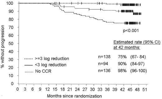
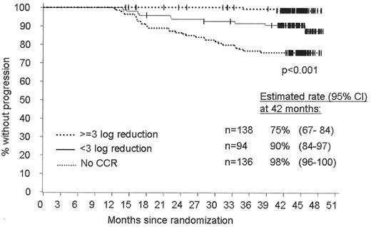
In patients with chronic phase CML who had failed IFN therapy the rate of CCR was 41% at 18 months and 52% at 40 months. Progression-free survival (PFS) at 48 months was 76%.1 In the IRIS study, 400 mg imatinib daily was compared with the combination of IFN and cytarabine (Ara-C) in recently diagnosed patients. The results of this study at 24 and 42 months are summarized in Table 2A .2,3 At 42 months 75% of patients in the imatinib arm continued on first-line therapy, while 25% had discontinued treatment because of disease progression (9%), toxicity (6%), or for other reasons (10%). In contrast, only 4% of patients randomized to IFN/Ara-C continued on their initial therapy. Estimated PFS in the imatinib group was 84%. Progression to accelerated phase or blast crisis had occurred in 6.1%, while 6.9% of patients had lost CHR or MCR. Notably, while the overall rate of progression had peaked in the second year of therapy (7.6%), the incidence of progression to advanced phase was practically constant over the years, at an average of 2% (Table 2B ). It remains to be seen whether this trend will continue. The risk of progression was inversely correlated with the depth of response: PFS was 98% in patients who were in MMR at 12 months, compared to 90% for patients with CCR only and 75% for patients without CCR (Figure 1 ). Late side effects (occurring after 18 months) were infrequent and included grade 3/4 neutropenia in 3.8%, thrombocytopenia in 2.1%, anemia in 1.0%, and other drug-related grade 3/4 adverse events in 5.8%.
Prognostic factors
Pretherapeutic disease features
Sokal score.
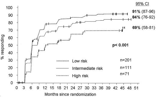
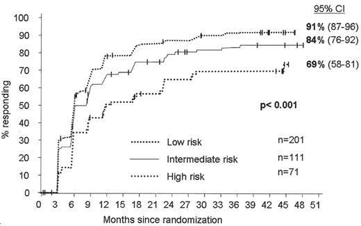
Analysis of the IRIS study showed that the Sokal score (which is based on age, spleen size, platelet and peripheral blood blast count) is well correlated with the likelihood of achieving CCR: 91% for low, 84% for intermediate and 69% for high-risk patients (Figure 2 ). A more detailed analysis of the IRIS data will be required to assess the association between individual risk factors and outcome.
Cytogenetics and fluorescence in situ hybridization (FISH).
Clonal cytogenetic evolution (CE) is defined as the acquisition of cytogenetic abnormalities in addition to Philadelphia chromosome (Ph). Only abnormalities that are demonstrable in at least two metaphases should be referred to as ‘clonal.’ It is not clear whether CE at diagnosis is an adverse prognostic feature in the absence of other features of advanced disease. In patients with myeloid blast crisis, CE is associated with shorter overall survival and in patients with accelerated phase, CE is associated with a trend toward shorter PFS, while no correlation between CE and MCR was found in patients in late chronic phase, previously treated with IFN. A small study in previously treated patients suggested that in the absence of other features of accelerated phase the adverse effect of CE can be overcome by treatment with 600 mg of imatinib4 but it is currently unknown whether this applies to newly diagnosed patients. Given the lack of data, it appears justified to start newly diagnosed patients with CE on 600 mg imatinib daily rather than on the standard dose of 400 mg. Some 10%–15% of CML patients have deletions flanking the breakpoints in ABL and less frequently BCR. When treated with IFN-based regimens these patients have a significantly poorer overall survival. The impact of chromosome 9 deletions on the outcome of patients treated with imatinib is a matter of controversy. While Huntly et al found universally lower response rates and a shorter PFS in patients with deletions,5 this was not confirmed in a subsequent study.6 This discrepancy may reflect the fact that in the latter study significantly more patients with deletions than without deletions were treated with high doses of imatinib. Ultimately, the question needs to be answered in a prospective study.
BCR-ABL mRNA levels.
Several studies in chronic phase patients failed to detect a significant association between pretherapeutic levels of BCR-ABL mRNA and cytogenetic response to imatinib.7,8 Given that BCR-ABL mRNA levels increase with disease progression, this is somewhat unexpected. One possible explanation is that unfractionated white blood or bone marrow cells, the cell population used for PCR analysis, are not representative of the cell population that determines response. It is also possible that protein levels could increase without changes in message levels. Alternatively, other disease features may override BCR-ABL levels as a determinant of response.
Gene expression profiling.
Microarray profiling of pretherapeutic whole blood samples from a subset of the IRIS patients was used to identify genes whose expression correlated with subsequent MCR.9 Although the gene list generated with this approach accurately predicted response in the cohort under study, no independent control group was included for validation, and similar studies either produced a completely different list of genes10 or found comparable expression profiles in responders and non-responders.11 Thus there is currently no established role for expression profiling to predict response to imatinib.
Ex vivo assays.
Phosphorylation of CrkL, a specific substrate of Bcr-Abl, was measured in mononuclear cells from newly diagnosed patients cultured ex vivo in graded concentrations of imatinib. The IC50, i.e., the concentration of drug at which phosphorylation was reduced to 50% of baseline, correlated with the reduction of BCR-ABL mRNA at 3 months and the likelihood of achieving MMR at 12 months. This correlation was, however, limited to patients in the low Sokal risk group.12 Similar results were seen in a pilot study that used flow cytometry to determine total phosphotyrosine levels in CD34+ cells incubated with imatinib ex vivo.13 Another study determined Bcr-Abl tyrosine phosphorylation, WT1 mRNA expression and myeloid colony formation in mononuclear cells treated with imatinib in vitro. All three parameters were positively correlated with each other. Patients with greater reduction of WT1 mRNA or colony formation had a higher chance of achieving a cytogenetic response.14 Whether any of these tests will eventually be clinically useful outside the research setting remains to be seen.
On-treatment features
Hematologic toxicity.
Grade 3/4 neutropenia or thrombocytopenia on therapy is associated with lower rates of MCR and CCR and shorter PFS in chronic phase patients after failure of IFN.15 It is likely that significant myelosuppression is a reflection of more advanced disease, with a smaller pool of residual normal progenitor cells capable of restoring Ph-negative hematopoiesis. In addition, dose reductions or treatment interruptions triggered by significant hematological toxicity may also contribute to the poorer outcome. Myeloid growth factors are capable of reversing imatinib-induced neutropenia and appear to improve cytogenetic responses.16 In contrast, anemia, a frequent finding in patients on imatinib, is not an adverse prognostic factor and is usually responsive to erythropoietin.17
Clonal cytogenetic evolution.
Another time-dependent variable associated with a poor prognosis is CE on therapy. The precise pathogenetic role of most abnormalities for disease progression is unknown, but it is conceivable that CE is a surrogate marker for genetic instability that predisposes to the accumulation of mutations. Consistent with this, there is a significant association between CE and the detection of kinase domain mutations in imatinibnaïve patients.18 It is likely that certain abnormalities will be more significant than others but no formal analysis is currently available.
Monitoring and Milestones
The fact that most patients with early CML respond well to standard dose imatinib has placed considerable responsibility on clinicians to identify those patients who have an inadequate response or show signs of relapse. There is no doubt that patients with signs of progression beyond chronic phase will benefit from early intervention. For example, the results of allogeneic stem cell transplantation (SCT) in frank blast crisis are extremely poor; thus, the decision to proceed to SCT should be made as early as possible. It is plausible that earlier intervention, for example with alternative Abl kinase inhibitors, will also benefit patients who are still in chronic phase, but this remains to be formally proven. Recommendations for monitoring are given in Table 3.
Hematologic monitoring.
Close to 90% of newly diagnosed patients with chronic phase enter CHR within 3 months of standard dose imatinib. This is the basis for the widely held view that CHR should be achieved within this time frame, although this has not been formally analyzed. One should bear in mind that lack of CHR encompasses a broad spectrum of incomplete responses, ranging from no response at all to perhaps a minimally elevated white cell count as the only feature that defines the lack of CHR. While in the first scenario therapeutic alternatives need to be explored urgently, watchful waiting may be justified in the second. The recommendation is to monitor patients with weekly blood counts until a stable CHR has been attained. This will identify both non-responders as well as those patients who develop significant cytopenias and may require dose adjustments or the addition of myeloid growth factor and/or erythropoietin.
Cytogenetics.
In patients with chronic or accelerated phase CML achievement of MCR or CCR at any time during imatinib therapy is associated with superior PFS. As a global measure of in vivo drug sensitivity the cytogenetic response appears to override pretherapeutic prognostic parameters, at least those that are currently available. At 3 months, MCR was associated with superior PFS in patients with late chronic phase and accelerated phase disease, making MCR at 3 months a desirable outcome. It is less obvious what constitutes an inadequate cytogenetic response at 3 months. A recent analysis of the IRIS study showed that 55% (95% CI, 37%–73%) of patients without any cytogenetic response at 3 months had achieved CCR at 42 months, while 66% (95% CI, 44%–88%) of patients with a “minimal” cytogenetic response (66%–95% Ph+ meta-phases) and 68% (95% CI, 51%–84%) with a minor response (36%–65% Ph+) had achieved CCR at 42 months (B Druker, personal communication).19,20 In contrast, 95% (95% CI, 91%–99%) with partial cytogenetic response (1%–35% Ph+) at 3 months had attained CCR at 42 months. These data suggest that while a partial response at 3 months is optimal, the majority of patients who do not achieve this landmark will still achieve a CCR. Thus, according to presently available information, failure to achieve any level of Ph negativity at 3 months does not require a change in the therapeutic strategy. However, this recommendation may change when more data become available. Of patients who remained ≥ 95% Ph+ at 6 months, only 22% (95% CI, 0%–44%) went on to achieve CCR at 42 months, compared to 45% (95% CI, 20%–70%) of patients with a minimal and 56% (95% CI, 32%–80%) of patients with a minor cytogenetic response. Based on these data, a minimal cytogenetic response (less than 95% Ph+) at 6 months can be defined as a milestone that patients on standard therapy should pass. Lastly, only 40% (95% CI, 0%–83%) of patients who are 36%–65% Ph+, but 80% (95% CI, 65%–94%) of patients with a partial cytogenetic response at 12 months achieved CCR at 42 months. Thus, MCR at 12 months is considered the third milestone. There are currently no data to rationally define the latest time point at which a patient should be in CCR, but a plausible limit could be 18 months. Due to the efficacy of imatinib, most patients pass these milestones. Thus, the recommendations are based on relatively small numbers of patients who fail to achieve the minimum responses and hence confidence intervals are large. It must be stressed that the recommendations emphasize conservative approaches. A more aggressive approach may be warranted in the appropriate clinical situation, for example in younger patients with a transplant option or in patients with a high Sokal risk.
Fluorescence in situ hybridization.
FISH for BCR-ABL instead of conventional karyotyping is used with increasing frequency to follow patients on therapy. There are several issues that need to be taken into account. First, it is important to distinguish between hypermetaphase FISH (HMF), which essentially increases the number of metaphase cells screened, and FISH on interphases, which analyzes cells regardless of proliferative status. While several studies found a good correlation between the two methods, this has not been universally confirmed.20 Generally, negative results with interphase FISH tend to correlate well with conventional karyotyping, but considerable discrepancies may be encountered in patients with some degree of Ph-positivity. This is due to the unpredictable contribution of B-lymphocytes whose Ph status varies greatly between patients.20 Secondly, unlike conventional karyotyping and quantitative PCR (qPCR), FISH has not yet been validated in a prospective fashion with clinical endpoints, and thus the results should be interpreted with caution. Thirdly, the gain of sensitivity over conventional karyotyping is only one order of magnitude (if 200 cells are analyzed and scored compared to 20 metaphases by conventional karyotyping). In a recent study, FISH detected residual leukemia in more than 50% of patients with CCR by conventional karyotyping, but all the FISH− patients were positive by qPCR.21 Since qPCR covers the entire range of residual disease, the value of performing FISH in addition to qPCR in patients with CCR is therefore questionable. In contrast to FISH, conventional cytogenetics provides information on chromosomal abnormalities other than Ph. This is relevant, as CE on therapy is an adverse prognostic indicator.22 In addition, clonal chromosomal abnormalities have also been detected in Ph− cells.23 The prognostic significance of this finding is currently unknown, but progression to acute myeloid leukemia has been seen in patients with dysplastic morphology.
Quantitative PCR.
qPCR has emerged as the method of choice for monitoring residual disease in patients with CCR. Given that the majority of patients in first chronic phase achieve CCR, qPCR is the only method that allows disease monitoring in these patients and should be regarded as state of the art for following patients with imatinib-induced CCR. In addition, analysis of a subset of patients from the IRIS trial revealed a significant correlation between the molecular response at 3 months and CCR at 12 months.8 At 12 months patients with CCR can be classified according to their molecular response into those with a reduction of BCR-ABL transcripts by at least 3-log (MMR) and less than 3-log.24 With 42 months of follow-up, PFS for patients with CCR and ≥ 3-log reduction was 98%, compared to 90% for patients with CCR but < 3-log reduction of transcripts, and 75% for patients without CCR.3 Importantly, the log reductions in this study were calculated with a pretherapeutic standard derived from a group of untreated patients. This implies that qPCR monitoring can be initiated at any time during imatinib therapy, without the need for an individual pretherapeutic specimen. As there is a very good correlation between BCR-ABL mRNA levels in the blood and bone marrow,7 qPCR analysis can be performed on peripheral blood, which is much more convenient for the patient. Current recommendations are to perform qPCR on peripheral blood every 3 months. Standardization of qPCR methodology is urgently required to allow for adequate monitoring of patients outside of clinical trials and academic centers.
Screening for kinase mutations.
Patients should be screened for mutations of the BCR-ABL kinase domain whenever there is an indication of loss of response at whichever level. The frequency of kinase domain mutations in patients with acquired resistance ranges between 50% and 90% in various studies, probably reflecting differences in assay sensitivity. A recent study detected kinase domain mutations in 61% of patients with a more than 2-fold rise in BCR-ABL mRNA but in only 0.6% of patients with stable or decreasing levels.25 These data still await confirmation as precise measurement of increases of BCR-ABL mRNA as subtle as 2-fold will be difficult in a non-research setting. Nonetheless, these results support the notion that screening patients without evidence of disease progression may not be effective. The same holds true for high sensitivity screening of patients prior to therapy, since mutant clones present at low levels do not guarantee that resistance to imatinib will develop.18 At present, mutation analysis is largely limited to academic institutions, but it is likely that a commercial test will become available soon. The main purpose of mutation screening in the non-academic setting may be to exclude mutations such as T315I that would not respond to alternative Abl kinase inhibitors.
Options for Patients with a Suboptimal Response to Standard-Dose Imatinib
Any patient with a suboptimal response or signs of resistance should be considered for a clinical trial, primarily with an alternative Abl kinase inhibitor. Outside the clinical trial setting the most straightforward approach is dose escalation of imatinib, which will be effective in 30%–50% of patients.26 However, these responses may not be as durable as those achieved with standard dose imatinib up-front. Another possibility is adding a conventional agent like cytarabine, although no data from controlled trials are available for this approach. Lastly, the option of SCT may be revisited in eligible patients. It is obvious that clinical judgment is critical for making these decisions. For example, while SCT should certainly be considered in a patient with low transplant risk and failure to achieve CHR, this would clearly be inappropriate in an elderly individual who fails to achieve MCR at 12 months.
Early Intensification of Therapy
Disease progression of CML and resistance to imatinib are caused by cell clones that acquire additional genetic abnormalities or specific mutations that interfere with drug binding. Thus, it is conceivable that a rapid reduction of the leukemic burden would reduce the risk of such events, providing a rationale for early intensification of therapy. Imatinib is synergistic with a number of conventional and experimental antileukemic agents, at least in vitro. Approaches to early intensification of therapy in newly diagnosed patients include high-dose imatinib and combinations with Ara-C or IFN, two drugs with significant single-agent activity in CML. Currently available data are limited to phase I/II studies and summarized in Table 4 . The results of these studies are somewhat difficult to compare due to differences in follow-up. In addition, the RT-PCR technology was not standardized. Nonetheless, the emerging picture is that the rates of MCR and CCR in the combination studies are comparable to the IRIS trial but higher in patients treated with 800 mg imatinib daily, while the rates of MMR and CMR are generally higher compared to standard dose imatinib. This increased efficacy comes at the cost of more toxicity. For example, the incidence of grade 3/4 neutropenia was 63% in patients treated with imatinib and pegylated IFN and 41% experienced grade 3/4 nonhematologic toxicity. As a result, only a fraction of the planned IFN dose was actually administered.
Caution needs to be taken when interpreting these early single-armed studies. Taken together, they clearly suggest that early intensification of therapy may increase the frequency of profound remissions, although at the price of more toxicity. The results are particularly promising for high-dose imatinib. However, it is possible that the rate of MMR and CMR in the IRIS study will catch up with longer follow-up, and it remains to be proven that achieving these responses early in the course of treatment is superior to achieving them later. Although it is likely that an MMR achieved with a high dose of imatinib or a drug combination confers the same excellent PFS as MMR achieved with standard-dose imatinib, even this point remains to be proven. Standard-dose (400 mg daily) and high-dose imatinib (800 mg daily) are currently compared in a phase III intergroup study in the US and are part of several multi-armed studies in Europe. It is also possible that alternative Abl kinase inhibitors with increased potency, such as BMS-354825 (dasatinib)27 and AMN107,28 may be superior to high-dose imatinib.
Outcome of Patients in CCR or MMR Who Discontinue Imatinib
Only limited data are available regarding discontinuation of imatinib in patients with CCR or CMR. Five of 6 patients published as case reports (5 with a least one PCR− sample prior to stopping imatinib) had reappearance of Ph+ metaphases after stopping imatinib. Three patients were retreated, and all responded. In a larger study on 23 patients in CCR, 12 of them after SCT, BCR-ABL transcripts increased in all but 3 patients (all of whom had been allografted), and cytogenetic relapse occurred in 53% after discontinuation of imatinib. Re-treatment induced responses in all 5 patients with available data.29 Together these data suggest that discontinuation of imatinib even in a state of CMR is usually followed by (drug responsive) relapse, consistent with persistence of leukemic stem cells with proliferative potential. The high rate of immediate relapse is in contrast to patients with CCR induced by IFN, a considerable proportion of whom maintain their response. Cytotoxic T-cell responses directed against peptides derived from myeloblastin (proteinase 3) have been demonstrated in patients with CCR to IFN but not to imatinib, which could be critical for maintaining remission after discontinuation of drug.30
Disease Persistence
The frequency of CMR varies widely between different studies (Table 5 ). For example, less than 4% of patients achieved CMR in the IRIS study, while a rate of 28% was reported in a phase II trial of newly diagnosed patients treated with 800 mg imatinib daily. This large variation may reflect the risk profile of the cohorts under study and treatment intensity but also differences in the sensitivity of the PCR methodologies used to diagnose CMR. There is an emerging consensus that CMR should be defined as negativity by nested RT-PCR for BCR-ABL. This takes into account that PCR is somewhat less sensitive than nested PCR in most laboratories and that in patients treated with SCT, negativity by nested PCR > 6 months after the transplant is a strong predictor of freedom from progression. With these stringent criteria applied it has become clear that only a small fraction of patients achieve CMR, and that responses are much less durable than in patients after SCT.31 Ex vivo studies suggest that non-cycling BCR-ABL+ hematopoietic progenitor cells with an immature phenotype (CD34+/CD38−) are resistant to imatinib.32 Since repopulation assays in immune-deficient mice have shown that these cells have self-renewal capacity, it is conceivable that they represent the residual leukemic population in patients with CCR. At present it is unknown whether imatinib inhibits Bcr-Abl kinase activity in these immature progenitor cells. This information will be critical for classification of persistence as Bcr-Abl-dependent or independent, which would have obvious implications for the development of strategies to eradicate residual leukemia. Several mechanisms have been proposed that could account for Bcr-Abl-dependent residual leukemia (reviewed in 33). A small study found kinase domain mutations in CD34+ cells of 5 out of 13 patients in CCR. Interestingly, mutations conferring only a modest degree of resistance were dominant in this study, in contrast to more highly resistant mutations observed in patients with full-blown relapse. This implies that in the presence of drug the mutant clones may be able to maintain viability but fail to expand. It is not clear, though, whether the cohort under study is representative of typical CCR patients, given that most patients were BCR-ABL+ by interphase FISH, and there was a high rate of relapse within a short period of follow-up. Another study suggested that lineage-negative progenitor cells express much higher levels of BCR-ABL mRNA than their more differentiated progeny. This would explain why imatinib fails to significantly impact the leukemic stem cell compartment, while it effectively targets the more differentiated cells. A third possibility is that the intracellular drug concentrations achieved in the leukemic stem cells are much lower than plasma levels suggest. Imatinib is a substrate for several drug transporters, including PGP and ABCG2 that are expressed by hematopoietic progenitor cells and may lower intracellular cellular drug levels below effective doses. The common feature of all three of these mechanisms is that they might be overcome with alternative Abl inhibitors that exhibit activity against Bcr-Abl kinase domain mutants, increased potency against wild type Abl or have a more favorable affinity profile to drug pumps present in the target cells. There is, however, the possibility that residual leukemic cells are Bcr-Abl–independent. The fact that BCR-ABL+ progenitor cells are not growth factor independent suggests that they are still responsive to physiological stimuli, although their threshold of response has been reset. Conversely, these cells may be able to use physiological signals if Bcr-Abl is inhibited. Consistent with this, growth factor-dependent activation of mitogen-activated protein kinase has been demonstrated in CD34+ progenitor cells from some CML patients treated with imatinib ex vivo. One could argue that elimination of such cells may require targeting stem cells rather than targeting Bcr-Abl.
What Is the Current Role of Allogeneic Stem Cell Transplantation?
Newly diagnosed patients.
There is current debate as to whether any sub-group of patients with newly diagnosed chronic phase CML should be treated by SCT as primary therapy. It has been suggested for example that patients classified as ‘poor-risk’ by Sokal and good risk for allografting should be transplanted without preceding treatment with imatinib. The same has been suggested for children regardless of their Sokal score. To complicate matters further the relative merits of a conventional SCT and an SCT using reduced-intensity conditioning are not yet clearly defined. There is no current consensus on these various issues. Moreover in some countries SCT is relatively cheap and may therefore be recommended in preference to imatinib for patients who would otherwise have to personally pay for life-long drug treatment.
In practice, almost all patients in the developed world do now receive imatinib as first-line therapy, which raises the question whether prior use of imatinib for a finite period may increase the risk of mortality associated with a subsequent transplant. Several retrospective studies addressed this issue, with partially conflicting results. Although this should ideally be studied in a prospective fashion, withholding imatinib from the control group would pose ethical problems.
Can one identify a subgroup of newly diagnosed patients in chronic phase who should be transplanted as first-line therapy, regardless of their response to imatinib? Given the strong predictive power of time-dependent variables and their dominance over pretreatment features, there is probably no such subgroup. Since SCT in accelerated phase or blast crisis carries a much poorer prognosis than in chronic phase, early detection of relapse is critical. However, even meticulous monitoring will not always detect relapse early, as some patients have progressed directly to accelerated phase or blast crisis, even from CCR. Thus patients with a low-risk transplant option, for example young patients with an HLA-matched sibling donor, should be informed that according to currently available data, imatinib does not eradicate leukemia, that life-long therapy is required and that there is small risk of progression to advanced phase even in those with an excellent response and sometimes without advance warning.
Imatinib refractory or resistant disease.
There is a widely accepted consensus that patients who progress beyond chronic phase on imatinib should be offered SCT if this is an option. Although alternative Abl kinase inhibitors show promising activity in such patients,27,28 the transplant recommendation for this group of patients is likely to remain valid for the foreseeable future. In contrast, if an effective salvage therapy were available, this would further raise the threshold for chronic phase patients with refractoriness or acquired resistance, particularly for those who have a suboptimal response at the cytogenetic or molecular level. It is presently impossible to give firm evidence-based recommendations and treatment decisions must be individualized, taking into account the patient’s risk of transplant-related mortality and personal preferences.
Conclusion
Imatinib at a dose of 400 mg daily is the current standard therapy for CML in chronic phase. Higher doses are promising, but results await confirmation in prospective studies. Discontinuation of treatment is almost invariably followed by recurrence, consistent with persistence of residual disease. Monitoring of patients on therapy should be done according to standardized guidelines, with qPCR for BCR-ABL emerging as the most important test. Time-dependent variables are the most powerful predictors of PFS, overriding baseline risk factors. Failure to achieve defined therapeutic milestones must trigger a reassessment of the overall therapeutic strategy.
Progression-free survival of newly diagnosed chronic myeloid leukemia patients treated with 400 mg imatinib daily according to molecular responses at 12 months.
Abbreviations: CCR, complete cytogenetic response
Progression-free survival of newly diagnosed chronic myeloid leukemia patients treated with 400 mg imatinib daily according to molecular responses at 12 months.
Abbreviations: CCR, complete cytogenetic response
Progression-free survival of newly diagnosed chronic myeloid leukemia patients treated with 400 mg imatinib daily according to Sokal risk at diagnosis.
Progression-free survival of newly diagnosed chronic myeloid leukemia patients treated with 400 mg imatinib daily according to Sokal risk at diagnosis.
The author is grateful to Brian J Druker, Howard Hughes Medical Institute and John M Goldman, NHLBI, for helpful comments and to Chris Koontz, Oregon Health & Science University, for editorial assistance.
Acknowledgments: Michael W.N. Deininger is a recipient of an American Society of Hematology Clinical/Translational Research Scholar Award.


