Abstract
Reactivation of fetal hemoglobin (HbF) expression is an important therapeutic option in patients with hemoglobin disorders. In sickle cell disease (SCD), an increase in HbF inhibits the polymerization of sickle hemoglobin and the resulting pathophysiology. Hydroxyurea, an inducer of HbF, has already been approved for the treatment of patients with moderate and/or severe SCD. Recent clinical trials with other pharmacological inducers of HbF, such as butyrate and decitabine, have shown considerable promise. In this chapter, we highlight the important clinical trials with pharmacological inducers of HbF, discuss their mechanisms of action and speculate about the future of this therapeutic approach in the treatment of patients with SCD.
β-Hemoglobinopathies are common genetic disorders that cause considerable morbidity and mortality throughout the world. Sickle cell disease (SCD) results from an amino acid substitution of valine for glutamic acid at position 6 of the adult β-globin chain, which results in the polymerization of hemoglobin upon deoxygenation, leading to deformed dense red blood cells. The predominant pathophysiological feature of SCD is vaso-occlusion, which leads to acute and chronic complications such as painful crises, acute chest syndrome and strokes. Patients with SCD have a markedly decreased life expectancy and their quality of life is greatly compromised by their disease.
The levels of fetal hemoglobin (HbF) in erythrocytes account for a large part of the clinical heterogeneity observed in patients with SCD and β-thalassemia.1 Patients with SCD in Saudi Arabia and India who inherit a genetic determinant for high fetal hemoglobin typically have a very mild clinical disorder. Moreover, the Cooperative Study of Sickle Cell Disease identified HbF as a major prognostic factor for several clinical complications including painful events,2 acute chest syndrome3 and death.4 These clinical and epidemiological observations provided important clues about the beneficial role of HbF in ameliorating the clinical complications of SCD and β-thalassemia. As a result, pharmacological induction of HbF production was proposed as a therapeutic strategy to decrease the severity of these disorders.
Pharmacological Agents that Induce Fetal Hemoglobin
The human β-like globin genes are expressed in vivo in a tissue- and developmental stage-specific manner. The mechanisms of regulation of globin gene expression have been the subject of intense investigation for many years. These studies led to the identification of a repertoire of cis-elements and trans-acting factors that regulate the expression of the genes of the β-globin cluster. Such studies also led to the appreciation of the crucial role of epigenetic modifications in the regulation of globin gene expression. DNA methylation and histone acetylation are two of the most important epigenetic modifications that are involved in the regulation of most eukaryotic genes, including the genes of globin family. Thus, targeting epigenetic silencing of the fetal globin genes became the focus of a novel therapeutic approach for patients with SCD and β-thalassemia.
During the last two decades, considerable efforts have been focused on the pharmacological induction of HbF in patients with hemoglobin disorders. Multiple drugs including 5-azacytidine (and decitabine), hydroxyurea, butyrate and erythropoietin were shown to induce HbF in vivo in animal models and in patients with these disorders. In spite of the incomplete understanding of the mechanisms of induction of HbF by these agents, considerable progress has been made in developing these agents as drugs that induce HbF in patients with SCD.
5-Azacytidine
5-Azacytidine was the first prototype of an agent that induces HbF by targeting epigenetic silencing. 5-azacytidine was first shown to induce very high levels of HbF in anemic baboons. Its ability to stimulate HbF production was also demonstrated in a small number of patients with SCD and β-thalassemia.5,6 Treatment with 5-azacytidine resulted in a significant increase in the level of HbF that was associated with a decrease in the fraction of dense cells in patients with SCD. Treatment with this drug also led to a partial correction of the non-α:α chain imbalance and a decrease in transfusion requirements in few patients with β-thalassemia. These clinical observations provided strong support for the hypothesis that expression of the γ-globin gene expression could be re-activated in adult life by DNA hypomethylation, resulting in high levels of HbF. In spite of these promising preliminary results, this agent was never tested in large-scale clinical trials due to concerns about its potential carcinogenic effects, since a previous study conducted in laboratory rats showed that 5-azacytidine increased the incidence of tumors.7 Interestingly, controversy over the mechanism of induction of HbF by 5-azacytidine led to the studies which showed that hydroxyurea, another cytostatic agent, can also result in a marked induction of HbF in Baboons.8
Hydroxyurea
Hydroxyurea is a potent inhibitor of ribonucleotide reductase that had been in clinical use for many years in the treatment of patients with myeloproliferative disorders. It is an orally available drug that is relatively well tolerated and easy to use. After the demonstration of its ability to induce HbF synthesis in baboons, hydroxyurea was tested in a number of small clinical trials in adults and children with SCD.9–18 A larger multicenter study of hydroxyurea showed a marked decrease in the frequency of painful crises and episodes of acute chest syndrome and a reduction in transfusion requirements and hospitalizations in adults with moderate to severe SCD.10,16 As a result, hydroxyurea was the first drug to be approved by the FDA for the treatment of patients with moderate and/or severe SCD. After 9 years of follow-up, patients treated with hydroxyurea were shown to have improved survival.15 Other studies demonstrated the clinical efficacy and short-term safety of hydroxyurea in children with SCD.11,12,17,18 The major short-term toxicity of hydroxyurea was reversible bone marrow suppression manifested as a decrease in white blood cell counts and/or platelet counts. Interestingly, higher baseline HbF predicted a better response to hydroxyurea in children18 but not in adults with SCD.16 Overall, the response to hydroxyurea was variable and about one third of patients with SCD did not respond at all to this treatment.
Decitabine
In 2006, the FDA approved the use of decitabine, an analogue of 5-azacytidine, for the treatment of patients with myelodysplastic syndromes. As a result of the recent introduction of decitabine as a new DNA hypomethylating agent and follow-up animal studies which showed that hypomethylating agents could reduce the incidence of tumors, there has been a renewed interest in the use of DNA hypomethylation therapy for inducing HbF in patients with SCD. Three clinical trials have been reported in which decitabine was administrated to patients with SCD by either intravenous or subcutaneous injections.19–21 This resulted in significant increases in mean γ-globin synthesis, HbF levels and the fraction of F cells in the treated patients. Moreover, decitabine treatment resulted in an increase in total hemoglobin levels of 2 g/dL while reticulocyte counts decreased, suggesting decreased hemolysis. The increase in the level of HbF was associated with significant improvement in several parameters that are important in the pathophysiology of vaso-occlusion such as red blood cell adhesion, endothelial damage and activation of the coagulation pathway. The increase in HbF levels was shown to be associated with decreased DNA methylation at the promoters of the γ-globin genes. Interestingly, 100% of patients with SCD responded with an increase in HbF levels, including patients who had previously failed to respond to hydroxyurea.
The major dose-limiting toxicity that was observed in studies of decitabine in patients with SCD was reversible neutropenia. Interestingly, decitabine treatment resulted in an increase rather than a decrease in platelet count. In spite of the encouraging findings of these short-term studies, the long-term risks of treatment with decitabine are not known at this stage. A relatively short-term study of decitabine in patients with leukemia did not show an increase in incidence of secondary tumors after 2 to 5 years of therapy.22 An early study reported that this agent was not carcinogenic in the rat model.7 More interestingly, recent studies have shown that treatment of mice with a genetic disposition for colon or lung cancer with decitabine results in a marked reduction in tumor formation.23,24 It is believed that this reduction in tumorogenesis may reflect demethylation of tumor suppressor genes. Thus, these studies suggested that decitabine may provide potential chemoprevention for certain cancers. Larger and longer-term studies are clearly needed to confirm the safety and efficacy of decitabine in patients with SCD.
Butyrate
Butyrates, well-known inhibitors of histone deacetylases (HDAC), are being tested in clinical trials as inducers of HbF in patients with SCD and β-thalassemia.25 When administered by continuous infusion, arginine butyrate resulted in a significant increase in mean γ-globin synthesis, HbF levels and F cells in patients with SCD. However this increase in HbF levels was not sustained with continuous therapy.26,27 The decrease in HbF levels after prolonged exposure to arginine butyrate, considered in the context of the well-known growth-inhibitory activity of butyrate, suggested that an intermittent dosing schedule may prevent toxicity and allow proliferation of the erythroid cells in which HbF is induced. Thus, when arginine butyrate was given intermittently for 4 days every 4 weeks, it led to sustained induction of HbF production in a majority of patients with SCD. Interestingly, baseline HbF levels above 2% predicted response to butyrate therapy in both the continuous and intermittent butyrate studies, while lower levels were associated with resistance to the HbF-inducing activity of butyrate. Interestingly, in one patient, resistance to butyrate was reversed by prior treatment with hydroxyurea. Although arginine butyrate has shown considerable promise in the treatment of patients with SCD, the difficulty in administering large volumes of the drug through central venous veins poses a major challenge. Sodium phenylbutyrate is an orally administered agent that was previously shown to induce a rapid increase in the number of F-reticulocytes in patients with SCD.28 However, poor compliance with a regimen that required the intake of 30–40 tablets/day (15–20 g/day) was a major factor that limited the effectiveness of outpatient therapy. The same group investigated the activity of this agent at lower doses (1 to 11 g/day) in children with SCD and found that treatment with sodium phenylbutyrate increased F-reticulocytes in all patients within the first weeks of therapy.29 However, this increase was dose-dependent and was not sustained with continuous therapy. Further trials of sodium phenyl-butyrate with more patients are warranted to determine the efficacy and the optimal dosing regimen.
Mechanisms of Pharmacological Induction of HbF
5-Azacytidine and decitabine inhibit DNA methylation at cytosine residues by DNA methyltransferases (DNMT). These two drugs serve as cytidine analogs that are incorporated into DNA, where they form covalent bonds with DNMT, leading to depletion of functional enzyme.30,31 More recent studies have also shown that decitabine induces a rapid and selective degradation of DNMT1 through the proteasomal protein-degradation pathway.32 Recent studies showed that induction of HbF by decitabine is associated with a decrease in DNA methylation at the promoters of the γ-globin genes in erythroid bone marrow cells in baboons and patients with SCD.21,33 We have recently investigated the effects of decitabine in progenitor-derived cells from a patient with sickle/β-thalassemia. The decitabine-mediated increase in γ-globin mRNA level was associated with a marked decrease in DNA methylation at the promoters of the γ-globin genes (unpublished data). Interestingly, Lavelle and colleagues reported that treatment of baboons with decitabine is also associated with increased histone acetylation at the promoters of γ-globin genes.33 These observations suggest that decitabine-induced DNA hypomethylation may result in secondary changes in histone acetylation, giving rise to an open chromatin structure that allows the binding of transcription factors, leading to de-repression and transcriptional activation of γ-globin gene expression in adult life.
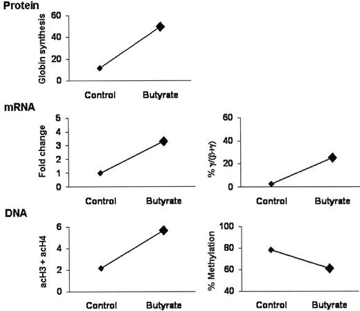
The leading hypothesis for the mechanism of induction of HbF by butyrate is that it increases the transcriptional activity of the γ-globin promoters by increasing the level of histone acetylation. We have investigated the effects of butyrate-mediated induction of γ-globin expression, histone acetylation and DNA methylation in progenitor-derived cells from a patient with SCD. As shown in Figure 1 , butyrate-mediated induction of γ-globin chains and mRNA is associated with an increase in the level of acetylation of histones H3 and H4 and a decrease in the level of DNA methylation at the promoters of the γ-globin genes. In a previous study from our laboratory, butyrate was also shown to increased HbF production in patients with SCD by increasing the efficiency of translation of γ-globin mRNA.34 Butyrate was also shown to activate signaling through the soluble guanylate cyclase35 and p38 MAP kinase36 pathways. It is not yet clear how these signaling activities lead to the transcriptional activation of the γ-globin genes. Earlier studies had suggested the involvement of the erythroid transcription factors GATA-1 and NF-E2 in the induction of erythroid differentiation and activation of globin expression by butyrate.37,38 Moreover, GATA-1 and NF-E2 were shown to be targets for histone acetyltransferases and their acetylation was shown to enhance their transcriptional activity. NF-E239 and GATA-240 were also shown to be associated with HDAC. Although the effects of these interactions on the level of acetylation of these transcription factors are still not known, it is tempting to speculate that the enzymatic activity of butyrate as an HDAC inhibitor may increase the acetylation of these transcription factors and result in the induction of γ-globin gene expression. More studies are necessary to fully elucidate the mechanisms of activation of fetal globin gene expression by butyrate.
The mechanism of induction of HbF by hydroxyurea is also not fully understood. It was originally proposed that hydroxyurea may elevate HbF levels by accelerating erythropoietic differentiation in the bone marrow, leading to the appearance of more “fetal-like” cells in the peripheral blood.41 More recent studies have suggested that hydroxyurea generates nitric oxide (NO) in vivo and that the resulting activation of the NO/cGMP signaling pathway might upregulate γ-globin expression in patients with SCD.42,43
Future Directions
The clinical studies summarized above support the hypothesis that the chromatin and DNA structures at the γ-globin gene promoters are very important for the regulation of HbF expression. These studies further demonstrate that the use of epigenetic modifiers in patients with SCD can reactivate γ-globin gene expression in adult life. Although these pharmacological agents have shown clinical efficacy in patients with SCD, the global impact of these new therapies on the natural history of these disorders, especially in developing countries where these disorders are much more common, has been modest. There is ample epidemiologic and clinical evidence that the higher the HbF levels, the better the amelioration of the clinical disorders.1 Thus, more effective and safer agents that can induce higher levels of HbF are clearly needed. Moreover, the complexity of the mechanisms of regulation of globin gene expression suggests that combination therapy consisting of two or more drugs with different mechanisms of action would induce higher levels of HbF than single agent therapy. As discussed above, induction of HbF by either DNMT inhibitors or HDAC inhibitors results in a change in both DNA structure (i.e., hypomethylated CpG sites) and chromatin configuration (i.e., hyperacetylated histones). These data emphasize the crucial role of coordinated epigenetic modifications in the regulation of γ-globin gene expression. Thus, the combination of a DNMT inhibitor, such as decitabine, with an HDAC inhibitor, such as butyrate, might result in more potent activation of γ-globin expression. Alternatively, the combination of DNMT and/or HDAC inhibitors with agents such as erythropoietin or hydroxyurea, which increase HbF by different mechanisms, may also be more effective than treatment with a single agent. Finally, the combination of agents that increase HbF levels with agents that target other aspects of the pathophysiology of hemoglobin disorders such as cell adhesion and/or cell dehydration may also provide more effective therapeutic strategies. After many decades of research to develop therapeutics for SCD, we may be at the dawn of a new era in which customized therapies with one or more agents might address the unique aspects of the pathophysiology in an individual patient.
Progenitor-derived cells from the peripheral blood of a patient with sickle cell disease were cultured in the absence (control) and presence of butyrate. Globin synthesis was assessed by polyacrylamide gel electrophoresis and fluorography and expressed as % γ/(γ+β). mRNA levels were measured by quantitative real-time PCR and are expressed as fold change relative to the control and as % γ/(γ+β). DNA methylation at 5 CpG sites in the γ-globin promoters was determined by pyrosequencing and expressed as the average of the methylation at all five sites. Acetylation of histones H3 and H4 was measured by chromatin immunoprecipitation combined with quantitative real-time PCR and expressed as sum of acetylated H3 and acetylated H4 (acH3+acH4). Larger symbols indicate that the changes from the control are statistically significant (i.e., p < 0.05, paired t-test).
Progenitor-derived cells from the peripheral blood of a patient with sickle cell disease were cultured in the absence (control) and presence of butyrate. Globin synthesis was assessed by polyacrylamide gel electrophoresis and fluorography and expressed as % γ/(γ+β). mRNA levels were measured by quantitative real-time PCR and are expressed as fold change relative to the control and as % γ/(γ+β). DNA methylation at 5 CpG sites in the γ-globin promoters was determined by pyrosequencing and expressed as the average of the methylation at all five sites. Acetylation of histones H3 and H4 was measured by chromatin immunoprecipitation combined with quantitative real-time PCR and expressed as sum of acetylated H3 and acetylated H4 (acH3+acH4). Larger symbols indicate that the changes from the control are statistically significant (i.e., p < 0.05, paired t-test).
Mount Sinai School of Medicine, Division of Hematology and Oncology, Box 1079, One Gustave L. Levy Place, New York, NY 10029.
Acknowledgments: We would like to thank Drs. Rona S. Weinberg and Yelena Galperin for their contribution to some of the unpublished observations described in the manuscript. This work was supported by the National Institutes of Health (RO1 HL-073438).

