Abstract
Patients with bone marrow failure syndromes are at risk for the development of clonal neoplasms, including paroxysmal nocturnal hemoglobinuria (PNH), myelodysplasia (MDS), and acute myelogenous leukemia (AML). Approximately 10% to 20% of those who survive acquired aplastic anemia will develop a clonal disease within the decade following their diagnosis. The relative risk of clonal neoplasms is very significantly increased in children and adults with inherited bone marrow failure syndromes as well. Until recently, the mechanisms underlying clonal evolution have been opaque, but a sufficient amount of evidence has now accumulated to support a model in which cells resistant to extracellular apoptotic cues are selected from the stem cell pool. Indeed, in the past two years this paradigm has been validated in preclinical models that are robust enough to reconsider new therapeutic objectives in aplastic states and to support the planning and development of rationally designed leukemia prevention trials.
Patients with bone marrow failure syndromes are at risk for the development of clonal neoplasms, including acute myeloid leukemia (AML), myelodysplastic syndromes (MDS), and paroxysmal nocturnal hemoglobinuria (PNH) (reviewed in Young1 and Bagby and Alter2). From 10% to 20% of survivors of acquired aplastic anemia will develop a clonal disease within the decade following their diagnosis,3–6 and the relative risk of clonal neoplasms is even more significantly increased in children and adults with inherited bone marrow failure syndromes as well.7 New insights on molecular mechanisms underlying clonal evolution, informed by advances in studies on evolutionary adaptation,8–10 now support the notion that at the stem cell level the process of clonal evolution is precisely adaptive.
Selection Coefficients and Determinants of Clonal Evolution
Principles of natural selection were developed in reference to studies on species but are perfectly applicable to asexual populations as well,9 so they can inform us about the processes of clonal evolution and carcinogenesis.11 Selective pressures from ecological niches that result in the emergence of adaptations in a species (coat color in rock pocket mice will be used as an illustration)12 are analogous to pressures that occur in stressful hematopoietic micro-ecosystems in which stem cells reside. In the latter case, the new genotype in adapted clones would result in an emergent phenotype of bone marrow cells more “fit” than their nonadapted progenitors. There is clear evidence that this is true not only for bacteria, in which adaptation to antibiotic challenge results in resistance,13,14 but for mammalian cells as well.15
To develop a clear picture of clonal evolution that occurs in the setting of bone marrow failure requires clarification of the relationships that exist between the target cells and the selective forces in the environment that determine fitness.16 Some mathematic models have even suggested that selection is a more important determinant in initiation of a tumor than is an increased baseline mutation rate,11 but it is intuitively more appealing to accept that variations in fitness in asexual populations (stem cells would be an example of such a population) increase not only as a function of the coefficient of selection but in proportion to the population size and mutation rate as well. The likelihood of clonal evolution depends upon the relative fitness differences between normal stem cells and mutant (potentially adapted) stem cells. This relative difference is expressed as the “selection coefficient.” A high selection coefficient exists when a somatic mutation accords to cells a uniquely strong advantage. Because any potential competitive advantage is relevant only in a head-to-head competition with the non-mutant with which the somatically mutated stem cell coexists in a given niche, the selectability of a specific mutation will be highest when the reference population is disadvantaged at the outset.
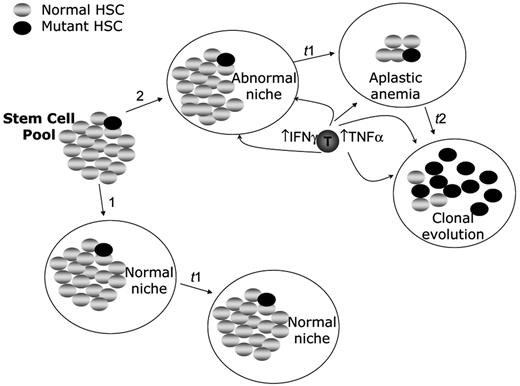
Hematopoietic stem cells assaulted by cytotoxic lymphoid populations17 represent perfect models of a disadvantaged population (Figure 1 ). This situation represents the generally accepted pathogenesis of acquired aplastic anemia. Unless the offending T-cell population is either eradicated or inactivated, the aplastic state would favor the evolution of new mutant stem cell clones in the hematopoietic tissues of patients with aplastic anemia who survive long enough. The three requirements for clonal evolution are that there is a sufficiently high coefficient of selection (ongoing selection against the non-adapted stem cells as would be the case with ongoing immune attack in acquired aplastic anemia), an initial stem cell population of sufficient size (as would be the case prior to the onset of aplasia), and an appreciable mutation rate (as would clearly be the case in many of the congenital aplastic states, particularly Fanconi anemia).
As shown in Figure 1 , selectable mutations might occur randomly in an otherwise normal stem cell pool, but because there is a low selection coefficient (the mutant cell exists in a large pool of stem cells that are not disadvantaged), clonal evolution would not occur (Figure 1 , pathway 1). This situation may explain how covert leukemic clones can be found during normal fetal development yet not raise their heads even later in life.18 That is, if there is no selective pressure on the stem cell pool, the potential advantage the covert leukemic clones might have is minimal. It is clear that in aplastic anemia, the stem cell pool is truly disadvantaged specifically because aberrant interactions take place involving them and their microenvironment.
Aberrant Interactions Between Stem Cells and Auxiliary Cells in Acquired and Inherited Aplastic Anemias
Acquired aplastic anemia
To discover pathways by which stem cells might adapt to a hostile environment in aplastic anemias first requires a clear understanding of the molecular mechanisms by which they are under stress. In the past two decades, experimental evidence from thoughtfully designed, truly translational studies places acquired aplastic anemia squarely into the category of autoimmune diseases. The evidence from a number of laboratories, recently summarized by Young et al, 1 has revealed that (1) aberrantly activated T-cell populations suppress hematopoiesis by releasing cytokines (including IFNγ and TNFα); (2) these cytokines and other factors induced by them cause apoptotic responses in hematopoietic stem cell and progenitor cells; (3) clinical responses to immunosuppressive therapy correlate directly with the capacity of the treatment to suppress T-cell function in individual patients; and (4) immune-mediated bone marrow failure can now be modeled in mice, and mono-clonal antibodies to TNFα and IFNγ prevent fatal aplasia in that model. In effect, acquired aplastic anemia can be viewed as a kind of microenvironmental cytokine storm in which overproduction of apoptotic cytokines by autoimmune T-cell populations results in high rates of apoptosis in hematopoietic stem cells and progenitors.
Inherited aplastic anemias
Aberrant interactions of IFNγ and TNFα with stem cells are also features of the most well studied of the inherited aplastic anemias, Fanconi anemia (FA). A number of years ago, based on bedside observations that viral infections sometimes profoundly suppress hematopoiesis in children with FA, we tested the notion that bone marrow failure in FA might also involve factors that evolve during the course of acute infections (e.g., IFNγ and TNFα). We also knew at the time that some of these factors might be involved in the pathogenesis of acquired aplastic anemia (reviewed by Young et al1). Indeed, studies on hematopoietic cells from children with FA and later in mice nullizygous for Fancc demonstrated not only that FA-C cells release more TNFαin the ground state,19 but that FA-C progenitor cells are inherently hypersensitive to apoptotic cues, including IFNγ, TNFα, mip1-α and TRAIL.20–22
Some of the mechanisms by which FA cells are hypersensitive to inflammatory cytokines are being clarified. For example, for TNFα hypersensitivity, at least two serine/threonine kinases are important. The first is the protein kinase PKR, a key molecular effector of the antiviral response. In normal cells, IFNγ and TNFα activate PKR. In normal cells, FANCA, FANCC, and FANCG proteins function to suppress PKR in the ground state.23 Functional loss of any one of those FA proteins results in high-level activation of PKR in the ground state and, as a consequence, high fractional apoptotic responses in the FA cell population not only at baseline but after stimulation with low doses of IFNγ and TNFα as well. In keeping with the importance of PKR activity in this disease, inhibition of PKR in FA cells reduces their apoptotic responses.23 The second, and likely related mechanism, involves the apoptosis signal-regulating kinase 1 (Ask1) known to be hyperactivated in Fancc−/− cells in response to TNFα. As was the case with PKR, inactivation of Ask1 normalizes the apoptotic response of murine FA-C cells exposed to TNFα.24 Interestingly, antioxidants also protected mutant cells from TNFα-induced apoptosis.
Not only are FA target cells hypersensitive to apoptosis-inducing cytokines, auxiliary cells that produce the cytokines have lower thresholds for induced production. Pang et al have noted that Fancc−/− mice treated with lps had higher mortality rates than wild-type mice, had higher serum levels of TNFα, IL-6, and MIP-2, and demonstrated hematopoietic suppression that could be directly attributed to TNFα (anti-TNF antibodies protected FA target cells).25 They also showed that transplantation of hematopoietic stem cells from wild-type mice protected Fancc−/− mice from endoxotin-induced mortality, clarifying the key role played by auxiliary cells of hematopoietic origin in the microenvironment.25
In summary, stem cells in patients with both acquired and certain inherited aplastic anemias are highly apoptotic, and the signaling pathways, both within and external to stem cells, which ultimately execute them, are highly related. In acquired aplastic anemia, the stress initially comes from rogue clones of T cells overproducing factors that result in stem cell death. In FA, the production of some of the same factors is increased because the normal FA proteins set thresholds for cytokine production responses to cytokine-inducing factors in auxiliary cells (e.g., in response to endotoxin25); in addition, the stem cell/progenitor pool is inherently hypersensitive to those factors because the normal FA proteins set their response thresholds for responses to some of the very apoptotic cytokines overexpressed in auxiliary cells.2 Although the experimental evidence supporting stem cell stress in other inherited marrow failure syndromes is not as robust, evidence is beginning to emerge supporting the idea that the heterogeneous inherited mutations result in higher rates of stem and progenitor cell apoptosis.26–28 These pools of damaged stem cells are all perfect breeding grounds for the development and selection of somatically mutated stem cell clones that have acquired the capacity to ignore completely those apoptotic cues. These clones will have a huge competitive advantage when compared with the highly disadvantaged reference population of stem cells. Therefore, it is likely that in all aplastic states the coefficient of selection is high in the stem cell pool.
Linking the specific type of insult with the adaptive tactic in stem cells
Once we learned some of the mechanisms by which fitness of stem cell populations can be reduced in aplastic states, it was a simpler task to test the clonal adaptation/selection model. Deductively, if an aplastic state puts stem cells under apoptotic stress that favors the outgrowth of neoplastic clones, clones selected in this way ought to be not only more fit, they should exhibit resistance to precisely the same factors that put the stem cell pool under pressure in the first place. For example, if an aplastic microenvironment packed with IFN-producing T cells provides the key selective pressure for the emergence of a fit clone of stem cells, the clonal progeny ought to be IFN resistant. Indeed, the evidence for this is overwhelming in both inherited and acquired aplastic anemia.
First, rare patients with FA exhibit a phenomenon (“mosaicism”) of genetic reversion in which a stem cell has corrected the inherited mutation and has taken over hematopoiesis by dint of its gain in fitness. In cases in which the entire hematopoietic organ includes progeny of a stem cell corrected in this way, the occurrence of AML/MDS is uncommon. In some patients whose hematopoietic tissues are a mixture of reverted and non-reverted clones, MDS and AML has been described. Consistent with the idea that the coefficient of selection is an important factor, the neoplastic clones in the only patient studied to date clearly arose from the non-reverted stem cell pool.29 In effect, the mosaic patient has two stem cell pools that perfectly match the scenario presented in Figure 1 . In reference to a potentially selectable mutant stem cell, the two stem cell pools have different selection coefficients, one of which favors clonal evolution, the other of which does not. These clinical observations are consistent with the importance of the selection coefficient in stem cells. Even stronger evidence exists from systematic experimental studies.
PNH
The most common somatic mutation in the context of acquired bone marrow failure is somatic inactivation of the PIG-A gene and, while the precise mechanisms involved are not completely known, genome-wide transcriptomal surveys of the PIG-A mutant progenitor cells indicate that they are less apoptotic than the non-mutant cells (reviewed in Young et al1).
MDS/AML
MDS or AML clones that evolve in the context of aplastic anemia are characterized by non-random chromosomal abnormalities, the most common of which are trisomy 8 and monosomy 7.30 Both trisomy 8 and monosomy 7 cells have a demonstrable advantage over the cells not bearing these rearrangements.31,32 We have observed cytokine hypersensitivity in progenitor cells of patients with FA but resistance in the progenitor cells from their affected siblings who have clonal evolution.33 That is, patients with FA in the aplastic phase have hypersensitive progenitors, but clonal progenitors from the same patients studied later during the MDS phase are resistant to precisely the same cytokines (TNFα and IFNγ). In murine models of FA, while stem cells and progenitors are hypersensitive to a variety of cytokines (reviewed in Bagby and Alter2), neoplastic clones are resistant.34–36
Two disclaimers are appropriate. First, while the adaptation/selection model of clonal evolution demands that the environmental factor damaging the cells is the one to which the new clone is resistant, the new clone may also be resistant to other factors as well,37 a phenomenon that would make the neoplastic clone even more capable of ascending in a competition against the disadvantaged stem cells. Second, somatic changes that lead to clonal evolution may not always be genetic, because epigenetic events have been described as factors in the evolution of hematologic neoplasms as well. In fact, genetic loss and epigenetic silencing may cooperate in some instances. For example, in myeloid leukemic clones with allelic losses due to chromosomal deletions, the retained allele can be suppressed epigenetically,38 resulting in a functional loss of heterozygosity.
How the Clonal Selection/Adaptation Model Influences Clinical Management
Choosing proper therapy and setting therapeutic objectives
The incidence of clonal evolution after immunosuppressive therapy is high,5,39 but is lower in patients treated only with matched-related donor bone marrow transplantation.5 In addition, in patients treated only with immunosuppressive therapy, more instances of clonal evolution have been found among those who had incomplete remissions and an ongoing requirement for immunosuppression.40 While loss of immune surveillance may play a causal role in clonal evolution in such instances, there is to date no direct experimental evidence in support of this. The experimental insights reviewed above do establish a physiologic basis for the superiority of stem cell transplantation (at least for patients under 41 years of age41) over immunosuppressive therapy for acquired aplastic anemia. Specifically, stem cell transplantation results in the replacement of both the offending auxiliary cells (the stem cell killers) and the depleted and vulnerable stem cell pool. Clearly, for patients with a suitable related donor the evidence-based path is bone marrow transplantation, and in such cases it is undesirable to use immunosuppressive therapy first.42
Therapeutic decisions in older patients are more difficult because complications of transplantation are substantial. Such patients should be treated using clinical trials focusing on improving complete response rates for immunotherapy or improving survival in recipients of bone marrow transplantation. For all patients with severe aplastic anemia ineligible for stem cell transplantation the goal must be to terminate the immune attack altogether and thereby (1) normalize hematopoiesis and (2) lower the coefficient of selection for stem cells bearing polymorphisms or mutations that make them much “more fit” than the more pressured wild-type stem cell population. The cocktail and doses of immunosuppressives should be evidence-based, and the therapeutic goals should be aggressive. For example, if a patient treated with antithymocyte globulin (ATG) and cyclosporine A has an improvement in peripheral blood counts sufficient to reduce their risk of intercurrent infections or bleeding but has not demonstrated normalization of blood counts, ongoing or alternative immunosuppressive therapy should be considered. This is because in such patients there is ongoing immune suppression of hematopoiesis, partial remission notwithstanding. That the coefficient of selection remains abnormal in patients in partial remission is clear from the observations that patients in complete remission have a lower incidence of relapse and clonal evolution than do patients with partial responses.40 Therefore, although in practice one must balance the risks of long-term immunosuppression with the risks of relapse and clonal evolution in individual patients, as an operating principle, hematologists should resist temptations to reduce the conventional doses of immunosuppressive agents used or to terminate immunsuppressive therapy early based on strictly theoretical concerns about potential adverse events associated with immunosuppression.
Conducting surveillance
For some of the inherited aplastic states, general surveillance guidelines are available (e.g., at www.fanconi.org); although they are helpful, the levels of certainty are not particularly high because they are such rare conditions. For some cytogenetic rearrangements, the best action is to do nothing because they are associated with a phenotype that rarely evolves to hematologic neoplasia. Such rearrangements include iso-chromosome 7q in patients with Shwachman-Diamond syndrome. On the other hand, other rearrangements such as duplications of chromosome 3q in patients with FA portend rapidly fatal leukemic transformations. In such cases, higher-risk stem cell transplantation options have to be strongly considered (reviewed in Guinan43).
For patients with acquired aplastic anemia, there are few formal surveillance guidelines. Although patients who underwent transplantations and complete responders to immunosuppressive therapy have fewer clonal events,40 there is no certain way to identify all patients at risk for clonal disease.39 Therefore, even stable transfusion-independent patients should be followed at least annually. In the population under surveillance, whether annual bone marrow biopsies provide sufficient information to warrant their use is debatable. Clearly because of technical considerations, bone marrow hypocellularity alone is not a meaningful data point. However, marrow aspirates should be obtained not only as a tool to seek morphologic evidence of trilineage dysplasia, but as a source of cells for the application of colony-forming unit or flow cytometric assays that can be used to identify ongoing evidence of hematopoietic inhibitory T cell activation17,44 and for cytogenetic analysis using conventional metaphase methods and interphase fluoresence in situ hybridization (FISH).
If there are signs that clones of potential significance are evolving (e.g., monosomy 7), medical management should be altered in as rational a way as possible. For example, if a patient with an evolving monosomy 7 clone is being treated with granulocyte colony-stimulating factor (G-CSF) along with immunosuppressive therapy, G-CSF should be discontinued and the clone followed with more frequent surveillance,32,45 and a novel transplantation trial should be considered if this approach doesn’t work. Patients with trisomy 8 can have a more stable clinical course and high-risk procedures may not be warranted on the grounds of this cytogenetic defect per se.39
The Future of Leukemia Prevention Research
Basic research
Much scientific and financial energy today focuses, for good reason, on developing targeted therapies for leukemic disorders. It is time to apply the same systems, molecular, genetic and chemical biology approaches to the problem of clonal evolution with the goal of preventing MDS and AML in patients at risk. Novel agents could, by reducing apoptotic stresses on HSC pool, lower the selection coefficient and thereby lower the risk of clonal evolution. While this would require matching the screening assay with the particular type of bone marrow failure syndrome, there is sufficient scientific evidence that a stressed stem cell population is a major factor in the evolution of preleukemic clones to warrant such a launch.
In both acquired and inherited types of aplastic anemia, novel approaches to transplantation ought to be developed because of the capacity of that approach to influence favorably both the activation state of auxiliary cells and the numbers and sensitivity of hematopoietic stem cells. Key limiting factors for transplantation are lack of perfectly matched donors, age over 40 years, and the high transplantation-related mortality for recipients of mismatched marrows. Therefore, increasing the safety of transplantation in the mismatch setting is an important priority. For patients ineligible for transplantation three strategic research paths ought to be considered. First, new immunosuppressive agents and combinations should be developed. The second investigative opportunity is to identify or develop agents that suppress discrete apoptotic pathways in stem cells ignited by the immune system (or by inherited mutations that reduce their fitness). The third opportunity is to do both things: combine new immunosuppressive agents with agents that lower the coefficient of selection by raising thresholds of adverse stem cell responses to the activated immune system. Robust preclinical models now exist35 that could facilitate this kind of approach and have even pointed the way to families of agents that ought to be tested first.
New preclinical models
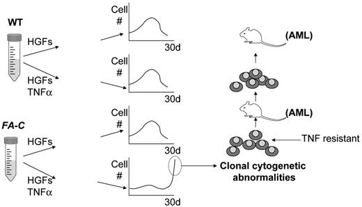
Reliable murine models will now permit the assessment of new agents. For acquired (autoimmune) aplastic anemia, the infusion of F1 mice with cells from parental lymph nodes results in marrow failure and increases in serum IFNγ. The F1 mice respond favorably to both immunosuppressive therapy and neutralizing antibodies to IFNγ and TNFα.46 For FA, at least two tractable models of clonal evolution exist now. The first is one in which murine Fanconi cells cultured ex vivo for a short period of time then transplanted into radiated recipients are at risk for clonal evolution,36 a complication prevented by correcting the FA defect.34 The second model is one in which clonal evolution can be initiated entirely in vitro by exposing Fancc−/− cells to TNF (summarized in Figure 2 ).35 This latter model is particularly attractive for two reasons. First, the differential responses of FA versus wild-type stem cells can be measured in just a few days, so the model is potentially translatable to a high-throughput screening environment. Second, the clear favorable impact of antioxidants on clonal evolution suggests that the first screening strategies ought to focus on libraries of antioxidant molecules. This kind of approach might allow identification of small molecules that normalize responses to stress factors such as TNFα. This approach seems likely to yield some fruit because potential molecular targets (e.g., TNFα, IFNγ, reactive oxygen species, p38, and JNK) have been identified in pre-clinical models for acquired aplastic anemia46 and FA.25 The ultimate goal would be to reduce the impact of unusual environmental stress factors on stem cells without reducing the responses of those cells to normal regulatory factors.
Designing clinical prevention trials
Clinical trials designed to increase the number of completely responsive patients are warranted. Achieving this goal will likely reduce the relative risks for clonal evolution. However, it is equally important that long-term interventional trials be developed for patients who have already received immunosuppressive therapy for aplastic anemia. The goal of these studies must focus on the goal of reducing late morbidity and mortality including the relative risk of clonal evolution. Along the way, given the limited sensitivity of FISH and Giemsa-banded cytogenetic analyses, more sensitive methods now available for quantifying genetic losses and gains ought to be exploited. Also, identification of potential molecular targets for prevention can be addressed using systems biology approaches. They would necessarily include attempts to define the emergent phenotypes associated with discrete cytogenetic rearrangements on a genome-wide scale (as has been done with 5q–cells38) and to establish sensitive target validation analyses for interventional studies using new molecularly targeted agents.
Summary
Approximately 10% to 20% of acquired aplastic anemia survivors will develop a clonal disease within the decade following their diagnosis, as will up to 40% of children and young adults with some of the congenital marrow failure syndromes. A good amount of recent evidence from the disparate fields of genetics, adaptation, stem cell biology, and hematopoiesis leads inescapably to the conclusion that clonal evolution in aplastic states arises in the context of ongoing stem cell damage through a process of clonal selection and adaptation. In the past year, this theoretical paradigm has been validated in clinical and preclinical models robust enough to inform surveillance strategies and reconsider therapeutic objectives in patients with aplastic states and to support the planning and development of rationally designed leukemia prevention trials in patients with bone marrow failure syndromes.
Coefficients of selection in normal bone marrow and in aplastic anemia. A hematopoietic stem cell (HSC) pool can reside in either a normal microenvironment (pathway arrow 1, “normal niche”) or within a hostile microenvironment (pathway arrow 2, “abnormal niche”). To emphasize the importance of the selection coefficient (low in pathway 1 and high in pathway 2), the pool of initially normal HSC contains one HSC with new alleles (bearing mutations of no consequence in the context of a normal niche). In the context of the hostile niche, the relative fitness of the same new alleles is massively amplified. Under the selective pressure of cytokines released by oligoclonal autoimmune T cells (“T”), the normal HSC are depleted over time (t1) and cytokine-resistant alleles are favored by selection because relative to the unfit wild-type stem cells, the mutant cells are much more fit. Under continued pressure from the aberrant T-cell population, the neoplastic clone preferentially expands over time (t2), while the less-fit normal HSC are selected against. In contrast, no evolution of the potentially neoplastic clone occurs in the supportive niche (pathway 1) because there is no selective pressure. That is, even though the phenotype of the mutant stem cell is the same as it was at the beginning of pathway 2, the fitness differences are not substantial enough to set the stage for clonal evolution. Not shown in this figure is a process by which a mutant stem cell arises in a population only after it is exposed to a hostile environment. This would meet strict genetic standards for an “adaptive mutation” in which the hostile environment per se induces mutations, some of which permit an adaptive response to the environment. The coefficient of selection idea would still be relevant here as well because relief of the environmental stress (e.g., effective immunotherapy of aplastic anemia) might lower the coefficient in time to prevent an outgrowth of adapted clonal progeny.
Coefficients of selection in normal bone marrow and in aplastic anemia. A hematopoietic stem cell (HSC) pool can reside in either a normal microenvironment (pathway arrow 1, “normal niche”) or within a hostile microenvironment (pathway arrow 2, “abnormal niche”). To emphasize the importance of the selection coefficient (low in pathway 1 and high in pathway 2), the pool of initially normal HSC contains one HSC with new alleles (bearing mutations of no consequence in the context of a normal niche). In the context of the hostile niche, the relative fitness of the same new alleles is massively amplified. Under the selective pressure of cytokines released by oligoclonal autoimmune T cells (“T”), the normal HSC are depleted over time (t1) and cytokine-resistant alleles are favored by selection because relative to the unfit wild-type stem cells, the mutant cells are much more fit. Under continued pressure from the aberrant T-cell population, the neoplastic clone preferentially expands over time (t2), while the less-fit normal HSC are selected against. In contrast, no evolution of the potentially neoplastic clone occurs in the supportive niche (pathway 1) because there is no selective pressure. That is, even though the phenotype of the mutant stem cell is the same as it was at the beginning of pathway 2, the fitness differences are not substantial enough to set the stage for clonal evolution. Not shown in this figure is a process by which a mutant stem cell arises in a population only after it is exposed to a hostile environment. This would meet strict genetic standards for an “adaptive mutation” in which the hostile environment per se induces mutations, some of which permit an adaptive response to the environment. The coefficient of selection idea would still be relevant here as well because relief of the environmental stress (e.g., effective immunotherapy of aplastic anemia) might lower the coefficient in time to prevent an outgrowth of adapted clonal progeny.
Formal proof of clonal adaptation/selection in a preclinical model of Fanconi anemia (FA). FA group C mutant cells are intrinsically hypersensitive to the apoptotic effects of TNFα, yet clonally evolved cells are resistant.33,34 To test the hypothesis that TNF exposure would change the fitness landscape significantly enough to permit rapid clonal evolution specifically in FA stem cells, Kit+, lin−, Sca+ marrow cells from wild-type (WT) and Fancc−/− mice (a murine model of FA of the C complementation group) were exposed for various periods of time (up to 35 days) to hematopoietic growth factors (HGFs) alone or to the same growth factors plus TNFα.35 Cell numbers rose then declined over a period of 30 days in wild-type cell cultures and in Fancc−/− cells exposed to HGFs alone. However, while, as expected, expansion of Fancc−/− stem cell progeny was markedly suppressed by TNFα for 2 to 3 weeks, by late in the third week, a population of proliferating cells appeared. These cells bore clonal cytogenetic defects, were resistant to TNFα, and when transplanted into recipient mice, led to the development of acute myelogenous leukemia (AML) within approximately 4 months. Transplantation of the leukemic cells led to the development of leukemia in a second recipient mouse with a much shorter latency period.35
Formal proof of clonal adaptation/selection in a preclinical model of Fanconi anemia (FA). FA group C mutant cells are intrinsically hypersensitive to the apoptotic effects of TNFα, yet clonally evolved cells are resistant.33,34 To test the hypothesis that TNF exposure would change the fitness landscape significantly enough to permit rapid clonal evolution specifically in FA stem cells, Kit+, lin−, Sca+ marrow cells from wild-type (WT) and Fancc−/− mice (a murine model of FA of the C complementation group) were exposed for various periods of time (up to 35 days) to hematopoietic growth factors (HGFs) alone or to the same growth factors plus TNFα.35 Cell numbers rose then declined over a period of 30 days in wild-type cell cultures and in Fancc−/− cells exposed to HGFs alone. However, while, as expected, expansion of Fancc−/− stem cell progeny was markedly suppressed by TNFα for 2 to 3 weeks, by late in the third week, a population of proliferating cells appeared. These cells bore clonal cytogenetic defects, were resistant to TNFα, and when transplanted into recipient mice, led to the development of acute myelogenous leukemia (AML) within approximately 4 months. Transplantation of the leukemic cells led to the development of leukemia in a second recipient mouse with a much shorter latency period.35
OHSU Cancer Institute, Oregon Health & Sciences University and Portland VA Medical Center, Portland, OR


