Abstract
Among inherited disorders, hemophilia has a number of characteristics that make it attractive as a model for gene transfer approaches. Several trials of gene therapy for hemophilia were carried out earlier in this decade; these trials were all first-in-class, i.e. the first use of a particular vector system in a particular target tissue, and thus yielded important safety data for the approaches under investigation. None, however, resulted in long-term expression of the clotting factor at therapeutic levels, and each encountered a critical issue, either in terms of safety, efficacy, or feasibility, that required further laboratory or clinical investigation. Ongoing trials of gene transfer for hemophilia include AAV-mediated gene transfer to liver using modified vectors (alternate serotypes, self-complementary constructs) or adjuvant therapies (transient immunosuppression). Preclinical studies using lentiviral vectors to transduce liver or hematopoietic cells have been promising, and genome editing and translational bypass strategies are also being investigated. Challenges to successful development of each strategy will be discussed.
The goal of gene therapy for genetic diseases is to bring about long-lasting expression of the missing or defective gene, i.e., to effect a “cure,” defined in the case of hemophilia as the ability to maintain hemostasis without significant ongoing medical intervention. Current management, by intravenous infusion of clotting factor concentrates, either prophylactically or on demand, is a highly effective treatment, but clearly falls short of this long-term goal. Advances that would likely be considered improvements by most consumers would include products that required less frequent infusions, those that could be taken orally rather than intravenously, and those that are less expensive than the currently marketed concentrates, which may cost in the range of $50,000 to $100,000/year for an adult with severe disease.
This chapter will review several (but by no means all) approaches to correction of genetic disease. These include gene addition therapy, gene correction therapy, and translational bypass therapy. For gene addition therapies, there are essentially two strategies for achieving long-term expression of a donated gene, as is required for genetic disease. The first is to introduce the gene into a stem cell with an integrating vector, so that all progeny carry the donated gene. The second is to introduce the gene into a long-lived (usually terminally differentiated) cell type; in this case, integration is not required, although duration of expression will then be determined by the lifespan of the transduced cell. Each approach has advantages and disadvantages, and both have been used successfully in the dog model of hemophilia. Neither has yet been successful in humans.
An advantage of hematologic disease in general and hemophilia in particular as models for developing gene transfer approaches is the ease of determining therapeutic endpoints. Two generations of experience have established that severity of disease correlates well with circulating levels of clotting factor, and these can be readily determined in most hospital coagulation laboratories. A second advantage is that it is clear from the natural history of disease in individuals with moderate (1% to 5%) or mild (> 5%) disease, and from the improved course of children with severe disease who are managed on prophylactic infusions,1 that even modest levels of transgene expression can ameliorate disease. The therapeutic window is wide in hemophilia, with levels as low as 3% and as high as 100% being acceptable endpoints, and biologically active factor can be synthesized in a range of tissues, allowing latitude in choice of target tissue for the donated gene. These characteristics favor hemophilia as a model for addressing problems of gene transfer.
Gene therapy products consisting of a vector and a transgene are arguably the most complex biologics yet developed as therapeutics. Given that immune responses to Factor VIII (F.VIII) or Factor IX (F.IX) are currently the leading complication of protein infusion therapy for hemophilia, it is perhaps not surprising that immune responses are proving to be a major issue for the more complex approach of gene therapy. Immune responses can occur either to the vector or to the transgene; the distinction is critical since it influences ongoing product development. A further complexity of gene transfer is that antigen presentation of vector and of transgene may vary depending on the gene transfer strategy, so that presentation by professional or nonprofessional antigen-presenting cells (tAPCs), or through the direct or indirect pathway, may be favored depending on the type of transgene, the identity of the target tissue, the type of vector, the levels of transgene expression, and the genetic background of the host. This means that problems of immune response may vary from one strategy to another and must be evaluated for each strategy undertaken.
The problems posed by gene transfer are similar for hemophilia A and B, but there are certain differences in the two diseases that may influence development of successful gene transfer strategies. The F9 cDNA is only 3 kb and thus fits easily into most vectors, whereas the F8 cDNA, at approximately 9 kb, exceeds the packaging capacity of some viral vectors. In addition, and perhaps partly as a consequence of the larger size of the protein, neutralizing antibodies to the protein (inhibitors) occur more frequently in hemophilia A than in hemophilia B. These confound analysis of endpoints and present a difficult problem clinically, favoring hemophilia B as a model for development of gene transfer. On the other hand, hemophilia A is approximately 6 times more prevalent, a consideration in prioritizing development of new therapies.
Beginning in 1999, several different gene transfer strategies for hemophilia A or B were investigated in phase 1 clinical trials involving 41 subjects with severe hemophilia (Table 1 ). These trials were sponsored by biotechnology companies, including Transkaryotic Therapies (ex vivo transduction of autologous fibroblasts by a plasmid expressing a truncated F.VIII molecule [B-domain deleted, BDD]),2 Chiron Corporation (intravenous infusion of a retroviral vector expressing BDD F.VIII),3 Avigen Inc. (adeno-associated virus [AAV]-mediated gene transfer into skeletal muscle [first trial] and liver [second trial] of a F.IX minigene),4–6 and GenStar Corporation (intravenous infusion of a gutted adenoviral vector expressing F.VIII). All of these trials were first-in-class, i.e., the first instance of a particular vector being used in a particular target tissue, and thus yielded important safety data for the approaches under investigation. None, however, resulted in long-term expression of the clotting factor at therapeutic levels. Each approach encountered a critical issue, either in terms of safety, efficacy, or feasibility, that required further laboratory or clinical investigation. In the wake of these disappointing initial results, more rigorous analysis of preclinical data in the hemophilic dog model has supported continued pursuit of some strategies and modification or abandoning of others. Circulating factor levels in the range of 5% to 25% have now been achieved in hemophilic dogs using three distinct approaches: a retroviral vector can be infused into neonatal dogs, where hepatocytes are still rapidly dividing, and yield high-level clotting factor expression;7 AAV vectors can be delivered to skeletal muscle via an intravascular route and can transduce a large number of muscle fibers, yielding therapeutic levels;8 or AAV vector can be delivered to liver via the hepatic artery, the portal vein, or in the case of AAV-8 vectors, intravenously, to yield therapeutic levels of clotting factors.9–11 This review will discuss approaches currently being pursued in clinical studies or being developed for clinical application.
Ongoing Trials
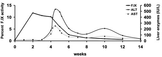
Of five original gene addition strategies for hemophilia that were analyzed in clinical trials in the US earlier in this decade (vide supra), only introduction of an AAV vector into the liver is currently being actively pursued. AAV vectors, or adeno-associated viral vectors, are arguably the simplest of all vectors, containing only a single-stranded DNA molecule encoding the therapeutic protein, packaged in an AAV capsid composed of three proteins designated VP1, 2, and 3. Once inside the cell the genome is converted to a double-stranded transcriptionally active form and is stabilized as a predominantly nonintegrated episomal form. In the initial published study, an AAV-2 vector expressing human F.IX was administered to 7 subjects, and in the highest dose studied (2 × 10¹² μg/kg) resulted in circulating F.IX levels of 10% to 12% for the first few weeks after vector infusion. Beginning at 4 weeks after vector infusion, however, F.IX levels began to fall, and gradually returned to baseline (<1%) at 10 weeks after vector infusion. Concurrently, serum transaminases, which had been normal for the first 3 weeks after vector infusion, began to rise, peaked at an ALT of 534 IU/L at 5 weeks, then slowly returned to normal without medical intervention (Figure 1 ). A subsequent subject infused at a 5-fold lower dose also experienced an asymptomatic, self-limited transaminase elevation, with a maximum ALT of 105 IU/L, over the same time course. Analysis of T-cell responses of peripheral blood mononuclear cells from this subject demonstrated by ELISPOT (enzyme-linked immunospot) assay IFN-γ production in response to AAV capsid, but not to F.IX.6 Subsequent mapping of the immunodominant epitope within the AAV capsid, and preparation of a pentamer that recognized the AAV capsid-specific TCR for this patient’s HLA haplotype, allowed direct detection of capsid-specific CD8+ T cells, which were shown to proliferate then disappear after vector infusion, with a time course that closely matched the rise and fall of serum transaminases.12
The absence of a detectable T-cell response to F.IX, or of antibody formation to F.IX in any of these subjects, eliminated F.IX as the target of the immune response. The question raised by this work is why human subjects manifest T cell responses to AAV capsid after vector infusion, while animal models do not. Prior exposure to AAV capsid probably underlies the difference in response. A high percentage of humans, the only natural hosts for wild-type AAV-2 infection, are infected by AAV-2 in childhood. Because AAV is naturally replication defective, this initial infection invariably takes place together with a helper virus infection such as adenovirus. Although AAV-2 on its own may not induce the inflammatory reactions needed for stimulation of a maximal adaptive immune response, in combination with the helper virus, which causes activation of the innate immune system, it is likely that CD8+ T cells directed to both the antigens of the helper virus and of AAV are formed. Upon controlling the infection, the frequency of AAV-specific CD8+ T cells would be expected to decline, leaving behind a small pool of memory T cells, which through homeostatic proliferation are maintained throughout the lifetime of an individual. On re-exposure to capsid, these memory CD8+ T cells are activated and eliminate the AAV capsid-harboring cells (the transduced hepatocytes). Because memory T cells are more readily triggered than naïve lymphocytes, human subjects undergoing reexposure have an outcome different from experimental animals undergoing what amounts to a primary infection with AAV.
Alternate hypotheses proposed to explain these findings have included packaging into the vector of residual plasmid expressing capsid, resulting in continuous expression of this viral protein in the transduced hepatocytes, or translation of alternate open reading frames within the F.IX expression cassette, leading to production of immunogenic foreign proteins.13 A third hypothesis has raised the possibility that a specific motif within the dominant protein of the AAV capsid favors receptor-mediated uptake of vector by human dendritic cells, with activation of capsid-specific CD8+ T cells occurring on that basis.14 This hypothesis, to be tested in an upcoming clinical trial of an AAV-8 vector expressing F.IX, predicts that alternate serotypes of AAV lacking this motif will avoid the capsid-specific CD8+ T-cell responses seen in the earlier study. Although the capsid sequences are fairly heavily conserved (60% to 98%), it is indeed possible that subtle differences in kinetics of vector uncoating, patterns of intracellular trafficking, or other aspects of antigen presentation may allow an alternate serotype to escape destruction by the immune response. The memory response hypothesis is being tested in a clinical study of AAV-2–F.IX co-administered with transient immunosuppression, to block the T-cell response to capsid. These studies should help to determine whether long-term correction of hemophilia B can be achieved with liver-directed gene transfer of an AAV vector.
If either of these strategies is successful, it is likely that the approach could be extended to hemophilia A. The major challenge for an AAV-based approach for hemophilia A is to design an expression cassette that can accommodate the larger F8 cDNA in a vector with a limited packaging capacity. Success has been achieved in animal models with a B-domain–deleted construct (approximately 4.4 kb of coding sequence). Couto and colleagues reported in 2003 sustained expression at 2% to 3% normal levels of a B-domain-deleted canine F.VIII in dogs with severe hemophilia A after portal vein infusion of an AAV-2–F.VIII vector,15 and Jiang et al subsequently showed similar circulating levels after infusion of AAV-6 and AAV-8 vectors expressing the same canine F.VIII cassette.16 These levels required a vector dose of approximately 1 × 10¹³ vg/kg, i.e., 10-fold higher than that required for comparable levels of F.IX expression in the hemophilia B dog model. Kazazian and colleagues have adopted another approach to the packaging problem by preparing 2 AAV vectors, 1 encoding the heavy chain, the other the light chain, of F.VIII.17 This approach has demonstrated similar results in the hemophilia A dog model, i.e., circulating levels in the range of 2% to 5% after portal vein infusion of approximately 1 × 10¹³ vg/kg (of two vectors, however, rather than one). Inhibitors were not seen in the dog model with either approach.
Strategies in Preclinical Stages
Therapies with integrating vectors
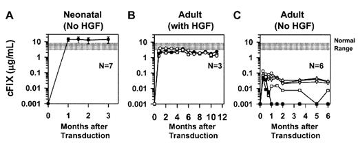
Since the goal of gene therapy for genetic disease is long-term expression, early efforts focused on the use of integrating vectors that could be passed to all daughter cells and thus ensure long-term expression of the donated gene. The most extensively investigated integrating vectors are retroviral vectors. Retroviral vectors are engineered from RNA viruses that use reverse transcription to generate a double-stranded DNA intermediate during replication. Replication-defective retroviral vectors contain an RNA genome that is reverse transcribed and integrated into the host genome. Most clinical experience with these vectors has been with ex vivo transduction of hematopoietic stem cells, followed by reinfusion of the genetically modified autologous cells back into the recipient. Indeed this strategy has accounted for the few clear-cut successes in the field of gene therapy to date, including reconstitution of the immune system in children with X-linked severe combined immunodeficiency (SCID) and with adenosine deaminase (ADA)-SCID,18 and in adults with chronic granulomatous disease.19 A key to success for these diseases is that the transduced cells and/or their progeny have a profound proliferative or survival advantage over non-transduced cells. Unfortunately, expression of coagulation factors confers no survival advantage for the cell, and thus is not easily amenable to conventional bone marrow-targeted approaches (but see below). However, there has been interest in developing integrating vectors that could target the liver, the normal site of synthesis of F.VIII and F.IX. Initial studies in adult subjects with hemophilia A3 demonstrated that a retroviral vector expressing B-domain–deleted F.VIII (driven by the viral 5′ long terminal repeat [LTR]) could be safely infused intravenously at doses up to 8.8 × 10⁸ transducing units (TU)/kg. These doses did not result in sustained F.VIII expression greater than 1%, nor was it clear which cells were being transduced; since retroviruses require a dividing target cell, one would speculate that hematopoietic cells and cells in the gastrointestinal tract would be targets, and indeed PCR analysis demonstrated persistence of vector sequences in peripheral blood mononuclear cells (PBMC) for more than 1 year in 3 of 4 patients tested. Preclinical studies by other groups clarified the conditions required for efficacy for retroviral vectors in hemophilia. Vandendriessche and colleagues demonstrated that intravenous infusion of a retroviral vector expressing B-domain–deleted F.VIII could result in high-level expression when infused at a dose of approximately 2 × 10¹⁰ colony-forming units/kg into newborn mice, where hepatocytes are still rapidly dividing. Ponder and colleagues extended this finding in several important ways (Figure 2 ). First, they constructed retroviral vectors with liver-specific promoters, which eliminated inhibitor development even when a canine F.IX construct was used in hemophilia B mice. Second, they demonstrated that injection of newborn animals gave positive results in a large animal model of hemophilia B, showing that infusion of a dose of approximately 1 × 10¹⁰ TU/kg into newborn puppies resulted in long-lasting expression of circulating levels of 2.5% to 6.7% F.IX activity. Finally they provided experimental proof-of-concept for a strategy by which this approach could be applied in adult animals, by demonstrating that adult mice pretreated with hepatocyte growth factor to induce hepatocyte replication could also achieve therapeutic levels of F.IX expression, albeit lower than the levels achieved in newborn mice.7 For several reasons, it may be problematic to take this strategy forward in patients with hemophilia, but this group has also demonstrated the success of this approach in the large animal model of mucopolysaccharidosis VII, a lysosomal storage disorder with much more limited therapeutic options.20
Lentiviral vectors would seem to offer a solution to the requirement of retroviral vectors for dividing target cells, since the former readily transduce nondividing cells. However, initial efforts to achieve long-term expression of clotting factors using lentiviral vector transduction of liver in animal models were disappointing, with some studies demonstrating formation of inhibitory antibodies to the transgene product.21–23 Indeed, Follenzi et al24 demonstrated that expression of a transgene from a ubiquitous promoter resulted in both humoral and cellular immune responses to the transgene product after introduction of the lentiviral vector into the livers of mice. They also documented that installation of a hepatocyte-specific promoter into the expression cassette resulted in long-term expression of F.IX in most mice, although the mechanism for this was not entirely clear, likely involving either elimination of antigen expression in APCs (lentiviruses efficiently transduce hematopoietic cells, including APCs) or an antigen-specific tolerogenic effect of hepatic gene expression.25 In elegant studies to probe the mechanisms of the failure to observe long-term expression in all mice, this same group determined that the “liver-specific promoter” was somewhat leaky in that it allowed some frequency of off-target expression in splenocytes. In response to this, these investigators re-engineered the vector to contain a target sequence for a hematopoietic-specific microRNA, mir-142-3p, and showed that inclusion of this target sequence in a vector prevented transgene expression in all hematopoietic cells. Intravenous administration of this modified lentiviral vector has allowed long-term expression of F.IX in all treated mice.26
Other than issues related to scale-up and manufacturing, the single remaining challenge for this approach is to minimize risk related to insertional mutagenesis. The level of risk represented by insertional mutagenesis in liver is not yet well understood; indeed, despite a great deal more work having been done, one could say the same for issues of insertional mutagenesis in hematopoietic stem cells. A series of studies have demonstrated preferential integration into actively transcribed genes for retroviral and lentiviral vectors (see first part of this chapter), but despite extensive analysis of patients treated in X-linked and ADA-SCID trials, it has been difficult to explain why 4 of 10 patients in one trial developed an ALL-like syndrome as a consequence of vector insertional events, while 0 of 18 subjects treated in two other studies have developed this complication.18 Possible roles of the transgene itself, and of subtle differences in the vectors and transduction protocols that might have resulted in transduction of slightly different target populations of cells, have been explored, without clear delineation of causative factors.
One solution to the risk of insertional mutagenesis has been proposed by Michele Calos and colleagues. These investigators have developed a nonviral gene delivery system based on the integrase of the Streptomyces phage ϕC31 (Figure 3; see Color Figures, page 511). The integrase is a serine site-specific recombinase that normally catalyzes a recombination event between two attachment (att) sites, attB and attP, one found in the ϕC31 phage and the other in the Streptomyces genome. Delivery of a plasmid containing a transgene of interest and an attB site, along with a second plasmid encoding the ϕC31 integrase, catalyzes the insertion of the donated gene into a restricted set of pseudo attP sites in the mammalian genome. This system was successfully exploited to achieve long-term F.IX expression in mice following plasmid delivery by hydrodynamic injection.27 The authors demonstrated that, even after removal of two-thirds of the liver by partial hepatectomy, mice previously infused with these plasmids showed return of F.IX levels to the original levels, establishing that the donated F.IX gene had been integrated into the hepatocytes. The characteristics of these integration sites in the mammalian genome have been defined; a recent study identified 101 unique integration sites out of 196 independent integration events.28 For human application, it will be critical to identify a safe and effective method for delivery of the therapeutic plasmids to the liver and to have a comprehensive understanding of the integration sites targeted by the plasmids. If these are indeed “safe” sites, then this vector will represent an improvement in safety over integrating vectors with random sites of insertion.
In a variation on the theme of integrating vectors, two groups have explored the possibility of using megakaryocytes as target cells for donated genes expressing F.VIII. The advantage of this strategy is that the platelet acts as a vehicle to transport F.VIII, stored in the platelet granules, to the site of a bleed, where, upon activation, it releases F.VIII at the site of the injury. Moreover, this strategy has the potential to extend the half-life of F.VIII, from as little as 12 hours for the infused protein, to up to 10 days for a platelet. Yarovoi et al, using a murine glycoprotein Ibα promoter to drive F.VIII expression in a transgenic mouse, showed that the ectopically expressed F.VIII was stored in the α-granules of platelets (i.e., co-localized with von Willebrand factor) and that it sufficed to correct a cuticular bleeding time and reduce blood loss in the mice, even though F.VIII was not detected in murine plasma.29 Shi et al, using mice expressing human BDD F.VIII under the control of the platelet-specific αIIb promoter, showed similar findings and also demonstrated that the bleeding phenotype could be corrected even in mice with high-titer anti-F.VIII inhibitors.30 The utility of this procedure in humans will depend to some extent on achieving high-efficiency transduction and engraftment of genetically modified hematopoietic stem cells, although it will certainly be difficult to match the efficiency achievable in a transgenic mouse model. The risks of this strategy will be related to the risks associated with the integrating vector system used to modify the autologous bone marrow cells.
Genome editing strategies
A long-term goal of gene transfer technology is to achieve correction of a mutant sequence, rather than insertion of the gene at another location (gene addition therapy). The advantage of a corrected sequence is that it allows expression under physiologic circumstances, i.e., from the endogenous promoter and subject to the control of other intrinsic regulatory elements. Early efforts to trigger a recombination event between a wild-type donated sequence and a mutant endogenous sequence were stymied by the low spontaneous rate of gene targeting in the mammalian genome. Introduction of a double-strand break in the target DNA stimulates homologous recombination by logs. In 2003, Porteus and Baltimore31 reported use of a novel synthetic nuclease, coupled with a donor template DNA, to effect site-specific correction of a mutant sequence at high efficiency. The synthetic nuclease consisted of a series of zinc finger elements, the most common DNA-binding motif in mammalian transcription factors, linked to an endonuclease (Figure 4; see Color Figures, page 511). Each zinc finger element recognizes a specific 3- to 4-bp sequence; several fingers can be linked to create a binding domain that recognizes a specific sequence with a high degree of specificity. Thus, the zinc finger elements guide the endonuclease to a specific site within the genome, and the endonuclease introduces a double-strand break at that site. Once the double-strand break has been introduced, the cell’s powerful repair mechanisms are engaged; homologous recombination repairs the damage by using similar sequences within the DNA as a template, but a donated wild-type sequence can suffice as template. Urnov et al32 demonstrated that they could correct 15% to 20% of mutated sequences in a cell line, and up to 5% in human T cells, using this approach. The potential for genome editing in hemophilia is clear; a requirement, however, will be to find a way to deliver both the zinc finger nuclease (ZFN)–encoding sequences and a normal donor template efficiently to hepatocytes. It should be noted that only short-term expression of the ZFN is required; once the sequence has been corrected, the change will be propagated in daughter cells. Several strategies exist for short-term expression, including integrase-defective lentiviral vectors33 or expression cassettes characterized by transcriptional shutdown of the promoter. Moehle et al recently reported an important extension of this technology to introduce an expression cassette of up to 8 kb into a precise location within the genome.34 Coupling of the ZFN technology with identification of “safe” insertion sites, i.e., sites not associated with insertional mutagenesis events, should lead to improved safety over vectors that integrate randomly within the genome.
Nonsense mutation suppression
The foregoing treatment strategies, in various stages of development, are designed to address genetic disease by altering the DNA. An alternative strategy addresses a specific type of mutation at the level of translation of RNA to protein. It was initially reported in animal models in 199935 that aminoglycoside antibiotics could promote read-through of premature stop codons by reducing the efficiency of translation termination at the premature stop codon (Figure 5, see Color Figures, page 511). Trials of these antibiotics in patients with muscular dystrophy, cystic fibrosis, and hemophilia showed some production of the missing proteins, but the renal and otic toxicities, the need for intravenous administration of the drugs, and the relatively poor potency all limited the clinical utility of the approach.36,37 High-throughput screens to identify small-molecule entities capable of promoting read-through of stop codons, followed by optimization for oral bio-availability, in vivo safety, and lack of in vitro off-target activity, resulted in identification of a small-molecule drug (an oxidiazole compound, PTC124) with no structural similarity to aminoglycosides, which has shown safety and tolerability in healthy humans and has yielded promising results in patients with muscular dystrophy with disease due to a stop codon.38,39 A major safety concern at the outset had been whether the drug would also promote read-through of normal stop codons, but a careful search for polypeptides corresponding to putative read-through products has been negative in rats, dogs, and humans. Approaches like this may also be useful in hemophilia, where 10% to 20% of severe cases are due to nonsense mutations. Readthrough of a premature stop codon results in substitution of a different amino acid at that site; at some residues nearly any amino acid will be tolerated, and a functional protein will result, whereas at others, only the wild-type amino acid will suffice for functional activity, and the yield of functional protein will be accordingly lower than the levels of antigen produced. This is a class of therapeutics that can treat only a subset of affected individuals, but correlation of underlying mutation to clinical response in early-phase testing should allow these individuals to be identified. The availability of treatment strategies such as translational bypass underscores the critical need for molecular diagnosis for all patients with genetic disease and heralds the onset of a new era of personalized molecular medicine.
Gene transfer strategies for hemophilia A and B.
| Trial . | No. patients . | Vector dose . | Endpoints . | Reference . |
|---|---|---|---|---|
| Intravenous infusion of retroviral vector expressing F.VIII | 13 | Up to 4.4 × 10⁸ TU/kg | Vector DNA detected in PBMCs of 8/8 subjects treated at ≥ 9.2 × 10⁷ TU/kg. No patients with consistent detection of F.VIII > 1%. | Powell et al3 |
| Ex vivo plasmid-F.VIII transfection of autologous fibroblasts | 12 | Up to 400 × 106 transfected autologous cells | F.VIII 1% to 2% in higher-dose cohort. | Roth et al2 |
| AAV-F.IX into skeletal muscle | 8 | Up to 2 × 10¹² μg/kg | Persistent F.IX expression in muscle biopsy. No patients with consistent F.IX levels > 1%. | Manno et al5 |
| AAV-F.IX into liver | 7 | Up to 2 × 10¹² μg/kg | F.IX levels > 5% × 4 weeks in top dose cohort. | Manno et al6 |
| Gutted adenoviral vector expressing F.VIII | 1 | 4.3 × 10¹⁰ particles/kg | Not published |
Factor IX (F.IX) activity levels and transaminases (AST, ALT) as a function of time in weeks after vector administration in a subject with severe hemophilia B infused with 2 × 10¹² vector genomes/kg of AAV-2-F.IX via the hepatic artery.
Factor IX (F.IX) activity levels and transaminases (AST, ALT) as a function of time in weeks after vector administration in a subject with severe hemophilia B infused with 2 × 10¹² vector genomes/kg of AAV-2-F.IX via the hepatic artery.
Levels of canine Factor IX (cFIX) in mice transduced with a retroviral vector. (A) Neonatal mice injected intravenously with a single dose of 1 × 10¹⁰ TU/kg at 2 to 3 days of age. The normal range of Factor IX levels is indicated by the gray bar. (B) Levels of cFIX in adult mice infused intravenously with the same vector following infusion with hepatocyte growth factor. (C) Levels of cFIX in adult mice infused intravenously with the same vector but without preceding hepatocyte growth factor infusion. Adapted with permission from Xu et al.7
Levels of canine Factor IX (cFIX) in mice transduced with a retroviral vector. (A) Neonatal mice injected intravenously with a single dose of 1 × 10¹⁰ TU/kg at 2 to 3 days of age. The normal range of Factor IX levels is indicated by the gray bar. (B) Levels of cFIX in adult mice infused intravenously with the same vector following infusion with hepatocyte growth factor. (C) Levels of cFIX in adult mice infused intravenously with the same vector but without preceding hepatocyte growth factor infusion. Adapted with permission from Xu et al.7
The Children’s Hospital of Philadelphia and Howard Hughes Medical Institute, Philadelphia, PA


