Abstract
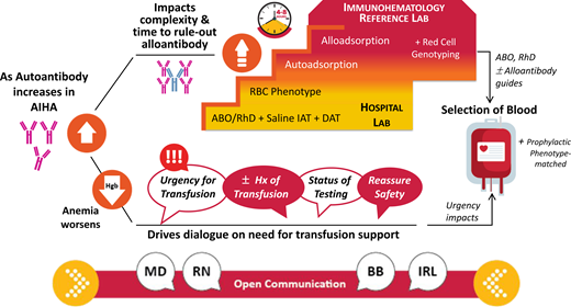
The serologic evaluation of autoimmune hemolytic anemia (AIHA) confirms the clinical diagnosis, helps distinguish the type of AIHA, and identifies whether any underlying alloantibodies are present that might complicate the selection of the safest blood for any needed transfusion. The spectrum of testing is generally dependent on the amount and class (immunoglobulin G or M) of autoantibody as well as the resources and methodologies where testing is performed. The approach may range from routine pretransfusion testing, including the direct antiglobulin test, to advanced techniques such as adsorptions, elution, and red cell genotyping. When transfusion is needed, the selection of the optimal unit of red blood cells is based on urgency and whether time allows for the completion of sophisticated serologic and molecular testing methods. From the start of when AIHA is suspected until the completion of testing, communication among the clinical team and medical laboratory scientists in the transfusion service and immunohematology reference laboratory is critical as testing can take several hours and the need for transfusion may be urgent. The frequent exchange of information including the patient's transfusion history and clinical status, the progress of testing, and any available results is invaluable for timely diagnosis, ongoing management of the patient, and the safety of transfusion if required before testing is complete.
Learning Objectives
Describe the difficulties in determining ABO group/RhD type and in identifying alloantibodies in patients with autoantibodies
Discuss strategies for the selection of blood for safe transfusion, keeping in mind patient history and completed laboratory testing
Explain the importance of communication among clinicians and laboratory professionals in the approach to transfusion
Introduction
Autoimmune hemolytic anemia (AIHA) is serologically defined as warm or cold based on the class of immunoglobulin (Ig), either IgG or IgM, causing hemolysis. Warm autoantibodies are IgG and primarily detected in vitro at 37 °C and in the indirect antiglobulin test (IAT), while cold autoantibodies (CAAs) are IgM and bind preferentially to red blood cells (RBCs) at colder temperatures in clinical testing (Table 1). Despite these simplified definitions, the serologic evaluation of patients presenting with these strong autoantibodies is a challenge for any laboratory. The extent of testing capabilities varies among hospital transfusion services and immunohematology reference laboratories (IRLs). An overview of the various tests employed and a reference for the clinical cases are provided in Table 2.
Summary of classic serologic findings in AIHA
| Type of AIHA . | Antibody class . | Antibody specificity . | Optimum test phase of antibody reactivity . | DAT result . | Eluate result . |
|---|---|---|---|---|---|
| Warm | IgG | Broadly reactive | IAT | 2-4+ IgG and C3 or IgG only | Positive—reactive with all/majority of panel RBCs |
| Cold | IgM | I/i | RT, 37 °Ca | 2-3+ C3 only | Not usually performed |
| Mixed Type | IgG and IgM | Broadly reactive | RT, 37 °C, IAT | 3-4+ IgG and C3 | Positive—reactive with all/majority of panel RBCs |
results after 15-to-30 minute incubation at 37 °C.
2-4+, agglutination strength; RT, direct agglutination at room temperature.
Immunohematology methods used in evaluating patients with warm and cold autoantibodies
| Method . | Description/purpose . | Comments/pitfalls . |
|---|---|---|
| Routine: | ||
| ABO group/RhD type | Determine patient's ABO group and RhD type | • Presence of high titer CAA may cause autoagglutination at room temperature. Steps to resolve this may include: o Warm/wash patient RBCs at 37 °C o Perform testing at 37 °C o Treat RBCs with thiol reagent (DTT/2-ME) to remove IgM coating patient RBCs |
| DAT • Polyspecific AHG | Detects IgG and/or C3 (indirectly detects IgM) on patient's RBCs | • Positive in >90% of cases of AIHA • Screening assay; when positive must repeat testing with anti-IgG and anti-C3d to define protein coating patient RBCs |
| DAT • Monospecific o Anti-IgG o Anti-C3d | Individual reagents to detect whether IgG, C3d, or both on patient's RBCs | • Both reagents plus control must be performed • False positives can occur due to autoagglutination (most common with IgM) |
| Antibody detection (screen) | Use of 2-3 reagent RBCs of known phenotype to screen for auto and/or alloantibody in patient's serum • Detects IgM CAA when performed at IS/RT (direct agglutination) using a test tube method • Detects IgG WAA at IAT with any method. | • Detection depends on test method used (test tubes, CAT, SP) • May be negative if all autoantibody bound to patient’s RBCs |
| Antibody identification | Use of panel reagent RBCs (10-16) of known phenotype to determine specificity of antibody. • Reactivity at IS/RT (direct agglutination) confirms CAA. Some show autoanti-I or -i specificity. • Confirms broad reactivity or specificity of WAA at IAT. | • Subsequent testing when antibody is detected to identify antibodies of clinical significance • Identifies specificity of any underlying alloantibody once autoantibody removed from patient serum |
| Crossmatch | Confirms ABO compatibility by either serologic (IS) or electronic/computer. | • If known clinically significant alloantibody, must perform serologic crossmatch at IAT (also known as AHG or full crossmatch) |
| Phenotype | Serologically type patient RBCs using known antisera. | • Performed when patient has not been transfused in last 3 months • Strong positive DAT may cause false positive; not all antisera able to give accurate results when DAT is positive • Partial antigens or variant antigens may not be detected |
| Advanced: | ||
| Elution | • Elution removes IgG bound to the patient's RBCs using a dilute acid solution. Resulting eluate is then tested against a panel of reagent RBCs to check for specificity of the RBC bound IgG. o Positive with all cells confirms WAA. o If negative, suspect drug-dependent antibody. | • Once diagnosis of AIHA, no need to perform with each subsequent pretransfusion test • Also used in suspected delayed transfusion reaction to detect binding of alloantibody to circulating transfused RBCs |
| Adsorption—autologous | • Removes IgG autoantibody (when incubated at 37 °C) from patient plasma using patient RBCs; then adsorbed plasma tested to identify underlying alloantibody. • Removes IgM autoantibody (when incubated at 4C) from patient plasma using patient RBCs; then adsorbed plasma tested to identify underlying alloantibody. | • Performed when patient has not been transfused in last 3 months • Patient RBCs are first treated to remove bound autoantibody to allow sites for autoantibody in plasma to bind • Requires sufficient quantity of patient RBCs for removal of autoantibody |
| Adsorption—allogeneic | • Removes IgG autoantibody (when incubated at 37 °C) from patient plasma using phenotyped donor RBCs; then adsorbed plasma tested to identify underlying alloantibody. • Removes IgM autoantibody (when incubated at 4C) from patient plasma using donor RBCs; then adsorbed plasma tested to identify underlying alloantibody. | • Typically, 3 donor RBCs of known phenotype (eg, R1R1, R2R2, rr) are selected • May miss antibody to high prevalence antigen |
| Advanced: | ||
| Genotype | • Molecular methods used to determine patient's probable phenotype. | • More accurate than serologic phenotyping; generally, provides more comprehensive antigen profile • Recommended when patient has been transfused in last 3 months • Unusual, rare genetic variants or partial antigens may not be detected |
| Prewarm technique | • Wash patient RBCs with 37 °C saline prior to testing. • Warm patient plasma and reagent RBCs at 37 °C separately prior to mixing and performing testing to avoid CAA reactivity. | • Avoids CAA from binding to patient or donor RBCs • Clinically significant, weakly reactive alloantibodies may not be detected in a prewarm IAT |
| Thermal amplitude study | • Incubate patient serum with reagent RBCs for 2 hours at 30 °C and 37 °C. • Determines if CAA has ability to bind to reagent RBCs at warm body temperature. | • Should be performed with adult RBCs (positive for I) and cord blood RBCs (positive for i) • Typically, only need to perform once to aid in diagnosis |
CAT, column agglutination technique; IS, immediate spin (direct agglutination at room temperature); 2-ME, 2-mercaptoethanol; RT, room temperature, direct testing; SP, solid phase technique.
CLINICAL CASE 1
A 35-year-old woman with a vague history of idiopathic thrombocytopenia presented to the emergency department with a complaint of fatigue, malaise, headache, and cough. Vital signs are temperature, 37.8 °C; blood pressure, 114/55 mmHg; heart rate, 98/min; respiratory rate, 20; and oxygen saturation, 100% on room air. Initial laboratory studies show a positive COVID test, a white blood cell count of 10 100/µL (4200-11 000), a hemoglobin (Hgb) level of 4.4 g/dL (12.0-15.5), and a platelet count of 6000/µL (140 000-450 000). Type and screen is ordered to prepare 2 units of RBCs and 1 unit of apheresis platelets for transfusion. The patient's blood type is O-positive, but routine antibody screening by a column agglutination test is strongly positive (4+) with all cells, including the patient's own cells. The direct antiglobulin test (DAT) is strongly positive (4+) with polyspecific reagent. Monospecific testing with anti-IgG and anti-C3d is not available at this laboratory. With further testing, the patient sample is strongly positive (3+) with all panel cells at saline IAT.
The clinical team is notified of the results and the need to send out patient samples to the IRL for further testing, with an anticipated delay of at least 6 to 8 hours. Subsequent labs show reticulocytes, 24.4%; absolute reticulocyte count, 278 000/µL (10 000-120 000); lactate dehydrogenase, 692 U/L (82-240); total bilirubin, 1.5 mg/dL (0.2-1.0); haptoglobin less than 8 mg/dL (30-200); and microspherocytes on peripheral smear, suggestive of a diagnosis of warm AIHA (WAIHA). Communication between the laboratory staff and clinical team reveals the patient is clinically stable, has a history of one missed abortion, and no prior transfusions. The safety of releasing RBC units before completion of testing and the low risk of any reactions were discussed with providers.
Pretransfusion testing in patients with WAIHA
For any RBC transfusion, initial routine tests include an ABO group, an RhD type, and an antibody detection test (also known as an antibody screen). The primary purpose of antibody detection testing is to detect and identify an antibody that could potentially react with transfused RBCs and cause hemolysis. Antibody detection involves the IAT to identify whether clinically significant alloantibodies or autoantibodies are present. For 90% or more of pretransfusion samples, the results are negative and interpretation, straightforward.1
The presence of an antibody that is broadly reactive, demonstrating similar agglutination with all reagent RBCs including the patient's own RBCs, is classified as an autoantibody. A salient point in the interpretation of autoantibodies is their correlation with clinical and other laboratory findings. An independent assessment of the presence or absence of hemolytic anemia is essential. “Benign” autoantibodies are fairly common in serologic workups but generally not associated with strong positive DAT or hemolysis.2
The case demonstrates one of the more perplexing problems for medical laboratory scientists (MLS) working in the hospital laboratory—distinguishing whether an underlying alloantibody is present in a sample containing a strong warm autoantibody (WAA). As seen in Table 3, all RBCs strongly agglutinate in the presence of a WAA at the IAT, easily masking the detection of an underlying alloantibody. The prevalence of RBC alloantibodies is higher in patients with WAIHA, ranging from 12% to 54% with a mean of 38% (Table 4). For this reason, further studies to evaluate for the presence of a clinically significant alloantibody are necessary for the selection of blood. Initially, a method that does not enhance antibody binding (known as the no-enhancement method or saline IAT) is undertaken. This step may omit or weaken the reactivity of a low-titer autoantibody and allow detection of any underlying alloantibodies.15
Typical antibody detection (screening) results for autoantibodies and alloantibodies
| Reagent red cells . | Cold autoantibody . | Warm autoantibody . | No antibody detected . | Alloantibody detected . | ||||||||
|---|---|---|---|---|---|---|---|---|---|---|---|---|
| RT . | 37C . | IAT . | RT . | 37C . | IAT . | RT . | 37C . | IAT . | RT . | 37C . | IAT . | |
| 1 | 4+ | 3+ | 1+ | 0 | 0 | 3+ | 0 | 0 | 0 | 0 | 0 | 0 |
| 2 | 4+ | 3+ | 1+ | 0 | 0 | 3+ | 0 | 0 | 0 | 0 | 0 | 3+ |
| 3 | 4+ | 3+ | 1+ | 0 | 0 | 3+ | 0 | 0 | 0 | 0 | 0 | 0 |
| Autocontrol* | 4+ | 3+ | 1+ | 0 | 0 | 4+ | 0 | 0 | 0 | 0 | 0 | 0 |
Results after mixing patient plasma with reagent RBCs. Agglutination is graded 1-4+; no agglutination is indicated as 0.
Autocontrol contains patient plasma plus patient RBCs.
Italics indicate typical optimum reactivity of cold autoantibody. Bold indicates typical optimum reactivity of warm autoantibody. Italics and bold indicate typical reactivity of alloantibody.
RT, room temperature (direct agglutination/immediate spin at room temperature); 37C, results after incubation at 37 °C for 10-30 minutes; IAT, results after addition of AHG.
Patients with WAIHA and underlying alloantibodies
| Reference . | Number of alloantibodies detected . | Number of samples . | % samples with underlying alloantibodies . |
|---|---|---|---|
| Morel et al3 (1978) | 8 | 20 | 40 |
| Branch and Petz4 (1982) | 5 | 14 | 36 |
| Wallhermfechtel et al5 (1984) | 19 | 125 | 15 |
| Laine and Beattie6 (1985) | 41 | 109 | 38 |
| James et al7 (1988) | 13 | 41 | 38 |
| Issitt et al8 (alloadsorptions) (1996) | 13 | 34 | 38 |
| Issitt et al8 (autoadsorptions) (1996) | 5 | 41 | 12 |
| Leger and Garratty9 (1999) | 105 | 263 | 40 |
| Maley et al10 (2005) | 39 | 126 | 31 |
| Das and Chaudhary11 (2009) | 7 | 23 | 30 |
| Yürek et al12 (2015) | 7 | 36 | 19 |
| Park et al13 (2015) | 87 | 161a | 54 |
| Shirey et al14 (2002) | 8 | 20 | 40 |
| TOTALS | 389 | 1013 | 38% |
124 were warm autoantibodies, 21 cold autoantibodies with wide thermal range, and 16 were cold-reactive only.
Direct antiglobulin test
The presence of an autoantibody in the plasma is the result of all antigen sites bound on the patient's RBCs and should trigger the MLS to perform a DAT. The DAT is a critical diagnostic tool for AIHA and is initially performed by testing the patient's washed RBCs with polyspecific antihuman globulin (AHG), which contains both anti-IgG and anti-C3d.
The use of a conventional test tube continues to be the primary method for the DAT, requiring manual observation for agglutination of the patient RBCs after the addition of AHG. It is reliable and often used as confirmatory testing for other DAT techniques. In addition, to detect C3d, a test tube method using anti-C3d is optimal. With the expanding role of automation in hospital laboratories, a DAT can be performed using column agglutination testing (known as gel or beads) or solid-phase methods. While these alternative methods for the DAT may be easier to perform and provide less subjectivity when interpreting the reaction results (mainly due to end point reading by automation), in the setting of AIHA the authors have not observed the other methods to be more sensitive than a well-performed DAT by tube technique. Correlation with patient history is paramount to assess the significance of any positive DAT result regardless of method.
When the DAT is positive with polyspecific AHG, further testing with monospecific reagents is required to differentiate whether IgG, C3d, or both are coating the patient RBCs and to help classify the type of hemolytic anemia (Table 1). In patients with WAIHA, more commonly both IgG and C3d coat the patient's cells.16 While the DAT alone is not diagnostic of hemolytic anemia, stronger reactions (2-4+) denote higher antibody density on the RBC surface and are more likely associated with hemolysis. Similarly, the presence and amount of C3 (C3b and/or C3d) on the RBC membrane is an important predictor of immune hemolysis.2,17 Conversely, a negative DAT does not rule out AIHA and may be seen in 5% to 10% of patients with hemolytic anemia.16,17
To confirm the positive DAT is due to an autoantibody, an elution procedure should be performed as part of the initial investigation. The predominant method utilized for eluate preparation removes cell-bound IgG autoantibody by the addition of a dilute acid solution to the patient's RBCs. The acid/RBC mix is centrifuged, and the supernatant (eluate) is buffered back to a normal pH. Antibody identification is performed on this eluate. The results are key in differentiating WAIHA from drug-induced immune hemolytic anemia. In WAIHA the eluate contains the IgG autoantibody, whereas with a negative eluate, a drug-dependent antibody would be suspected.
CLINICAL CASE 1 (Continued)
Testing at the IRL confirms a strongly positive DAT, including 4+ reactivity with both anti-IgG and anti-C3d. Strongly reactive autoantibody is present in the patient serum and eluate. The autoantibody remains strongly positive with all panel cells despite no enhancement at the IAT, necessitating the performance of specialized adsorption techniques. RH and K phenotyping show that the patient's RBCs are E− and K− and C+, c+, and e+. RBC genotyping is ordered to determine the extended phenotype.
Adsorption—allogeneic and autologous
Adsorption studies are the optimal method to remove autoantibody from the patient's plasma and leave alloantibody behind, if present. When a sample is referred, the first question asked by the IRL MLS is whether the patient has been transfused and when. If transfused within the last 3 months, allogeneic (using donor cells of known phenotypes) adsorption is performed over autologous (using the patient's own RBCs) adsorption. Even a small amount of transfused RBCs in the patient's sample can adsorb out alloantibodies.18 Another factor for the use of alloadsorption is the patient's degree of anemia. For patients with an Hgb level less than 5 g/dL or small pediatric patients, it is unlikely that an adequate quantity of patient RBCs can be collected to successfully perform repeated autoadsorptions.
Autologous adsorptions are logistically easier to perform and preferred over alloadsorptions for 2 reasons. One, removing antibody using the patient's own RBCs proves the autoimmune nature of the autoantibody. Second, autoadsorptions will not remove an alloantibody to a high prevalence antigen. Patient RBCs must first be chemically treated to remove autoantibody. Chemicals such as ZZAP (dithiothreitol (DTT)/2-mercaptoethanol + ficin/papain) or enzyme treating (ficin/papain) alone can remove autoantibody to free up antigen sites for more autoantibody binding. Repeat adsorptions (1 to 4) based on the strength of the autoantibody present (1+-4+) in the patient's serum are required, generally taking 3 to 4 hours.
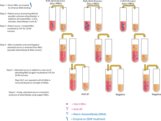
Allogeneic adsorptions are arguably one of the most complicated immunohematology tests to perform and are confusing to interpret. They are labor-intensive, generally requiring 4 to 8 hours to complete. An adequate inventory of phenotyped blood donors, typically only available in IRLs, is required for the proper antigen makeup and volume of donor RBCs to perform the adsorptions. Generally, 3 different donor RBCs are selected, and the antigen makeup of each must be carefully considered to reveal any clinically significant alloantibody since all donor RBCs adsorb the autoantibody. If an alloantibody is present, as depicted in Figure 1, it will bind to donor RBCs that express the corresponding antigen but remain behind if donor RBCs lack the antigen. Typically, 2 or 3 rounds of adsorptions deplete the autoantibody.
Adsorption procedure to remove WAA using allogeneic RBCs. Example of the multistep process for allogeneic adsorptions to remove WAA to identify whether an underlying alloantibody is present (eg, anti-Jka). Three donor RBCs of known phenotypes (ie, R1R1, R2R2, and rr) are selected and treated with enzyme or ZZAP. The patient's serum (or plasma) is added to an aliquot of each of the 3 donor RBCs and incubated for 10 to 60 minutes at 37 °C. Three separate adsorptions are performed, each undergoing 1 to 4 rounds of adsorption in which the patient's serum is repeatedly incubated with a new set of donor RBCs. Following an adequate number of adsorptions, adsorbed serum samples are tested against reagent RBCs. If 1 or more aliquots are positive, an alloantibody can be identified based on the pattern of reactivity and knowledge of the antigens expressed on each aliquot of donor RBCs used for adsorption.
Adsorption procedure to remove WAA using allogeneic RBCs. Example of the multistep process for allogeneic adsorptions to remove WAA to identify whether an underlying alloantibody is present (eg, anti-Jka). Three donor RBCs of known phenotypes (ie, R1R1, R2R2, and rr) are selected and treated with enzyme or ZZAP. The patient's serum (or plasma) is added to an aliquot of each of the 3 donor RBCs and incubated for 10 to 60 minutes at 37 °C. Three separate adsorptions are performed, each undergoing 1 to 4 rounds of adsorption in which the patient's serum is repeatedly incubated with a new set of donor RBCs. Following an adequate number of adsorptions, adsorbed serum samples are tested against reagent RBCs. If 1 or more aliquots are positive, an alloantibody can be identified based on the pattern of reactivity and knowledge of the antigens expressed on each aliquot of donor RBCs used for adsorption.
Regardless of which adsorption method is used, the adsorbed plasma must be retested to detect and identify any alloantibodies. If testing is negative, one can be relatively assured that no alloantibodies are present. In our case, 4 autoadsorptions and 4 alloadsorptions both failed to completely remove the WAA. Any further adsorptions would be impractical and likely increase the risk of missing weakly reactive alloantibodies. These rare situations illustrate the importance of RBC phenotyping/genotyping in patients with WAIHA.
Patient RBC phenotyping and genotyping
Knowing the patient's RBC phenotype can be a powerful resource to predict which alloantibodies the patient can make and to guide the selection of blood. At a minimum, the patient should be typed for RH (C, E, c, e) and K. Extended typing for additional blood group antigens (Fya/Fyb, Jka/Jkb, M/N, and S/s) is possible today with the use of monoclonal reagents. However, when the patient has a strong positive DAT, determining the extended phenotype can be problematic due to autoagglutination. The patient's RBCs can be chemically treated with EDTA-glycine hydrochloric acid or chloroquine to remove IgG prior to testing. These pretreatment methods are often tedious, and the treated RBCs cannot be phenotyped for all blood group antigens as EDTA-glycine hydrochloric acid denatures KEL blood group system antigens, and chloroquine treatment reduces RH antigen expression.16 More importantly, phenotype by serologic method should not be performed if the patient has been recently transfused.
RBC genotyping (RCG) is becoming an increasingly valuable, cost-effective tool in transfusion practice. As a send-out test for most laboratories, results are unlikely to be available for 48 to 72 hours. RCG allows testing of more blood group systems and avoids the technical difficulties that may occur with serologic testing when the patient's RBCs are coated with antibody. In addition, recent transfusion does not pose a problem, as DNA for molecular analysis is isolated from the patient's mononuclear cells. Using the patient's predicted phenotype from RCG, a strategy of prophylactic antigen-matched RBC units (RH [D, C, E, c, e], K, and where feasible JK, FY) may prevent alloimmunization, simplify subsequent pretransfusion testing, and possibly reduce the frequency of adsorption studies.14,19 In a survey of academic hospital transfusion services and IRLs (n = 54) on the testing practices for patients with WAA, 68% performed genotyping and 75% provided prophylactic antigen-matched RBC units, although policies varied on indications for use.20
Transfusion support in patients with AIHA
Generally, the decision to transfuse AIHA patients should be based on similar indications as for anemic patients without AIHA. In asymptomatic patients, a restrictive transfusion strategy (ie, Hgb <7 g/dL) should be practiced.21 The use of bed rest and supplemental oxygen in patients with severe anemia (ie, Hgb <6 g/dL) could help defer transfusion until testing is complete. The patient's clinical status, comorbidities, and symptoms should guide the decision to transfuse regardless of Hgb level and compatibility test results. When symptoms of significant hypoxia, confusion, angina, or hemodynamic instability are present, RBC transfusion should not be withheld, even when testing is incomplete. The presence of reticulocytopenia in an AIHA patient with severe anemia should be regarded as a medical emergency. Reticulocytopenia is a poor prognostic indicator representing an inadequate erythropoietic response and higher transfusion requirements.16,22
When transfusion is medically necessary, the patient's physician should be assured that transfused RBCS are unlikely to cause an acute hemolytic transfusion reaction. The risk for a transfusion reaction in patients with AIHA appears to be no higher than in other patients requiring transfusion. Chen et al.23 found that reactions occurred in 1.6% of 450 transfused AIHA patients, with febrile and allergic reactions the most frequent and no reports of hemolytic adverse events. Most patients with AIHA tolerate serologically incompatible RBC transfusions quite well, without a significant increase in their underlying hemolysis.12,13,23
Selection of blood for transfusion
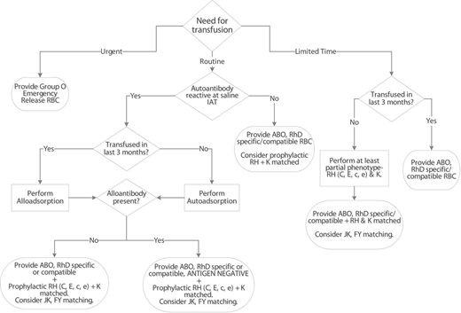
Selection of the optimal RBC unit for transfusion depends on the urgency, capabilities and resources of the hospital transfusion service, and the status of the pretransfusion testing. When there is life-threatening anemia and no time for completion of testing, emergency-release, group O RBC units should be given (Figure 2). The avoidance of hemodynamic collapse and clinical deterioration from profound anemia far outweighs the risk of hemolytic reaction from an alloantibody. If the patient has no history of transfusions or pregnancy, the probability that underlying alloantibodies are present is quite low, and one can be reassured of the safety of the transfused RBCs.
Initial selection of blood for patients with WAIHA. The algorithm is a suggested approach for the selection of RBC units in patients with WAIHA who are initially evaluated by the transfusion service. Serologic phenotyping of the patient's RBCs and provision of prophylactic antigen-matched RBC units are dependent on the available antisera at the institution and the patient's recent transfusion history. If it is discovered that the patient has a known history of alloantibodies, provide corresponding antigen-negative RBC units unless it would delay transfusion in urgent situations.
Initial selection of blood for patients with WAIHA. The algorithm is a suggested approach for the selection of RBC units in patients with WAIHA who are initially evaluated by the transfusion service. Serologic phenotyping of the patient's RBCs and provision of prophylactic antigen-matched RBC units are dependent on the available antisera at the institution and the patient's recent transfusion history. If it is discovered that the patient has a known history of alloantibodies, provide corresponding antigen-negative RBC units unless it would delay transfusion in urgent situations.
Extended phenotyping of the patient's RBCs beyond ABO/Rh D can be done relatively quickly and guides the selection of prophylactic antigen-matched blood for urgent transfusions (Figure 2). Even partial phenotyping, at a minimum RH (C, c, E, e) and K, and selecting the “best-matched” RBC unit from the available inventory may provide some measure of safety and reduce the risk of development of clinically significant alloantibodies.20,24 In patients with WAA and concomitant alloantibodies, at least 50% are directed against the RH and K antigens.25,26
Providing prophylactic phenotype-matched RBCs beyond RH and K may be particularly advantageous for patients with ongoing transfusion needs or previously identified alloantibodies.20,25 Delaney et al found that patients with WAA and preexisting alloantibodies had a higher incidence of new alloantibody formation (51.8% vs 37.0%), suggesting that the use of prophylactic antigen-matched RBC units in these patients could prevent additional alloimmunization.25 The degree of antigen-matching depends on the extent of phenotyping performed, when RCG results are available, the supply of appropriate phenotyped/genotyped donor RBCs, and the urgency of transfusion.
The technique used for crossmatching blood for patients with WAA varies based on local policies.20 One critical point that cannot be overemphasized is that all blood is serologically incompatible. Crossmatching RBC units to search for “least incompatible” should be discouraged as there is no clinical benefit for the patient. Likewise, the use of this term does not properly convey the most important aspect of pretransfusion testing—the exclusion of alloantibodies and/or the selection of RBCs based on extended phenotype matching. Discussion with the clinical team should include the extent of compatibility testing performed, assurance of the safety of the transfusion, and the fact that the survival of transfused RBCs are comparable to the patient's own RBCs.27
CLINICAL CASE 1 (Continued)
As the patient was stable and did not have rapidly dropping Hgb, her transfer to a tertiary hospital was arranged. She was started on intravenous Ig and methylprednisolone. Repeat labs 20 hours after her initial presentation showed an Hgb level of 3.6 g/dL. Assessment by the team noted that the patient started her menses and was complaining of mild headache and dyspnea. The adsorption studies by the IRL were not yet complete. In collaboration with the clinical team, a decision was made by the transfusion team physician to release partially antigen-matched RBC units (E− and K−) for transfusion. The patient received 4 RBC units over 3 days. RCG results were available at the time of the fourth transfusion and indicated no additional antigen matching was required.
Throughout a case, such as the one described, the need for frequent, effective dialogue between the clinical team and transfusion service for shared decision-making on any transfusion needs cannot be overemphasized. Information from the clinical team should focus on the patient's clinical status, urgency for transfusion, and any history of previous transfusions, pregnancies, or prior hospitalizations to assess the likelihood of underlying alloantibodies. Clear communication to the provider about the extent of required testing, time for completion, and options for blood selection is imperative to enhance understanding of the risks and benefits when transfusion is necessary.28,29
CLINICAL CASE 2
A 73-year-old woman with a history of hypertension and cerebrovascular disease presented for further evaluation of chronic anemia, with a current Hgb level of 8.0 g/dL and normal iron studies. She denied any significant chest pain, shortness of breath, or headache and had no episodes of bleeding or dark stools. Vital signs were blood pressure, 130/70 mmHg; heart rate, 71/min; temperature, 36.1 °C; oxygen saturation, 100% on room air. Further laboratory studies showed the following: white blood cell count, 6300/µL (4100-10 000); platelet count, 347 000/µL (150 000-450 000); reticulocytes, 6.6%; absolute reticulocyte count, 156 000/µL (22 000-84 000); haptoglobin lower than 3 mg/dL (50-220); lactate dehydrogenase, 270 U/L (88-207); total bilirubin, 5.4 mg/dL (0-1.0); and indirect bilirubin, 4.6 mg/dL. Routine antibody detection by solid phase was negative. Blood grouping showed an ABO discrepancy with extra positive reactions in the patient's reverse grouping. The DAT showed strong autoagglutination in all tubes including the saline control. After multiple washes of the patient's RBCs with 37 °C saline, the DAT with anti-IgG was negative and anti-C3d was 3+. CAA was identified at room temperature (RT) and below.
Cold AIHA (CAIHA), although serologically presenting with unexpected test results, is not as difficult or time-consuming to work up in the transfusion service or IRL. Automated methods for pretransfusion testing are designed to detect IgG antibodies, and with their growing use in hospital transfusion services, CAA may not be initially detected in the antibody screen.
As noted in the case, potent CAAs often cause ABO and Rh typing discrepancies. Patient RBCs that are heavily coated with IgM CAA autoagglutinate at RT, causing unexpected positive results in forward ABO/RhD typing. Washing patient RBCs with warm 37 °C saline prior to testing usually removes the autoagglutination. Rarely, if warming is unsuccessful, RBCs must be chemically treated with DTT, a thiol reagent, to remove IgM autoantibody. DTT treatment breaks disulfide bonds of IgM antibodies and therefore dissipates autoagglutination in the ABO forward group. The bigger challenge with CAA is unexpected positive results in the reverse ABO group when the patient's plasma is mixed with A1 and B reagent RBCs. To eliminate this interference, the patient's plasma is warmed to 37 °C before adding to prewarmed A1 and B cells. In rare cases, autologous or allogeneic adsorptions are required to remove CAA to obtain a valid reverse group.
Classically, in CAIHA the DAT is positive with only C3d, which infers IgM antibody binding. Similar to WAA, the evaluation of other laboratory studies for hemolysis is vital to determine the significance of the CAA. Autoagglutination can occur with a strong CAA and lead to false-positive results when performing a DAT, emphasizing the importance of simultaneous testing of controls. Resolution and removal of IgM coating the patient's cells follow similar steps as for ABO group discrepancy, with first warm washing of the cells and, if needed, DTT treatment.
CAAs rarely cause a challenge in ruling out underlying alloantibodies because IgM autoantibody binds to RBCs preferentially at colder temperatures but can carry over to 37 °C and the IAT, while clinically important IgG alloantibodies are reactive only at the IAT (Table 3). Eliminating the use of potentiators in traditional test tube methods and performing testing only at 37 °C (to avoid autoantibody binding at RT) is often sufficient to reduce reactivity by the CAA at the IAT. Negative reactions at the IAT indicate that no IgG alloantibodies are present. Rarely, 1 or 2 autologous or allogeneic adsorptions with treated RBCs incubated at 4 °C remove enough CAA to avoid interference in the IAT and allow alloantibodies to be ruled out.
Once the underlying alloantibodies have been excluded, the selection of RBCs for transfusion typically follows standard practice. The provision of prophylactic RH and K matching is generally recommended if the patient has an ongoing need for transfusion. If ABO grouping results cannot be resolved due to a potent CAA, group O RBCs should be selected. In patients with CAA with severe brisk hemolytic anemia, the use of an in-line blood warmer as well as warming blankets may mitigate any further hemolysis.16
As part of the diagnostic evaluation, a cold agglutinin titer may be ordered; however, the titer result is not reflective of the ability of the CAA to cause immune hemolytic anemia as the testing is performed at 4°C. In the authors' opinion, it is an overused test that provides little clinical relevance. A better test to determine the pathological nature of a CAA is a thermal amplitude study. This testing assesses the ability of the CAA to bind to RBCs at 30 °C or warmer, reflecting body temperature. Some may argue that neither is necessary if standard serologic testing and clinical signs and symptoms support a pathological CAA.
CLINICAL CASE 2 (Continued)
Our patient was asymptomatic and did not require transfusion at this time. Neither a cold agglutinin titer nor a thermal amplitude study was performed as the findings from her serologic testing, including a DAT, and other laboratory studies were consistent with CAIHA.
Conclusion
Both WAIHA and CAIHA present unique challenges for pretransfusion testing. The provision of RBCs for transfusion is based on the clinical status of the patient and the extent of testing affected by the class (IgG or IgM) and concentration of autoantibody. Testing for patients with acute WAIHA is the most difficult, requiring complicated, lengthy procedures such as adsorption studies and genotyping that may not be completed prior to needing transfusion. Thus, early and frequent communication is essential for the exchange of information to effectively guide the best approach for immediate and future transfusions. This extra effort provides assurance for the clinical and laboratory teams that units released for transfusion, although incompatible, have a high likelihood of being safe and efficacious for the patient.
Conflict-of-interest disclosure
Susan T. Johnson: book royalties: AABB Press.
Kathleen E. Puca: no competing financial interests to declare.
Off-label drug use
Susan T. Johnson: nothing to disclose.
Kathleen E. Puca: nothing to disclose.


