Abstract
This review addresses various aspects of HIV infection pertinent to hematology, including the consequences of HIV infection on specific aspects of hematopoiesis and an update on the current biologic, epidemiologic and therapeutic aspects of AIDS-related lymphoma and Hodgkin's disease. The results of the expanding use of progenitor cell transplantation in HIV infected patients are also reviewed.
In Section I, Dr. Scadden reviews the basis for HIV dysregulation of blood cell production, focusing on the role of the stem cell in HIV disease. T cell production and thymic function are discussed, with emphasis placed upon the mechanisms of immune restoration in HIV infected individuals. Results of clinical and correlative laboratory studies are presented.
In Section II, Dr. Levine reviews the recent epidemiologic trends in the incidence of lymphoma, since the widespread availability of highly active anti-retroviral therapy (HAART). The biologic aspects of AIDS-lymphoma and Hodgkin's disease are discussed in terms of pathogenesis of disease. Various treatment options for these disorders and the role of concomitant anti-retroviral and chemotherapeutic intervention are addressed.
Drs. Zaia and Krishnan will review the area of stem cell transplantation in patients with AIDS related lymphoma, presenting updated information on clinical results of this procedure. Additionally, they report on the use of gene therapy, with peripheral blood CD34+ cells genetically modified using a murine retrovirus, as a means to treat underlying HIV infection. Results of gene transfer experiments and subsequent gene marking in HIV infected patients are reviewed.
I. Stem Cells in HIV Infection
David T. Scadden, MD*
Massachusetts General Hospital, 149 13th Street, Room 5212, Boston MA 02129
Dr. Scadden is a consultant for Cell Science Therapeutics.
Fundamental to the pathophysiology of acquired immunodeficiency syndrome (AIDS) is the inability of the immune system to compensate for the depletion of specific immune effector cells induced by HIV-1 (HIV). By targeting the cells responding to it, HIV undermines the immune response favoring viral spread in a self-accelerating manner. Yet, for some individuals the immune system remains vigorous and capable of controlling HIV.1 The balance between host response and viral replication is critical in determining whether HIV persists silently or progressively erodes immune function. Anti-retroviral medication can diminish the rapidity of viral spread but does not appear able to restore critical, virus-controlling immunity. Except in cases where anti-retrovirals were begun during acute infection,2 there is little evidence that the immune recovery observed with anti-retrovirals is sufficient to permit immune control of HIV without medications. Strategies to overcome this problem and regenerate vigorous HIV-specific immunity focus on two basic goals: 1) To provide additional anti-HIV protection to developing cells; or 2) To enhance generation of specific T cell subsets. Potent, new anti-viral drugs and vaccines are respectively regarded as leading methods to achieve these goals. Autologous cells manipulated ex vivo and adoptively provided to alter the balance in favor of host control of HIV is a plausible alternative. Achieving any of these in a chronically infected host is in part contingent upon understanding the impact of HIV on both stem cells and thymic function influencing immune reconstitution.
Cell Kinetics in HIV Induced Immune Deficiency and Regeneration
T cell kinetics have been directly measured in HIV disease using a deuterium labeled glucose technique that has demonstrated a markedly shortened half-life of peripheral blood T cells from approximately 82 days to 23 days.3 A compensatory increase in CD8+ cell production occurred, but this increase was restricted to the CD8+ fraction with no such increase not evident in the CD4+ cell pool, thereby accounting for the gradual attrition in CD4+ cells during progression to AIDS. With initiation of anti-retroviral therapy in the same study, surprisingly no improvement in lymphocyte half-life was observed (rather a decrease for both CD4+ and CD8+ cells), but a dramatic increase in T cell production occurred. The increase in T cell numbers in the peripheral blood of treated patients appears to be dominated by improved production, a process that may be due to one of several mechanisms: expansion or redistribution of existing subsets of cells or de novo generation of newly minted T cells from the thymus.
The increase in T cells following initiation of HAART is biphasic. In the interval immediately following the start of therapy, there is a prompt increase in both CD4+ and CD8+ cells that is composed predominantly of cells of a memory phenotype (CD45RO+ or CD45RA+ CD62L-). This increase is slightly different for CD4+ cells, which increase more briskly (0.027/day) and plateau at ~3 weeks compared with CD8+ cells (increase of 0.008/day), which plateau at 8 weeks.4 This increase is thought to be due largely to a redistribution from peripheral tissues perhaps related to a changing level of activation of the cells with declining viral antigen stimulation. This initial increase in circulating cell numbers does not achieve normal blood levels of lymphocytes.5,6 The secondary, much slower phase of T cell increase tends to be sustained for months to years with a greater contribution of cells with a naïve phenotype (CD45RA+ CD62L+). The naïve population rises along with cells bearing the T cell receptor excision circle (TREC), an indicator of recent T cell receptor rearrangement that accompanies early T cell differentiation.7 It is this population that is generally regarded as thymus dependent and that is capable of truly expanding the immune repertoire.
Thymic Function in HIV Disease
The changes seen in the thymus of an HIV infected individual are partly dependent upon the stage of HIV disease and age of the person. Those with early HIV infection have an expanded population of perivascular thymocytes in regions of the cortex where early T cell differentiation and proliferation occur.8,9 In more advanced HIV infection, the thymus begins to take on a morphologic appearance of severely atrophic thymic architecture comparable to that seen in the elderly.10 The perivascular space is generally replaced with adipose tissue, there is disruption of cortical medullary junction, and the epithelial swirls making up Hassall's bodies where negative selection occurs are reduced and, at times, calcified. Epithelial cells in the thymus may be directly infected by HIV, partially accounting for the disturbances seen.10 However, despite these multiple abnormalities, residual functioning thymic tissue remains.
McCune and colleagues have shown that 50% of HIV-infected adults between ages 20-40 continue to have detectable thymic tissue by radiographic imaging and that the extent of tissue correlates with the abundance of naïve circulated T cells.11 The ability to define cells recently undergoing T cell receptor rearrangement as a surrogate for de novo T lymphopoiesis has further indicated that this residual thymic tissue functions and its activity improves with anti-retroviral therapy. The reduced production of TREC+ cells seen with active HIV disease can reverse with effective virus suppression.7 In addition, in vivo models have further defined that T cell generation from precursor populations both endogenous and exogenous to the thymus accompanies control of viremia.12,13 Thus thymic dysfunction is reversible and does not appear to restrict immune reconstitution with effective anti-HIV therapy. Rather, the stem cell pool or early steps in primitive cell differentiation may provide the difference between those patients who improve to normal or near normal T cell levels and those who do not.
Alterations in hematopoiesis
The presence of cytopenias in addition to the signature CD4+ T lymphopenia of HIV disease has long suggested that the suppressive effects of HIV on the hematopoietic compartment are far more broadly based than just a select subset of T cells. A number of studies have assessed the bone marrow microenvironment, the cytokine milieu, and the number and the function of primitive hematopoietic elements in HIV disease. Each of these has supportive evidence suggesting it as a mechanism in suppressing normal cell production.14
The potential for HIV infection of primitive hematopoietic cells themselves, directly suppressing hematopoiesis has been addressed in multiple different experimental settings. Progenitor populations such as those yielding megakaryocytes or monocytes are infectable;15,16 however, a very different picture has emerged for stem cells. An in vivo model in which human fetal stem cell containing fetal liver and thymic tissue are co-implanted and engrafted into an immunodeficient mouse (SCIDhu mouse) has been particularly informative. Using this system in conjunction with a reporter gene encoding recombinant HIV, Zack et al demonstrated that primitive cells are not directly infected though their function is markedly disturbed by the presence of the virus.17 This has been further supported by data from other laboratories evaluating the potential for adult human stem cell populations to be directly infected. Weichold and Young found that long term culture initiating cells assays (LTC-IC) were not affected by prior exposure of cells to HIV and no virus could be detected in the culture system. We assessed whether stem cells bore the molecular receptor and co-receptors necessary to permit HIV infection and found evidence of low level mRNA expression and surface protein production of CD4 and chemokine receptors, CXCR-4, and, to a lesser extent, CCR-5. While these receptors were functional in response to native ligands as evident by an intracellular calcium flux, they did not function as viral co-receptors.18 The cells themselves could sustain HIV infection and reverse transcription if an alternative (VSVg) viral envelope was used, but the wild type HIV envelopes were unable to gain entry to the cell. The block to infection was either at receptor binding or virus fusion, and the mechanism remains undefined.
While direct infection of stem cells does not occur, alterations in stem cell number and function have been documented. Indirect effects on hematopoietic cells due to infection of cells other than the stem/progenitor fraction have been documented in the SCID-hu mouse model17 and perhaps most definitively confirmed to be due to stromal elements by the studies of Bahner and Kohn.19 They documented that stromal support of long-term bone marrow culture in the presence of HIV was highly dependent upon the susceptibility of the stromal layer to HIV infection. When stroma endogenously or genetically altered to be uninfectible was used, hematopoiesis proceeded unimpaired, but HIV susceptible stroma resulted in diminished hematopoietic output by human or mouse primitive cells. How the microenvironment induces these alterations is unknown, but inhibitory cytokines have been implicated by studies such as that by Gradstein et al where TNF-α binding protein reversed in vitro hematopoietic defects.20 The relationship of virus replication to inducing the hematopoietic defects is most readily apparent in the clinical changes seen when patients initiate potent anti-retroviral therapy.
Among patients beginning HAART, Huang and McCune noted a significant increase in WBC, PMNs and platelets in addition to CD4+ T cells as plasma HIV RNA levels declined.21 In addition, Isgro and Aiuti documented increases in marrow mononuclear cells as well as functional improvement in progenitor (CFC) and stem cell (LTC-IC) assays.22 The basis for these improvements is most likely due to the reversal of inhibitory effects of HIV replication, but data by Sloand and Young suggest that the HIV protease inhibitors may have a direct effect on hematopoietic cells.23 They noted inhibition of caspase 1 by the protease inhibitor, ritonovir, resulting in a reduced apoptotic rate and improved colony forming capacity of bone marrow cells derived from HIV infected individuals. Therefore anti-viral medications may have a supplemental effect enhancing their primary role in suppressing viral replication.
While cell production is reduced, the effect of HIV on the quantity of stem cells has been difficult to discern in vivo. The absence of a reliable method of quantitating primitive cell pools in humans restricts such an analysis. In an effort to address this issue, the AIDS Clinical Trials Group sequentially assessed the concentration of CD34+ cells in the circulation following G-CSF mobilization and generated an area under the curve analysis for patients at various stages of HIV infection. The results indicate an inverse relationship of mobilizable CD34+ cells with the baseline CD4+ cell count. While patients with lower CD4+ cell counts had lower concentrations of CD34+ cells following G-CSF mobilization, the total number of harvestable CD34+ cells for transplant was sufficient for clinical use even among those with CD4+ cell counts below 200 cells/mm3.24 Whether the effect of HIV in reducing the stem cell pool is reversible with anti-retroviral therapy is at present unknown and is the subject of an ongoing follow-up study.
Implications for Future Therapies
The absence of HIV in stem cells and their ability to be highly resistant to HIV infection excludes them as a long-lived reservoir of virus. The ability to transduce the cells with pseudotyped lentivirus indicates the potential for genetic modification of the cells in a therapeutic context. A number of efforts are ongoing using retroviral transduction of primitive cells from which much will be learned about the potential for such an approach. By transduction of genetic constructs to protect cells from HIV infection, enhanced immune reconstitution is hypothesized and currently being tested. Provided levels of viremia can be controlled during the transplantation, the ability of cells to mature and generate effective immunity should be preserved.
Of concern in the setting of stem cell gene therapy is the ability to achieve transduction of a population that will ultimately be the most relevant for affecting the pathophysiology of HIV disease. In vitro systems to induce stem cell expansion are critical for permitting gene transfer into these cells, and some of the systems used in this context may affect the lineage outcome of the cells. For example, it has been noted that the presence of serum in the culture, flt-3 ligand or IL-3 may influence stem cell and lymphoid versus myeloid outcomes.25–,27 Alternative strategies are being considered using the stem cell effects of other agents such as Notch ligands.28 Activation of Notch influences T lymphoid differentiation29,30 and may affect stem cell lymphoid lineage choice if used in stem cell expansion techniques. Enhancing T lymphoid regeneration is an obviously desirable goal that stem cell expansion strategies may ultimately be tailored to achieve. In addition, ex vivo T differentiation systems are being developed.
The ability to achieve T cell neogenesis ex vivo has been difficult except by using organ culture or, with limited success, co-culture systems. There are currently other efforts using three-dimensional matrices that permit single positive CD4 and CD8 cells to emerge from CD34+ or AC133+ bone marrow cells.31 While de novo T cell generation has been documented in these systems by T cell receptor excision circle analysis and a broad profile of TCR Vβ chains are represented, the ability to expand this system to a clinical scale is untested. Such strategies will also require rigorous testing to assure proper T cell selection to avoid autoimmune attack and to demonstrate that the cells may be useful in a host defense context. If successful, however, such efforts could potentially lead to the ability to generate HIV-specific immune reactivity for subsequent adoptive transfer.
Alternative strategies to achieve improved immune function using ex vivo T cell manipulation are also being tested. One such method involves the ex vivo expansion of existing circulating T cells in HIV individuals using a method developed by June et al that results in HIV-free population of CD4+ and CD8+ cells.32 A modification of this approach is to transduce the cells during the expansion phase with a chimeric T cell receptor gene. This gene is a fusion of the T cell receptor ζ chain with CD4. The TCR ζ chain is critical for signal transduction and activation of T cells during receptor engagement. By combining the extracellular and transmembrane portions of CD4 with the TCR ζ, the molecule will bind HIV envelope glycoprotein, gp120.33 In the setting of the hybrid gene with intracellular TCR ζ, the cell will then be activated and has been shown to result in robust effector cell function.34 Clinical studies using this approach have demonstrated successful transduction and survival of the transduced T cells in vivo for up to 6 months.35 Further, the cells traffic to mucosal sites and demonstrate a modest antiviral effect. This alternative means of enhancing HIV-specific immune function is promising though complicated by substantial technical challenges.
In sum, immune regeneration is only partially successful with available anti-retroviral strategies, providing protection from most opportunistic infections but failing to achieve the immune control of HIV now considered possible. Achieving immune control without the need for chronic anti-HIV medications is clearly a goal of enormous value and will require regeneration of the robust HIV-specific immune response seen with acute HIV infection. Further definition of events restricting immune regeneration of that response and strategies to overcome these restrictions are an ongoing challenge with tremendous therapeutic potential. Hematologically based therapies using gene modified cells or ex vivo generated cells offer a possible approach.
II. AIDS-Related Lymphoma and Hodgkin's Disease
Alexandra M. Levine, MD*
University of Southern California, Norris Cancer Hospital, 1441 Eastlake Avenue, Room 3468, Los Angeles CA 90033
Dr. Levine is a consultant for Medscape and Ortho and receives grant support from Elan (The Liposome Company), Chiron, Inex, Ilex, Novuspharma, Ortho, and Amgen.
Worldwide Epidemiology of AIDS
Worldwide, approximately 36 million people are currently living with AIDS, of whom 25 million reside in sub-Saharan Africa.1 In the year 2000, approximately 15,000 new infections occurred each day, and 5.4 million new HIV cases were diagnosed throughout the world. Among adults, over 50% of these new cases occurred in women, and over 50% were diagnosed in individuals aged 15 to 24 years. UNAIDS estimates that approximately 11.3 million people died from AIDS in 1999, with cumulative deaths approaching 21.8 million people, worldwide. Almost 75% of these deaths have occurred in sub-Saharan Africa, where HIV/AIDS remains the most common cause of death. Globally, HIV/AIDS is now the fourth leading cause of death, accounting for 4.8% of all mortality, and surpassed only by heart disease, cerebrovascular disease, and lower respiratory infections.1
Epidemiology of AIDS in the US
The peak of the AIDS epidemic in the US occurred in 1993, a year in which new cases were also added on the basis of an expanded case definition, which included cervical cancer, recurrent bacterial pneumonia, tuberculosis, and others. Conversely, a decline in the incidence of new AIDS cases was first documented in 1995, clearly as a result of the widespread use of highly active anti-retroviral therapy (HAART).2 Nonetheless, the prevalence of HIV continues to increase in the US, indicating that new infections have not declined and that increasing numbers of HIV infected individuals will require care in the decades ahead.
Epidemiology of AIDS-Related Lymphoma
Lymphoma has traditionally been considered a late manifestation of HIV infection, more likely to occur in the setting of significant immune suppression,3 with CD4 cells below 200/mm3, and prior history of an AIDS defining illness. Thus, following an earlier diagnosis of AIDS, the relative risk of immunoblastic lymphoma is approximately 627-fold increased, while that of diffuse large cell lymphoma is 145-fold increased, over that expected in the general population.4,5 Of interest, when linking cancer and AIDS registries, even low grade lymphoma was found to be increased 14-fold over that expected in individuals who had already been diagnosed with an AIDS defining illness.4,5 while the incidence of T cell lymphoma has also increased among patients with AIDS.6
While HAART therapy has been associated with a significant decline in the incidence of various opportunistic infections and Kaposi's sarcoma,2,7 such a major and significant decline has not yet been uniformly described in regards to systemic AIDS-lymphoma. In a cohort of 6,636 HIV infected individuals from Switzerland, reflecting over 18,000 patient-years of follow-up, no decrease in lymphoma was seen when comparing the periods 1992-1994 (prior to the widespread use of HAART) with the period from July 1997 to June 1998 (7). A recent report of over 7,300 HIV infected patients from 52 European countries compared data on AIDS defining illnesses diagnosed during 1994, prior to the HAART era, with those diagnosed in 1998, after widespread use of HAART in these regions.8 The incidence of AIDS defining conditions declined from 30.7/100 patient-years in 1994 to 2.5/100 patient-years during 1998 (p < 0.0001). However, while the proportion of new AIDS cases due to various opportunistic infections decreased, the proportion of new AIDS secondary to lymphoma increased significantly, with lymphoma representing less than 4% of all AIDS diagnosed in 1994 and 16% of all AIDS diagnosed in 1998 (p < 0.0001). In contrast, there was no evidence for an increase in the proportion of AIDS diagnoses due to primary central nervous system lymphoma.8
An international collaborative study, including cancer incidence data from 23 prospective studies that included 47,936 HIV seropositive individuals from North America, Europe and Australia, sought to determine the adjusted incidence rates of various AIDS defining conditions since the advent of HAART.9 In terms of lymphoma incidence, the rate ratio showed a significant reduction when cases diagnosed in 1992-1996 were compared with those from 1997-1999. Of interest, however, the rate ratio for immunoblastic lymphoma and primary central nervous system lymphoma declined significantly during these two time intervals, while that of Burkitt's lymphoma and Hodgkin's disease (HD) showed no such decline.9
Taken together, these data would suggest that the incidence of primary central nervous system and systemic lymphoma have decreased since the widespread use of HAART. However, the decline in lymphoma is far less impressive than that observed for opportunistic infections or Kaposi's sarcoma, resulting in a proportionate increase in lymphoma as an initial AIDS defining illness. Furthermore, while initial controlled clinical trials have indicated that approximately 80% of treated subjects will achieve a non-detectable HIV viral load after HAART therapy, only approximately 40% will achieve this end-point in “real world” conditions.10 The effect of HAART on the incidence of AIDS lymphoma will clearly be dependent upon the long-term efficacy of combination anti-retroviral therapy when assessed at the population level. Issues of access, compliance, drug resistance and underlying host and environmental factors will all likely be operative. Further time will thus be required to elucidate the full impact of HAART on the incidence of AIDS-related systemic and primary CNS lymphoma.
Genetic Epidemiology of AIDS-Related Lymphoma
In distinction to Kaposi's sarcoma, which occurs primarily in men who have sex with men, lymphoma is seen in all population groups at risk for HIV.11 Similar to de novo lymphoma occurring in HIV negative individuals,12,13 AIDS lymphoma is more common in men than in women. All age groups are affected, and lymphoma is the most common malignancy in HIV infected children.14 Epidemiologic studies have failed to identify major environmental factors associated with AIDS lymphoma among HIV infected individuals.15–,17 However, host genetic factors may be operative. Thus, HIV infected patients who are heterozygotes for the CCR5D32 deletion are statistically less likely to develop lymphoma,18 while those with SDF-1 mutations (3′A) are statistically more likely to develop lymphoma.19
Changing Characteristics of Patients with AIDS-Lymphoma in the Era of HAART
At this time, there is some inconsistency regarding potential changes in the clinical or pathologic characteristics of patients with AIDS-lymphoma since the widespread use of HAART. These inconsistencies may be related to differing patient populations, access to HAART, or other unknown factors. Levine et al reviewed records of 369 patients diagnosed with AIDS-lymphoma at a single institution from 1982 through 1998 and compared these data to population-based information from the County of Los Angeles.20 Significant changes in the demographic characteristics of AIDS-lymphoma occurred in both populations, with the latter time period characterized by statistically significant increases among women, Latino/Hispanic individuals, and those who acquired HIV heterosexually. The median CD4+ lymphocyte count at the time of lymphoma diagnosis decreased significantly over the years, with a median count of 177/mm3 in the earliest time period and 53/mm3 in the latest. A decrease in small non-cleaved (Burkitt or Burkitt-like) lymphomas occurred over time, while the prevalence of diffuse large cell lymphoma increased. Despite changes in the use of anti-retroviral and anti-neoplastic therapy, the median survival did not change appreciably over time.20 Similarly, Matthews et al, reporting on experience in London, UK, with 7840 HIV positive patients, representing over 43,000 patient-years of follow-up, noted no change in the median survival of patients with AIDS-related lymphoma, diagnosed between 1988-1995 and 1996-1999.21 While the incidence of AIDS-lymphoma did not change over time, lymphoma became more common as an initial AIDS-defining illness. On multivariate analysis, characteristics statistically associated with development of AIDS-lymphoma included lower CD4 lymphocyte counts (both at baseline and at nadir), older age, and lack of HAART therapy.21 In a study of HIV infected patients followed in Paris, France, the incidence of AIDS-lymphoma has decreased since the advent of HAART, and the median CD4 cell count at lymphoma diagnosis has increased significantly, from 63/mm3 in the earliest to 191/ mm3 in the latest time interval.22 Coincident with these changes, the median survival of 145 patients with AIDS lymphoma statistically increased over time. Of great interest, while the overall incidence of AIDS-lymphoma decreased, when evaluated by specific CD4 lymphocyte count or strata, no change in the incidence of lymphoma was apparent. Thus, the decrease in overall incidence of AIDS-lymphoma was driven by the fact that CD4 lymphocyte counts had increased, presumably due to the widespread use of HAART.22 These studies would indicate that the successful use of highly active anti-retroviral therapy may be associated with higher CD4 lymphocyte counts and an increased survival of patients with AIDS-lymphoma. At the same time, the improvement in immune function has also been associated with a decrease in the over-all incidence of lymphoma.
Prognostic Factors in Patients with Systemic AIDS-Related Lymphoma
The factors associated with shorter survival of patients with AIDS-related lymphoma include CD4 cells < 100/mm3, stage III or IV disease, age > 35 years, history of injection drug use, and elevated LDH.23,24 The International Prognostic Index (IPI) for aggressive lymphoma has also been validated in patients with AIDS-lymphoma.25
Therapy of Patients with Systemic AIDS-Related Lymphoma
Standard versus low dose chemotherapy: The AIDS Clinical Trials Group (ACTG) evaluated the use of standard versus low dose m-BACOD (methotrexate, bleomycin, adriamycin, cyclophosphamide, vincristine, dexamethasone) chemotherapy in patients with newly diagnosed AIDS-lymphoma (Table 1 ).26,27 While standard dose therapy was associated with statistically greater likelihood of severe hematologic toxicity, neither response rates nor overall or disease-free survival were influenced by dose intensity. It is important to note that this study was conducted prior to the widespread use of HAART. It is certainly possible that toxicity could have been ameliorated and survival prolonged if HAART had been available and if patients had initiated chemotherapy at higher CD4 lymphocyte counts.
Use of concomitant HAART plus chemotherapy: The use of dose-reduced and standard dose CHOP (cyclophosphamide, adriamycin, vincristine, prednisone) was studied by the National Cancer Institute (NCI)-sponsored AIDS Malignancy Consortium, with chemotherapy administered along with HAART in a cohort of 65 patients with newly diagnosed AIDS-lymphoma.28 HAART therapy consisted of indinavir, stavudine and lamivudine. Grade 3 or 4 neutropenia was more common among patients receiving full-dose CHOP (25% versus 12%), but there were similar numbers of patients with other toxicities. Doxorubicin clearance and indinavir concentration curves were similar in patients on this study when compared to historical controls, while cyclophosphamide clearance was decreased 1.5-fold when compared to controls; no clinical consequence of this change was apparent. With complete remission rates of 30% in the low dose CHOP group and 48% among those who received standard dose CHOP, the authors concluded that either regimen, when delivered with HAART, was effective and tolerable.28 Despite these results, however, caution should be used when using chemotherapy together with zidovudine, which may cause significant bone marrow compromise in itself and should be avoided in this setting.29
Infusional Cyclophosphamide, Doxorubicin and Etoposide (CDE): Sparano et al have developed and tested the “CDE” regimen30,31 in patients with newly diagnosed AIDS-lymphoma (Table 2 ). A large, multi-institutional ECOG trial of 107 patients received the 4-day infusion, including 48 patients who received concomitant anti-retroviral therapy with didanosine (ddI), while 59 received HAART regimens.30,31 For the group as a whole, the rate of complete remission was 44%, with partial responses in 11%. While there was no difference in complete remission rate among patients who received HAART versus ddI, the median overall survival was longer in those patients who received combination anti-retroviral therapy. This series of trials would indicate that while response rates to infusional CDE appear similar to those achieved with either low-dose or standard dose m-BACOD, survival appears superior in those patients who receive concomitant HAART. Further, since failure-free survival was also improved with CDE plus HAART, the increase in overall survival may also be due to better control of the lymphoma itself.
Infusional, risk-adjusted EPOCH regimen: Wilson et al at the NCI have developed the EPOCH regimen (Table 3 ), consisting of a 4-day infusion of etoposide, vincristine and doxorubicin, with risk-adjusted bolus dosing of cyclophosphamide on day 5, and prednisone given orally on days 1 through 5 of each 22 day cycle.32 Granulocyte colony-stimulating factor (G-CSF) is used uniformly, beginning at day 6, and all anti-retroviral therapy is withheld until day 6 of the last dose of chemotherapy. With a total of 33 patients reported thus far, a complete remission rate of 79% was achieved, including 67% in those with CD4 lymphocyte counts < 100/mm3 and 86% among patients with CD4 lymphocyte counts > 100/mm3. A total of 73% of the group as a whole remain alive at 33 months; notably, no patient has yet experienced relapse of lymphoma. The HIV viral load rose by 1000-fold by cycle 4 of therapy but returned to pre-treatment levels within 3 months of restarting anti-retroviral therapy. Likewise, although CD4 lymphocyte counts fell during chemotherapy, they returned to baseline values by 12-24 months post EPOCH. No increase in opportunistic infections was noted, despite the fact that anti-retroviral therapy was withheld during the six months of chemotherapy.
Supportive therapy: Serious bacterial infection may occur in HIV infected patients with absolute neutrophil counts < 500/mm3, and the risk is increased in patients receiving cancer chemotherapy.33 In this setting, both G-CSF and granulocyte-macrophage colony stimulating factor (GM-CSF) have been shown to decrease the number of febrile episodes and hospitalizations without significant toxicity, although no change in median survival has been reported.34,35 Aside from G-CSF or GM-CSF, routine use of prophylaxis against Pneumocystis carinii is advised, in addition to other prophylactic anti-infective agents, dependent upon the baseline CD4 lymphocyte count in the individual patient.
Current questions regarding optimal therapy of AIDS-lymphoma: The studies discussed above leave several important questions unanswered. First, the relative importance of HAART used together with combination chemotherapy in patients with newly diagnosed AIDS-lymphoma remains unanswered. The value of dose-reduced versus standard dose intensity chemotherapy remains unknown, in the current era of highly active anti-retroviral therapy, when patients have higher median CD4 lymphocyte counts and may be more able to tolerate dose-intensive therapy. The optimal regimen of chemotherapy remains uncertain, as does the value of rituximab, when added to standard chemotherapy regimens such as CHOP. In this regard, CHOP plus rituximab has recently been shown to be statistically more efficacious than CHOP alone in a group of elderly patients with aggressive lymphoma.36 All of these questions are currently being addressed within the context of ongoing clinical trials, and results are awaited with great interest.
Therapy of patients with relapsed or primary resistant AIDS-lymphoma: Treatment options for patients with relapsed or refractory AIDS-related lymphoma are extremely limited. The infusional CDE regimen has been associated with a complete remission rate of 4% in a group of 24 patients with relapsed/refractory disease and with a median survival of 2 months.37 A regimen consisting of etoposide, prednimustine and mitoxantrone resulted in complete response in 8 of 21 patients (38%) but a median survival of only 2 months.38 While associated with uniform grade 4 neutropenia, the ESHAP (etoposide, cytarabine, cisplatinum, prednisone) regimen, when given to 13 patients with relapsed or refractory AIDS-lymphoma, led to complete remission in 31%, overall response in 54%, and median survival of 7.1 months from the time of ESHAP.39 Clearly, additional work will be required to ascertain the optimal therapy for patients who fail first line therapy for AIDS-lymphoma, or relapse after initial response.
Hodgkin's Disease in the Setting of HIV Infection
While not considered an AIDS-defining illness, the incidence of HD is clearly increased among HIV infected individuals.40–,42 Unusual clinical and pathologic characteristics of HD have been described in this setting.43 Thus, systemic “B” symptoms are almost always present, mixed cellularity HD is the predominant pathologic sub-type of disease, and advanced, extra-nodal disease is expected in the majority.43 Bone marrow involvement has been documented in 40-60% of patients at initial diagnosis, and patients often undergo the initial diagnostic bone marrow examination for the evaluation of fever of unknown origin in the setting of HIV infection and pancytopenia.43,44 While standard multiagent chemotherapy may be curative in most HIV negative patients with stage III or IV HD, the median survival for HIV infected patients has been in the range of 1 to 2 years.43 A recent prospective multi-institutional trial evaluated the use of the standard dose ABVD (adriamycin, bleomycin, vinblastine, dacarbazine) regimen45 with hematopoietic growth factor support in a group of 21 HIV infected patients.44 Anti-retroviral therapy was not used. Neutropenia to levels < 500 cells/mm3 developed in almost 50%, and median survival for the group was only 18 months. It is possible that results would have improved with concomitant use of highly active anti-retroviral therapy (HAART), as was demonstrated with the Stanford V regimen,46 employed in 50 HIV infected patients from Italy.47 In this study, complete remission was attained in 78%, and 68% of these (i.e. 53% of all patients treated) are estimated to remain disease free at 2 years.47 Grade 3 or 4 neutropenia occurred in 82%, despite use of G-CSF. Further work will be required to define the optimal therapy for such patients.
III. Progenitor Cell Transplantation for AIDS-Related Lymphoma: Response in Terms of Lymphoma and HIV
Stem Cell Transplantation in HIV/Non-Hodgkin's Lymphoma
Influence of HAART on Feasibility Trials for Lymphoma Treatment. As HAART has improved immune function in HIV-infected patients and can be administered concomitant with chemotherapy with minimal toxicity, new approaches to the treatment of HIV-associated non-Hodgkin's lymphoma (HIV-NHL) such as myeloablative chemotherapy with stem cell transplantation have been explored. Initial case reports of allogeneic and autologous transplant in HIV-infected individuals not on HAART were notable for multiple infectious complications but did demonstrate that engraftment following myeloablation was possible in an HIV-infected patient.1,2 In a recently reported case of liver transplantation in an HIV-infected individual, the patient was maintained on FK506, lamivudine, stavudine, and nelfinavir post-transplant and had an undetectable HIV viral load. He did develop transient CMV antigenemia that responded to ganciclovir therapy. Of note, he also had a petit mal seizure from high FK506 levels, in part due to the interaction of nelfinavir and FK506. This emphasizes the continued need for monitoring of pharmacokinetic interactions between HAART and chemotherapy.3
Several investigators have demonstrated the feasibility of using G-CSF for mobilization of stem cells from HIV-infected individuals without lymphoma. G-CSF treatment was found to cause no significant changes in the HIV viral load. In addition, the clonogenic potential of CD34+ Thy 1+ stem cells collected through apheresis from HIV-infected patients has been demonstrated.4 Hence, using G-CSF-mobilized peripheral blood progenitor cells from HIV-infected individuals with lymphoma for stem cell rescue following myeloablative chemotherapy seemed feasible.
Stem Cell Transplantation for AIDS Lymphoma. In the HIV-negative setting, high dose chemotherapy and autologous stem cell transplantation is the optimal therapy for relapsed NHL.5 This approach is also being explored for patients in first remission with high-risk disease as defined by the International Prognostic Index.6 Given the high-risk features of HIV-NHL as well as the improvement in immune function with HAART in HIV-infected individuals, investigators at the City of Hope explored the use of stem cell transplantation in poor-risk or relapsed HIV-NHL. Twelve patients with HIV-NHL or HD on HAART received a combination of chemotherapy and G-CSF (10 μg/kg) for stem cell mobilization (see Table 4 ). Nine patients had either relapsed disease or were in partial remission (7 NHL, 2 HD) and 3 were in the first remission, with high-risk features as defined by the International Prognostic Index. A median of 10.9 x 10⁶ CD34+ cells/kg were collected. Eleven patients received CBV (cyclophosphamide 100 mg/kg ideal body weight, BCNU 450 mg/m2, etoposide 60 mg/kg), and 1 patient received fractionated total body irradiation (FTBI; 1200cGy, cyclophosphamide 100 mg/kg ideal body weight, etoposide 60 mg/kg) as the conditioning regimen. All patients were maintained on HAART, but 2 were unable to be compliant with the regimen because of gastrointestinal toxicity. Routine antibiotic prophylaxis was administered to all patients in a fashion similar to the HIV-negative autologous stem cell transplant setting. All patients engrafted, and white cell engraftment, defined as an absolute neutrophil count > 500/μL, occurred at a median of 10.5 days; a time course of engraftment similar to the HIV-negative setting.
Pre-engraftment infectious complications were the same as those seen in HIV-negative autologous transplants, with gram positive bacteremias predominating. All subjects responded to conventional treatment. The most serious septic episode was in a patient with culture-negative febrile illness, manifesting as gastrointestinal bleeding, skin erythema, hypotension and hypoxia requiring several days of intensive care before recovery. Post-engraftment complications in the first two years were primarily respiratory, and included lobar pneumonia in 1 patient and interstitial pneumonia in 2 patients between days +40-60 without documented infection. Most notably 1 patient developed an influenza-like syndrome with presumed bacterial pneumonia at +21 months for which he required temporary ventilatory support.
Opportunistic infections were seen in 3 patients. An uncomplicated varicella zoster infection occurred at two months in 1 patient not on acyclovir prophylaxis. Another patient developed cytomegalovirus viremia and fevers at day +37. One patient who stopped pneumocystis prophylaxis and anti-retrovirals developed pneumocystis pneumonia at 8 months and cytomegalovirus retinitis at 17 months. All patients responded to treatment.
Conditioning regimen-related complications were seen in 2 patients. The oldest patient (age 69) developed a cardiomyopathy at day + 10 and subsequently died with multiorgan failure. One other patient presented with interstitial infiltrates at day +55 and was ultimately diagnosed with BCNU-related pneumonitis, which responded to prednisone.
CD4 counts reached nadir at a median of 4.5 months in 8 patients who recovered their CD4 counts to pre-transplant levels by a median of 9 months. One patient had extremely high pre-treatment CD4 counts (> 1000) and has not returned to this level. Three patients were not evaluable due to early death. Of the 9 evaluable patients, 7 had a transient rise in their HIV viral load (VL) in the first 2 years following transplant. The main reason for this rise was non-compliance with HAART in 6 patients. One patient required multiple changes in his antiretroviral regimen. The median VL has returned to undetectable levels in the majority of these patients. Three patients died, 1 of regimen related complications and 2 of relapsed lymphoma. All others are alive and in remission with median follow-up of 18.5 months. The outcome of the first 2 subjects has been published showing remission at > 24 months in each.7
Other Experiences using High-Dose Chemotherapy for AIDS Lymphoma. A previously published experience regarding feasibility of autologous stem cell transplantation has been reported by investigators in France.8 This series included 8 patients with either relapsed or refractory HIV associated lymphoma: 4 with HD (first relapse in 3, second relapse in 1) and 4 with NHL (2 primary refractory, 2 first relapse). Seven of the 8 patients received HAART during the period of stem cell collection and throughout the transplant. A median of 7 x 10⁶ CD34+ cells/kg were collected. As conditioning therapy, 3 patients received high dose chemotherapy alone and 5 received chemotherapy plus FTBI. One patient died early post-transplant and was not evaluable, and the others engrafted white blood cells at a median of 12 days. Follow-up in terms of viral load and CD4 count recovery was incomplete; nevertheless, 3 patients had an increased viral load at 1 month post-transplant while 3 had undetectable viral loads. Four patients were alive in complete remission at the time of reporting and 4 had died, 3 from lymphoma and 1 from opportunistic infection.
Gene Transfer into Stem Cells as an Adjunct to Potent Anti-HIV Therapy
HAART Limitations. The complexities of HIV-1 pathogenesis, the high mutation rate of the viral genome, and its ability to persist in lymphoid and other tissues, all allow HIV-1 to evade many therapies.9 HIV-1 integrates into the cellular genome, which facilitates persistence and acts as a reservoir for reactivation and replication. Cells non-productively infected with HIV-1 have been identified following infection in vitro and in vivo, and these cells may shelter the virus from anti-viral therapy.9,10 Patients with HIV infection live longer and healthier lives using HAART.11–,13 Yet, there are limitations to this antiviral chemotherapy. Many patients do not have a complete antiviral response to HAART, especially, but not only, those who have had prior therapy.14,15 HAART regimens are costly and can have major unpleasant or toxic side effects.16;,17 In addition, dosing regimens are complex and difficult to follow. With therapy, overall immunity improves, but the increase in peripheral blood CD4+ T cells often does not reflect new naive CD4+ cells but rather mobilization of committed CD4+ cells from reserves.18,19 Although some increases in naive cells may be seen over time, specific immune function may or may not improve;20,21 thus, despite continued objective improvement with therapy, HIV-1 persists,22 probably for life,23 and may still be transmitted to others.24 Most disturbingly, HAART-resistant strains of HIV-1 arise during therapy,25 and, for any specific patient on HAART, it is likely that resistance will become increasingly important in the management over a lifetime. Thus, there is a need for additional therapeutic options in treating AIDS patients. Therapies that are less toxic, less expensive, and/or more specific, would be desirable adjuncts to HAART.
Gene Transfer as an Anti-HIV Approach. A diverse array of transgenes has been developed to suppress HIV-1 functions or block the infectious cycle, and these can be categorized into two types: RNA elements and proteins. Among the RNA-based suppressors are various antisense molecules designed to target such critical HIV genes as tat, rev, and integrase.9,26,27 In addition, RNA decoys serving as RNA homologues such as TAR and RRE, can recognize and bind viral proteins and compete with native ligands necessary for HIV replication.28–,30 Another category is ribozyme, an RNA molecule that can cleave RNA at specific sequences and can be designed to target HIV at critical sites such as tat, rev, and gag.31,32
The protein structures developed for targeting of HIV by gene transfer include transdominant negative mutants, intrakines, toxins, and single chain antibodies. RevM10, a protein that retains two Rev functions—the ability to bind RRE and the ability to form Rev multimers, was the first transdominant protein to be evaluated in human trials.33,34 Other examples include tat and a fusion of tat and rev transdominant genes coding for Tat/Rev fusion protein called Trev.35 Intracellular toxins or conditionally toxic proteins, such as herpes simplex thymidine kinase,36 HIV-dependent diphtheria toxin,37 and even modified lytic viruses have been designed for anti-HIV activity.38 Since HIV-1 uses the cellular CD4 receptor and a chemokine co-receptor to infect cells, systems utilizing intracellular expression of either SDF-1, the ligand for CXCR4, or RANTES and MIP-1a, the ligands for CCR5, or CD4 itself have all been shown to inhibit HIV-1 infection in vitro.39–,41 Finally, intracellular HIV-specific single-chain antibodies (intrabodies) can target and redirect essential HIV proteins away from required subcellular compartments, and block the function or processing of essential proteins, such as HIVgp120,42 Rev,43 gag,44 reverse transcriptase,45 and integrase.46
Hematopoietic Stem Cells (HSC) as Target for Gene Transfer. HSC are attractive candidates for gene therapy since the targeting of a self-renewing population could provide them and their progeny with a selective advantage over non-transduced cells in the setting of HIV infection and could potentially provide a reservoir of HIV-resistant cells.47 HSC proliferate rapidly and produce numerous progeny of several lineages. Turnover of CD4+ T cells in HIV-1-infected individuals is rapid, and if even a small fraction of such stem cells is protected from HIV-1 infection, the selective survival advantage of their progeny might allow expansion of a population of cells resistant to HIV-1. Pioneering studies of therapy of adenosine deaminase (ADA) deficiency and other diseases showed that genetically engineered HSC may persist in the bone marrow and that their derivatives may persist in the peripheral blood for years.48,49 Recent successes notwithstanding,50 levels of cell marking in large animals and in human clinical trials have generally been disappointing.51 Experience with stem cell gene therapy for ADA deficiency, AIDS and other diseases have underscored the limitations of our understanding of these cells and how to transduce them.49 A significant limitation of retroviral gene delivery vectors for CD34+ stem cell gene delivery is that murine retroviruses cannot traverse the nuclear membrane effectively and so require active cell division for transduction. This restriction is generally addressed by transducing cells ex vivo, using cytokines to stimulate mitosis,52 and this stimulation can induce them to differentiate. Thus, this process can result in transgene delivery to lineage-committed cells rather than to a self-replenishing pool of multipotent hematopoietic progenitor cells. Furthermore, retroviral vectors are prone to have their promoters inactivated resulting in diminishing expression over time.53,54 As a consequence of these limitations, levels of genetically modified derivative cells in the peripheral blood have been well below the targeted range.51,52,55 Other vectors that apparently do not require CD34+ cell division for transduction are recombinant adeno-associated virus (AAV) and lentivirus vectors (LV). AAV gene delivery to bone marrow progenitor cells has been reported,56 but differences in AAV gene delivery to CD34+ cells may reflect wide variability in levels of AAV receptor and co-receptors by CD34+ cells.57 Gene delivery to HSC by lentiviruses has been described, and these viral vectors, whether based entirely on HIV or involving other lentiviruses as well, do not require active cell division for effective transduction.58,59 They appear to be more efficient than murine retroviruses in delivering transgenes to CD34+ cells60 but are still susceptible to promoter inactivation and thus declining transgene expression with time. A major limitation to lentivirus availability in clinical studies is the absence of safety studies. Much more work needs to be done before the full potential of lentivirus gene delivery to these cells becomes clear.
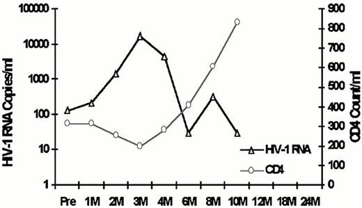
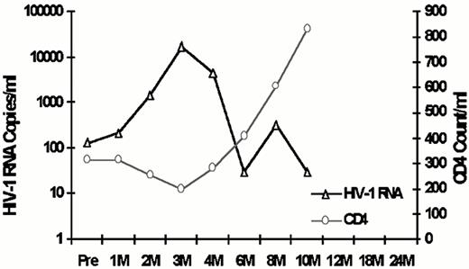
Stem Cell Transplantation for AIDS Lymphoma: A model for gene marking. At City of Hope, 5 volunteers with AIDS lymphoma underwent autologous stem cell transplantation and received, in addition to unmanipulated stem cells, selected CD34+ cells transduced with a retrovirus encoding ribozymes targeted to tat and rev. These patients all received the City of Hope CBV conditioning regimen and are included in Table 4 as UPN 202-204, UPN208, and UNP209. As a safety study, the regimen-related side effects, the CD4 counts, and HIV RNA levels were followed. As with the larger group, all subjects engrafted early and no significant toxicity occurred in the first 3 months post-transplant. These patients showed a transient increase in blood HIV levels, with a rise in HIV load immediately after the transplantation and return to baseline within 10 months (Figure 1 ). Subjects were maintained on HAART during the transplant period, and, despite the CBV-regimen-related nausea and vomiting, there were few missed doses of antiviral drugs. In the first 3 months post-transplant, the CD4 counts decreased, returned to baseline within 8 months post-SCT, and have risen above baseline by 10-12 months. Thus, the gene manipulation appeared to be safe and without long-term effect on HIV or on the incidence of significant opportunistic infection (data not shown).
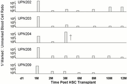
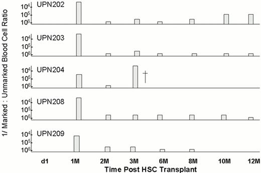
Murine retroviruses used for stem cell transduction in published studies have not resulted in high levels of genetically marked peripheral blood cells.51,52,55 In a prior study at City of Hope, the effect on genetic marking in healthy HIV+ adults who received gene modified stem cells without prior myeloablative conditioning showed minimal transient engraftment of marked cells (data not shown). However, the 5 subjects with AIDS lymphoma undergoing CBV conditioning before stem cell transplantation showed a 10- to 50-fold increase in gene-marked cells post-transplant compared to this group.61 As shown in Figure 2 , the durability of this engraftment was short-lived. When genetic marking was performed in these subjects, there was observable marking in multiple cell lineages during the first 6 months post-transplant, and this declined over the next 6 months to minimum levels of detection. The conclusion from this study is that the retrovirus system used did not efficiently transduce uncommitted stem cells but only committed progenitors, although the late (> 1 yr) observation points continue to be collected. It is unclear whether this was due to the viral vector or to the conditions of the transduction (interleukin-3, interleukin-6, stem cell factor) or to both.
Model for Stem Cell Gene Therapy. With this initial experience, there is the possibility for comparative gene marking using other candidate gene transfer systems as they become available. The setting of AIDS lymphoma and the use of anti-HIV gene systems provides a situation that is both ethical and scientifically sound for development and evaluation of more efficient gene transfer. In this model (see Figure 3; color page 541), the conventional AIDS lymphoma chemotherapy proceeds until demonstration of responsiveness to therapy, and then, following the last course of standard therapy, peripheral blood stem cell are collected and divided into fraction A (unmanipulated cells) and fraction B (CD34 selected cells). The model shows two potential conditioning regimens currently in use. At present, this model has only been used by combining transduced and unmanipulated stem cells (fraction A and B in Figure 3) following conditioning therapy, but eventually, based on future results, it should be possible to transplant only the transduced cells, with the unmanipulated stem cells frozen as back-up material. It is proposed that use of this approach with the next generation of viral vectors will provide an efficient and practical method for comparison of vectors that could have general application to stem cell gene transfer for other hematologic diseases.
Conclusion
While the overall effects on lymphoma free survival from these studies is yet unknown, they do demonstrate that the improvements in antiretroviral therapy and supportive care for HIV-infected individuals now allows the treatment of HIV related lymphoma to be approached in a manner similar to the HIV-negative setting. This includes consideration of autologous stem cell transplantation for those patients with high risk or relapsed lymphoma. In addition, this treatment modality is a model system for evaluating candidate gene vectors for stem cell transplantation.
CD4 count/human immundeficiency virus (HIV) load after stem cell transplantation.
CD4 count/human immundeficiency virus (HIV) load after stem cell transplantation.
DNA PCR detection of transgene from MuLV transduced PBPC transplanted into patients with AIDS lymphoma. Results expressed as reciprocal of the ratio of gene marked cells to unmarked cells in peripheral blood during the first year after transplantation.
DNA PCR detection of transgene from MuLV transduced PBPC transplanted into patients with AIDS lymphoma. Results expressed as reciprocal of the ratio of gene marked cells to unmarked cells in peripheral blood during the first year after transplantation.


