Key Points
We report unique familial cases of CAMT presenting with a novel MPL W272R mutation in the background of the activating MPL K39N mutation.
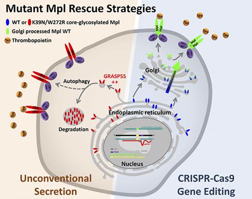
Function of mutant Mpl receptor can be rescued using 2 approaches: autophagic cell surface delivery and CRISPR-Cas9 gene editing.
Abstract
Thrombopoietin (Tpo) and its receptor (Mpl) are the principal regulators of early and late thrombopoiesis and hematopoietic stem cell maintenance. Mutations in MPL can drastically impair its function and be a contributing factor in multiple hematologic malignancies, including congenital amegakaryocytic thrombocytopenia (CAMT). CAMT is characterized by severe thrombocytopenia at birth, which progresses to bone marrow failure and pancytopenia. Here we report unique familial cases of CAMT that presented with a previously unreported MPL mutation: T814C (W272R) in the background of the activating MPL G117T (K39N or Baltimore) mutation. Confocal microscopy, proliferation and surface biotinylation assays, co-immunoprecipitation, and western blotting analysis were used to elucidate the function and trafficking of Mpl mutants. Results showed that Mpl protein bearing the W272R mutation, alone or together with the K39N mutation, lacks detectable surface expression while being strongly colocalized with the endoplasmic reticulum (ER) marker calreticulin. Both WT and K39N-mutated Mpl were found to be signaling competent, but single or double mutants bearing W272R were unresponsive to Tpo. Function of the deficient Mpl receptor could be rescued by using 2 separate approaches: (1) GRASP55 overexpression, which partially restored Tpo-induced signaling of mutant Mpl by activating an autophagy-dependent secretory pathway and thus forcing ER-trapped immature receptors to traffic to the cell surface; and (2) CRISPR-Cas9 gene editing used to repair MPL T814C mutation in transfected cell lines and primary umbilical cord blood–derived CD34+ cells. We demonstrate proof of principle for rescue of mutant Mpl function by using gene editing of primary hematopoietic stem cells, which indicates direct therapeutic applications for CAMT patients.
Introduction
Thrombopoietin (Tpo) and its receptor (Mpl) are the principal regulators of early and late thrombopoiesis and hematopoietic stem cell (HSC) maintenance. Germline or somatic mutations in MPL are thus contributing factors in multiple hematopoietic diseases. Gain-of-function mutations in MPL are associated with myeloproliferative neoplasms (essential thrombocythemia, primary myelofibrosis) and hereditary thrombocytosis, whereas loss-of-function mutations can be directly linked to bone marrow failure syndromes such as congenital amegakaryocytic thrombocytopenia (CAMT). CAMT is a rare inherited syndrome characterized by thrombocytopenia at birth that rapidly progresses to bone marrow failure and pancytopenia. Since the first description of a disease-associated MPL mutation in CAMT in 1999,1 more than 50 different genetic events have been reported for MPL,2,3 and they are sometimes associated with defects in surface presentation.4-6 Notably, the MPL Baltimore substitution (K39N) is associated with high platelet counts in patients of African American descent, despite incomplete processing and reduced Mpl protein levels.7
Although cell surface expression of Mpl is required for stimulation by its ligand (Tpo), complex relationships between mutant and wild-type (WT) forms of Mpl, Jak2, and the endoplasmic reticulum (ER) chaperone calreticulin govern both the intracellular trafficking of receptors and signal propagation.8-10 Prior work has shown that the canonical ER-Golgi route for trafficking of Mpl to the cell surface is aberrant in myeloproliferative neoplasms, linked in part to requirements for WT Jak2 acting as a chaperone.11,12 An alternative pathway to the surface for Mpl is provided by an unconventional autophagy-linked secretory pathway.13 It is important to determine the functional impact of CAMT mutations on Mpl signaling and trafficking, because clinical presentation, disease progression, and treatment options reflect the underlying cellular mechanisms.6,14,15
In this study, the severity of CAMT type I disease in 3 siblings was associated with a homozygous double MPL K39N/W272R mutant that resulted in complete blocking of Mpl trafficking to the plasma membrane. Currently, HSC transplantation is the only curative option for pediatric patients with life-threatening CAMT.15,16 We show that CRISPR-Cas9 gene editing methods17 could be used to correct abnormalities in the MPL gene.
Methods
Patient and healthy donor material
All material from healthy donors (HDs) and patients was obtained after written informed consent, according to institutional guidelines.
Culture of primary cells and cell lines
Murine Ba/F3 and human UT-7 cells were obtained from DSMZ (Braunschweig, Germany) and maintained according to the supplier’s recommendations. CD34+ cells were maintained and expanded in StemSpan SFEM-II media (STEMCELL Technologies) supplemented with thrombopoietin, interleukin-6 (IL-6), Flt-3, and stem cell factor, all at 100 ng/mL (Peprotech).
MPL complementary DNA subcloning and sequencing
Messenger RNA molecules were purified from cord blood (CB) cells isolated from patient II.4. Reverse transcription was performed by using random hexamer primers and Moloney murine leukemia virus reverse transcriptase (Thermo Fisher Scientific), followed by primer-specific complementary DNA (cDNA) amplification, subcloning, and sequencing (for primers sequences, see supplemental Table 1).
cDNA constructs and transfection
Human MPL cDNA fused to mNeonGreen was generated by gene fusion polymerase chain reaction (PCR) using Kapa HiFi Hotstart DNA Polymerase (Kapa Biosystems) and was cloned into pcDNA3.1 (Life Technologies). K39N and W272R mutations were inserted by site-directed mutagenesis (for primer sequences, see supplemental Table 1). The same cloning technique was used to generate the TagRFP-T–tagged human calreticulin cDNA (Sino Biological) into pcDNA6.2. All constructs were checked by sequencing. Transient transfections of Ba/F3 and UT-7 cells were performed by using the Amaxa Nucleofector device (Lonza). Cells (10 × 106) were transfected with 25 µg of plasmid DNA following the supplier’s recommendations. Cells were harvested for experiments 24 hours later. To generate stably expressing Mpl-mNeonGreen (MplmNG) variants, cells were selected by using geneticin and fluorescence-activated cell sorting.
Immunoblots
Cells (10 × 106) were harvested and treated, or not, with recombinant human Tpo (PeproTech) at 50 ng/mL for 10 minutes at 37°C, then lysed in radioimmunoprecipitation assay (RIPA) buffer. Then, 25 μg of total proteins was loaded on 4% to 15% polyacrylamide gradient gels (Bio-Rad) and transferred to nitrocellulose membranes (iBlot 2 device, Life Technologies). After blocking, membranes were probed with primary antibodies for Mpl or β-actin (Millipore), phosphorylated (p)-Jak2, p-Stat1, pStat5, p-Akt, p-Erk1/2, calreticulin (CRT), or calnexin (CANX) (Cell Signaling Technology). In some cases, membranes were stripped once (Thermo Fisher Scientific) and then re-probed. After incubation with horseradish peroxidase–conjugated secondary antibodies, reactive bands were revealed with a chemiluminescent substrate (Thermo Fisher Scientific) and imaged with a Bio-Rad ChemiDoc XRS+ imaging system (Bio-Rad) equipped with Image Lab 4.0.1 software.
Co-immunoprecipitation assay
Cells (25 × 106) were lysed in 1 mL of RIPA buffer (without vortexing) for 20 minutes on ice, then spun down at 13 000g for 20 minutes at 4°C. mNeonGreen-tagged Mpl proteins were immunoprecipitated by using mNeonGreen-nAb–conjugated agarose beads (Allele Biotechnology), following the manufacturer’s instructions, before denaturation in Laemmli buffer and western blot analysis.
Biotinylation assay
For each condition, 25 × 106 parental or transfected UT-7 cells were harvested, washed 3 times in ice-cold phosphate-buffered saline (PBS), and re-suspended in 1 mL of PBS containing 1.5 mg of Sulfo-NHS-Biotin (Thermo Fisher Scientific). Biotinylation reactions proceeded for 30 minutes on ice, followed by 3 ice-cold washes in PBS supplemented with 100 mM glycine. Cells were lysed in RIPA buffer, and biotinylated proteins were immunoprecipitated by using streptavidin-agarose beads (Life Technologies). Samples were evaluated by sodium dodecyl sulfate polyacrylamide gel electrophoresis and western blotting.
Confocal imaging and processing
Confocal images were acquired on a Zeiss LSM 510 META microscope equipped with a 405-nm laser diode, argon and helium-neon 543-nm lasers, a 63× differential interference contrast oil or water objective and ZEN software.
Statistical analysis
Statistical analysis was performed and diagrams were made by using GraphPad Prism 5. Statistical tests are described in the figure legends. Pearson’s pixel intensity correlation over space coefficient, R, of 2D bicolor confocal images was calculated by using the FIJI Coloc_2 plugin.
Genome editing
For editing Ba/F3 cells, a high-scoring guide RNA (gRNA) (gRNA#1WR) was designed by using the CRISPR portal (http://crispr.mit.edu/) and then subcloned into a modified PX458 plasmid (Addgene #48138). For subcloning, 2 partially complementary oligonucleotides (Integrated DNA Technologies) were assembled by PCR. Gel-purified PCR products were cloned into a BbsI-digested PX458 plasmid by using Gibson Assembly (New England Biolabs). After sequencing, the final plasmid with the repair template single-stranded oligo donor DNA (ssODN) W272R>WT #1 was used to electroporate Ba/F3 cells expressing Mpl W272R mutant. Red fluorescent protein (RFP)–expressing cells were selected by flow cytometry and transferred to an IL-3–deprived/Tpo-supplemented media before performing a cell proliferation assay. Editing of UT-7 cells expressing Mpl W272R mutant was conducted by using a double nickase approach. Two complementary gRNAs (#2 and #3) were designed and built similarly to gRNA #1WR. gRNAs #2 and #3 were subcloned into PX461 vectors (Addgene #48140), modified in-house for expression of a cyan fluorescent protein (CFP) or RFP reporter. UT-7 cells were transfected with both gRNAs and the repair template ssODN W272R>WT #2. Doubly transfected cells were flow sorted and plated in the absence of granulocyte-macrophage colony-stimulating factor and in the presence of Tpo before proceeding with a cell proliferation assay. For K562 and primary CD34+ cell editing, a WT-specific version of gRNA #1WR was built (gRNA #1WT) and was subcloned as described before. gRNAs #1WT and #1WR were also produced as in vitro transcribed RNA and then complexed with Sp. Cas9 to form ribonucleoprotein (RNP) complexes as described.18 See supplemental Table 1 for gRNAs, primers, and repair template sequences.
In vitro cutting assay
Sp. Cas9 protein (50 ng; New England Biolabs) was mixed with 250 ng of in vitro–transcribed gRNA (either #1WT or #1WR) and incubated at 37°C for 5 minutes in a PCR device to form RNPs. RNPs were then combined with 150-ng target templates, incubated for 1 hour at 37°C, and then denatured for 5 minutes at 80°C. Reaction products were loaded on a 1.2% agarose gel, stained with GelGreen, and quantified with a Bio-Rad ChemiDoc XRS+ imaging system (Bio-Rad) equipped with Image Lab 4.0.1 software.
Megakaryocytic colony assay
Colony assays were conducted as described.19 Starting materials were CB-derived CD34+ cells from HDs (StemExpress) or study patient II.4, with or without editing.
Flow cytometry
Five days after gene editing, control, II.4, and II.4-edited CD34+ cells were harvested and incubated with human Fc block. Cells were then incubated with an anti-Mpl (CD110)-AlexaFluor 647 dye or an isotype control also coupled to AlexaFluor 647 following the manufacturer’s recommendations. Fc block reagent and antibodies were from BD Biosciences. Data were acquired on a Becton Dickinson LSRFortessa flow cytometer.
Proliferation assay
Parental, stably transfected, or CRISPR-edited cells (Ba/F3 or UT-7) were plated in 96-well plates at 5000 cells per well and incubated for 0 to 120 hours with 10 ng/mL Tpo. Cell proliferation was assessed by using an XTT II colorimetric assay (Roche) following the manufacturer’s recommendations.
Amplicon sequencing by next-generation sequencing
gDNA purified from gene-edited CD34+ cells from patient II.4 was used as a PCR template using MPL exon 5 sequencing primers (for sequences, see supplemental Table 1). Purified PCR products were sequenced using next-generation amplicon sequencing by Genewiz Inc., and results were analyzed by using Integrative Genomics Viewer software (http://software.broadinstitute.org/software/igv).
Results
Study family
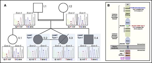
In a French family of Moroccan descent, consanguineous parents (first-degree cousins) and their eldest daughter were found to be heterozygous for 2 MPL mutations: the activating K39N mutation and a previously unreported W272R mutation of unknown function. These 3 individuals had normal platelet counts and were asymptomatic (Figure 1A). Monozygotic twin daughters presented at birth with severe thrombocytopenia (platelet counts: 12 000/L and 14 000/L), low hemoglobin levels (104 g/L and 78 g/L), and very high Tpo levels (3650 pg/mL and 3115 pg/mL) but normal white blood cell counts (13.4 × 10⁹/L and 9.7 × 10⁹/L). Bone marrow smears performed 19 days after birth showed only a single megakaryocyte, indicative of severe megakaryocytopenia. Bone marrow colony formation assays yielded 3 and 9 megakaryocytic colonies vs 84 megakaryocytic colonies per 105 mononuclear cells for the control, leading to a diagnosis of CAMT type I. Whole-blood sequencing revealed the presence of homozygous MPL K39N/W272R mutations. The twins underwent bone marrow transplantation and 1 survived. A younger male sibling was subsequently found to be homozygous for the MPL K39N/W272R mutations. He was diagnosed with CAMT type I and received bone marrow transplantation. Additional follow-up was not possible because the family relocated.
Novel in cis double MPL mutation is associated with familial CAMT type I. (A) Pedigree tree that illustrates the autosomal recessive transmission pattern in this family. Circles represent females and squares represent males. Open symbols indicate healthy family members, filled symbols indicate family members with CAMT type I, single horizontal line connecting 2 symbols indicates monozygotic twins, and slashes represent deceased family members. The genotypes of all family members are presented as genomic DNA sequencing chromatograms. Family members I.1, I.2, and II.1 are heterozygous for the 2 G117T mutation in exon 2 and the T814C mutation in exon 5. Family members II.2, II.3, and II.4 are homozygous for both mutations. (B) Schematic representation of the functional domains of the Tpo receptor and the location of extracellular G117T (K39N) polymorphism and T814C (W272R) mutation. C, cysteine residue; FN III, fibronectin III domain; SP, signal peptide; TM, transmembrane domain; Tyr, tyrosine residue.
Novel in cis double MPL mutation is associated with familial CAMT type I. (A) Pedigree tree that illustrates the autosomal recessive transmission pattern in this family. Circles represent females and squares represent males. Open symbols indicate healthy family members, filled symbols indicate family members with CAMT type I, single horizontal line connecting 2 symbols indicates monozygotic twins, and slashes represent deceased family members. The genotypes of all family members are presented as genomic DNA sequencing chromatograms. Family members I.1, I.2, and II.1 are heterozygous for the 2 G117T mutation in exon 2 and the T814C mutation in exon 5. Family members II.2, II.3, and II.4 are homozygous for both mutations. (B) Schematic representation of the functional domains of the Tpo receptor and the location of extracellular G117T (K39N) polymorphism and T814C (W272R) mutation. C, cysteine residue; FN III, fibronectin III domain; SP, signal peptide; TM, transmembrane domain; Tyr, tyrosine residue.
Novel CAMT-causing W272R MPL mutation is associated in cis with MPL K39N
Sequencing of genomic DNA isolated from peripheral blood from both parents and their 4 children showed the presence of the K39N (G117T) polymorphism in exon 2, along with an exon 5 mutation at W272R (T814C) (Figure 1A). Sequencing of cDNA, generated by reverse transcription PCR of CB-derived messenger RNA, revealed that mutations were associated in cis (13 of 13 cDNA clones). The asymptomatic parents and 1 sibling were heterozygous, whereas the 3 symptomatic children were homozygous for the double MPL mutation. The schematic in Figure 1B shows that both amino acid substitutions are located in the Mpl extracellular domain. The W272R mutation lies within the conserved WGSWS motif.20
Mpl mutants do not respond to ligand stimulation
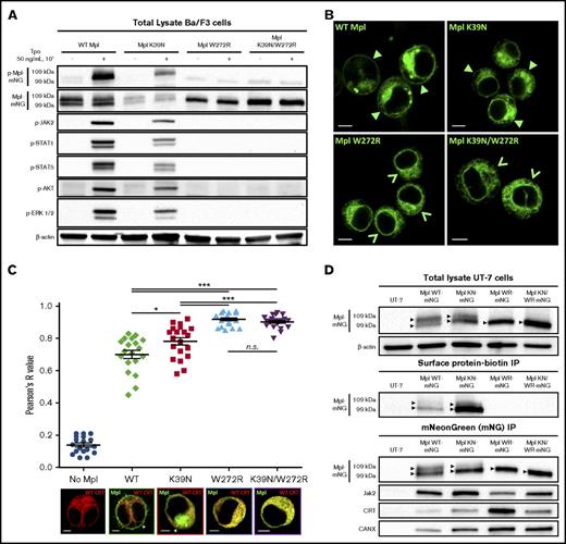
Functional effects of Mpl mutations were investigated in murine Ba/F3 cells and in human megakaryoblastic UT-7 cells. Ba/F3 cells are an IL-3–dependent cell model; UT-7 cells depend on granulocyte-macrophage colony-stimulating factor.21 The relative contributions of the variants upon Mpl trafficking were revealed in live cells expressing Mpl-mNeonGreen fusion proteins (MplmNG) as WT, single substitutions, or double substitutions in both cell lines. Western blotting results in Ba/F3 cells (Figure 2A) showed that the WT and K39N forms of Mpl are signaling competent, based upon elevated levels of phosphorylated signaling proteins after stimulation of cells with Tpo. In contrast, cells expressing Mpl bearing either the single W272R or the double K39N/W272R mutation did not respond to ligand stimulation. Note that WT Mpl migration during gel electrophoresis resolved the receptor into 2 different forms: the upper band represents mature glycosylated Mpl (indicating passage through the Golgi), and the lower band represents core-glycosylated Mpl (ER pattern).13 Both forms were seen in cells expressing WT or K39N Mpl, but only the lower form was seen for Mpl mutants bearing the W272R mutation, indicating failure to progress from the ER to the Golgi.
Mpl W272R and K39N/W272R are absent from the cell surface and do not respond to ligand stimulation. (A) Western blot results for total Mpl protein and phosphorylated (p) signaling partners in cell lysates prepared from transfected Ba/F3 cells with and without Tpo stimulation (50 ng/mL, 10 minutes, 37°C). Labels at the top indicate WT or mutant MplmNG constructs for each cell line tested. (B) Representative confocal images of the live Ba/F3 cells used for western blot characterization in panel A. Closed arrowhead symbols indicate the presence of surface Mpl in cells expressing WT or K39N Mpl. Open arrowhead symbols point to the absence of a clearly defined plasma membrane outline in cells expressing W272R or K39N/W272R mutant Mpl. (C) Image analysis of human UT-7 cells co-expressing MplmNG WT or mutant proteins and the ER-resident protein calreticulin (CRT) fused to TagRFP-T (CRTTagRFP-T). Co-localization of both fluorescent markers was assessed by using Pearson’s analysis of dual-channel confocal images from at least 20 cells for each condition. Means ± standard error of the mean are shown, and pairwise statistical analyses using unpaired Student t test are represented by horizontal bars. Representative images of each cell population are shown at the bottom of the panel. (D) Co-immunoprecipitation (IP) of mutant Mpl proteins with ER-resident proteins CRT and calnexin (CANX) in stably transfected UT-7 cells. Upper bands in the WT and K39N (KN) lanes represent fully glycosylated receptors, indicative of maturation in the Golgi. Scale bars = 5 µm. *P < .05; ***P < .0001. n.s., not significant.
Mpl W272R and K39N/W272R are absent from the cell surface and do not respond to ligand stimulation. (A) Western blot results for total Mpl protein and phosphorylated (p) signaling partners in cell lysates prepared from transfected Ba/F3 cells with and without Tpo stimulation (50 ng/mL, 10 minutes, 37°C). Labels at the top indicate WT or mutant MplmNG constructs for each cell line tested. (B) Representative confocal images of the live Ba/F3 cells used for western blot characterization in panel A. Closed arrowhead symbols indicate the presence of surface Mpl in cells expressing WT or K39N Mpl. Open arrowhead symbols point to the absence of a clearly defined plasma membrane outline in cells expressing W272R or K39N/W272R mutant Mpl. (C) Image analysis of human UT-7 cells co-expressing MplmNG WT or mutant proteins and the ER-resident protein calreticulin (CRT) fused to TagRFP-T (CRTTagRFP-T). Co-localization of both fluorescent markers was assessed by using Pearson’s analysis of dual-channel confocal images from at least 20 cells for each condition. Means ± standard error of the mean are shown, and pairwise statistical analyses using unpaired Student t test are represented by horizontal bars. Representative images of each cell population are shown at the bottom of the panel. (D) Co-immunoprecipitation (IP) of mutant Mpl proteins with ER-resident proteins CRT and calnexin (CANX) in stably transfected UT-7 cells. Upper bands in the WT and K39N (KN) lanes represent fully glycosylated receptors, indicative of maturation in the Golgi. Scale bars = 5 µm. *P < .05; ***P < .0001. n.s., not significant.
Mpl W272R and K39N/W272R are retained in the ER
Confocal microscopy studies (Figure 2B) confirmed that only WT MplmNG and Mpl K39NmNG reached the cell surface in transfected Ba/F3 cells (solid arrowheads). In contrast, the chimeric MplmNG proteins bearing the W272R mutation, alone or together with the K39N mutation, showed no detectable surface expression of the Tpo receptor. Open arrowheads (Figure 2B, lower panels) indicate the absence of a clearly labeled plasma membrane in these cells. Instead, we observed a reticular distribution of W272R and K39N/W272R mutant Mpl that is consistent with retention in the ER. After biotinylation of surface proteins, anti-biotin immunoprecipitation confirmed the absence of Mpl at the surface of cells expressing W272R mutants (Figure 2D, center panel). Further proof of the trafficking defect of mutant Mpl was obtained by co-expressing WT and mutant MplmNG in UT-7 cells, together with WT calreticulin fused to RFP as an ER marker (Figure 2C). We applied spatial statistics methods (Pearson’s R value) to show a significantly higher colocalization of mutant Mpl W272RmNG proteins with the ER marker compared with either WT or K39N receptors. A statistically significant difference in colocalization of Mpl K39NmNG with the ER compared with WT MplmNG was also noted. Finally, results show increased co-precipitation of the mutant Mpl’s with the ER resident chaperones CRT and CANX, particularly those receptors with a single mutation at W272R (Figure 2D).
Autophagy-based functional rescue of mutant Mpl surface expression
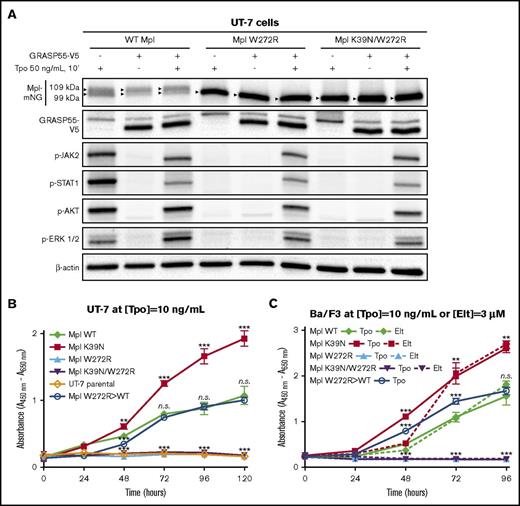
In a previous study that focused on the complex intracellular trafficking pathways of Mpl, we discovered that immature, core-glycosylated Mpl proteins were able to traffic from the ER to the plasma membrane by using an autophagy-based secretory pathway.13 This unconventional route bypasses the canonical ER-to-Golgi-to-plasma membrane delivery route for glycosylated proteins and can be promoted in cells by overexpressing Golgi reassembly stacking protein 55 (GRASP55). Because autophagy provides an alternative pathway for misfolded proteins to reach the cell surface,22 we tested the hypothesis that overexpression of GRASP55 might partially restore Tpo-induced signaling in cells expressing the doubly substituted Mpl K39N/W272R. Figure 3A shows results in UT-7 cells stably expressing either WT or mutant Mpl, followed by transient overexpression of V5-tagged GRASP55. GRASP55 overexpression overrode the block in signaling for the double Mpl mutant, resulting in phosphorylation of Jak2, STAT1, AKT, and Erk after Tpo challenge. Levels of stimulation were comparable to that of cells expressing WT Mpl. Results show that the doubly mutated, core-glycosylated Mpl can bind ligand and initiate signaling if it is forced to the cell surface through the autophagic secretory pathway.
Gene editing or autophagic delivery of mutant Mpl to the cell surface rescue receptor function in vitro. (A) Transient overexpression of GRASP55 tagged with a V5 epitope results in accumulation of the lower-molecular-weight core-glycosylated form of Mpl regardless of WT or mutant status. Receptors are shown to be signaling competent on the basis of phosphorylation of key signaling proteins in the Jak/STAT and PI3K pathways in response to Tpo. (B-C) XTT-II proliferation assays performed on UT-7 or Ba/F3 cell lines expressing WT or mutant MplmNG and selected for growth in the presence of Tpo (panel C, solid lines) or eltrombopag (Elt) (panel C, dotted lines). CRISPR-Cas9–edited cells that were reverse-engineered to restore the WT sequences in MPL exon 5 from the mutated W272R sequence (labeled Mpl W272R>WT) are represented by blue open circles. UT-7 cells were edited by using the D10A Cas9 mutant and 2 single gRNAs in a double nickase approach. A classical WT Cas9 approach (ie, coupled to a unique single gRNA) was used to edit Ba/F3 cells. **P < .005; ***P < .0001.
Gene editing or autophagic delivery of mutant Mpl to the cell surface rescue receptor function in vitro. (A) Transient overexpression of GRASP55 tagged with a V5 epitope results in accumulation of the lower-molecular-weight core-glycosylated form of Mpl regardless of WT or mutant status. Receptors are shown to be signaling competent on the basis of phosphorylation of key signaling proteins in the Jak/STAT and PI3K pathways in response to Tpo. (B-C) XTT-II proliferation assays performed on UT-7 or Ba/F3 cell lines expressing WT or mutant MplmNG and selected for growth in the presence of Tpo (panel C, solid lines) or eltrombopag (Elt) (panel C, dotted lines). CRISPR-Cas9–edited cells that were reverse-engineered to restore the WT sequences in MPL exon 5 from the mutated W272R sequence (labeled Mpl W272R>WT) are represented by blue open circles. UT-7 cells were edited by using the D10A Cas9 mutant and 2 single gRNAs in a double nickase approach. A classical WT Cas9 approach (ie, coupled to a unique single gRNA) was used to edit Ba/F3 cells. **P < .005; ***P < .0001.
Tpo analog eltrombopag fails to promote proliferation of mutant Mpl-bearing cells
The ability of UT-7 and Ba/F3 cell lines to proliferate in the presence of Tpo (10 ng/mL) was evaluated (Figure 3B-C). UT-7 and Ba/F3 cells expressing gain-of-function Mpl K39NmNG (red filled squares) showed a significant increase in proliferation compared with WT MplmNG- expressing cells (green filled diamonds). Consistent with results in Figure 2A, cells expressing Mpl W272RmNG or Mpl K39N/W272RmNG remained unresponsive to Tpo (cyan and purple filled triangles, respectively). Data in Figure 3C show that Ba/F3 cells expressing WT and K39N Mpl displayed similar growth curves when treated with the membrane-impermeable Tpo analog eltrombopag (dotted lines) compared with Tpo (solid lines). Ba/F3 cells expressing the W272R mutants were unresponsive to eltrombopag.
Genome engineering restores Tpo response in MPL W272R–transfected cell lines
CRISPR-Cas9 engineering methods were used to correct the W272R mutation in transfected cells and restore the WT sequence. As an initial proof of principle, expression plasmids in Ba/F3 cells were edited by using a unique single gRNA coupled to the WT Cas9 enzyme and an RFP reporter. Expression plasmids in UT-7 cells were edited by using a multiplex double nickase approach, which consists of 2 separate single gRNAs, each paired with either a CFP or an RFP reporter and coupled to a mutated Cas9 enzyme (Cas9 with the D10A mutation). For both editing strategies, an asymmetric ssODN was used as a repair template to maximize homology-directed (HDR) editing.23 Figure 3 shows results for the restoration of growth competence to Tpo in edited cells (labeled Mpl W272R>WT; blue open circles). Figure 3B shows data for UT-7 cells, and Figure 3C shows data for Ba/F3 cells. Successfully edited cells recovered growth competence in the presence of Tpo, with growth rates similar to those of WT MplmNG cells (green filled diamonds; Figure 3C-D).
Efficient editing of the MPL locus in K562 and CD34+ cells
To test our gene editing tools, we next applied our gRNA/WT Cas9 editing approach to human K562 cells and primary HSCs. Our goals were to confirm targeting of the MPL locus and to demonstrate the potential to restore megakaryocytic progenitor capabilities in CAMT patient–derived CD34+ cells.
Editing strategies in K562 cells
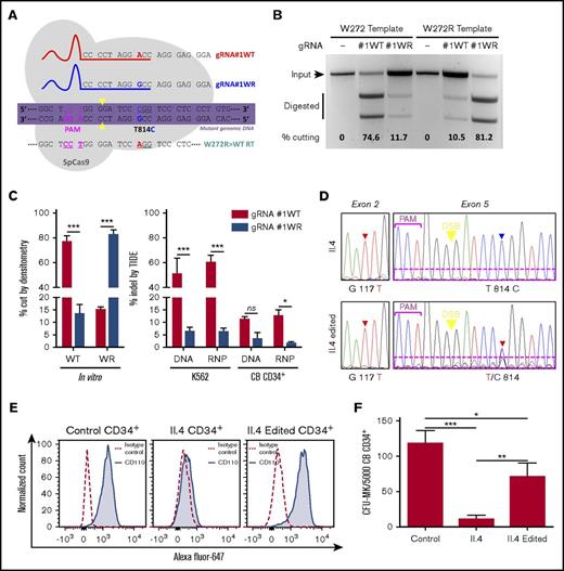
Because human K562 cells are homozygous for WT MPL, we designed a WT version of gRNA#1WR (named gRNA#1WT). Both gRNAs differ by only 1 bp corresponding to the T814C mutation on MPL that is spanned by the gRNAs, thus creating a WT-specific and a mutant (WR)-specific gRNA. The schematic in Figure 4A represents gRNA#1WT and gRNA#1WR, the W272R>WT repair template, and the W272R mutated genomic MPL sequence. Cas9 WT, the proto-adjacent motif (PAM), and cleavage sites are also represented. Although they are still difficult to predict, single bp mismatches located 1 to 12 bp in the PAM-proximal region of a gRNA can drastically impact its specificity.24 To test the specificity of gRNA#1WT and gRNA#1WR, we conducted an in vitro cutting assay using WT or WR templates. Figure 4B shows that both gRNAs in the presence of their respective templates exert a very good and similar cutting efficiency nearing 80% (quantified in Figure 4C, left panel). However, we also noticed that both gRNAs were able to cut their mismatch target to a nonnegligible level (about 15%), indicating the potential for a strong off-target effect of gRNA#1WR on the WT MPL sequence if it is used in a heterozygous cell-editing setup.
Gene editing in K562 cells and primary CD34+ cells. (A) Schematic of sequence-specific gRNA#1WT and gRNA#1WR and their target genomic MPL sequence representing the protospacer-adjacent motif (PAM), double-strand break site (yellow arrowheads), and the W272R single point mutation site (T814C). DNA codons are underlined, and the repair template (RT) used to convert the W272R mutation to the WT sequence (W272R>WT) is also represented. (B) Example of in vitro digestion assay with gRNA#1WT or gRNA#1WR in the presence of their match or mismatch target sequences. Quantification of cutting efficiency was performed by using densitometry analysis. (C) Left panel shows quantification of in vitro cutting capabilities of gRNA#1WT and gRNA#1WR. Right panel shows quantification of the percentage of indel formation obtained with gRNA#1WT and gRNA#1WR when delivered as plasmid DNA or RNP complexes in K562 or CB CD34+ cells. (D) Control, unedited, and edited CD34+ cells isolated from patient II.4 were sequenced at day 5 after editing. G117T represents the K39N mutation and T814C represents the W272R mutation. Dotted magenta rectangles highlight the presence of additional overlapping sequences in edited cells for the T814C locus, indicating an off-target effect. (E) Flow cytometry analysis of anti-Mpl (CD110)-AlexaFluor-647 binding on control CD34+ cells, unedited patient II.4 CD34+ cells, or edited II.4 CD34+ cells at day 5 after editing. (F) In vitro megakaryocytic colony formation assay conducted in the presence of Tpo with the same cell samples used in panel E. *P < .05; **P < .005; ***P < .0001. CFU, colony-forming unit; DSB, double-strand break.
Gene editing in K562 cells and primary CD34+ cells. (A) Schematic of sequence-specific gRNA#1WT and gRNA#1WR and their target genomic MPL sequence representing the protospacer-adjacent motif (PAM), double-strand break site (yellow arrowheads), and the W272R single point mutation site (T814C). DNA codons are underlined, and the repair template (RT) used to convert the W272R mutation to the WT sequence (W272R>WT) is also represented. (B) Example of in vitro digestion assay with gRNA#1WT or gRNA#1WR in the presence of their match or mismatch target sequences. Quantification of cutting efficiency was performed by using densitometry analysis. (C) Left panel shows quantification of in vitro cutting capabilities of gRNA#1WT and gRNA#1WR. Right panel shows quantification of the percentage of indel formation obtained with gRNA#1WT and gRNA#1WR when delivered as plasmid DNA or RNP complexes in K562 or CB CD34+ cells. (D) Control, unedited, and edited CD34+ cells isolated from patient II.4 were sequenced at day 5 after editing. G117T represents the K39N mutation and T814C represents the W272R mutation. Dotted magenta rectangles highlight the presence of additional overlapping sequences in edited cells for the T814C locus, indicating an off-target effect. (E) Flow cytometry analysis of anti-Mpl (CD110)-AlexaFluor-647 binding on control CD34+ cells, unedited patient II.4 CD34+ cells, or edited II.4 CD34+ cells at day 5 after editing. (F) In vitro megakaryocytic colony formation assay conducted in the presence of Tpo with the same cell samples used in panel E. *P < .05; **P < .005; ***P < .0001. CFU, colony-forming unit; DSB, double-strand break.
We then compared 2 different CRISPR-Cas9 delivery methods (plasmid DNA and RNPs) to edit K562 cells and HD CB CD34+ cells. In both cases, we used electroporation to deliver either 0.5 µg of plasmid DNA or 200 pmol of RNPs per 200 000 cells. Editing efficiency was measured at 24 to 48 hours posttransfection by using the tracking of indels by decomposition (TIDE) Web tool. This tool performs Sanger sequencing trace file deconvolution to measure the percentage of indel formation after nonhomologous end joining repair of a Cas9-induced double-strand break. Figure 4C (right panel) shows that gRNA#1WT performed equally well when delivered as either a plasmid DNA or an RNP in K562 cells. Both delivery systems were also found comparable in inducing indel formation when editing CB CD34+ cells, with a four- to sixfold decrease in efficiency compared with editing in K562 cells (∼50% and ∼10% of indels, respectively). As noted in the in vitro cutting assay, a significant 2% to 9% of off-target cutting was measured for gRNA#1WR when delivered to WT MPL cells.
Gene editing rescues double K39N/W272R Mpl mutant function in primary hematopoietic CD34+ cells
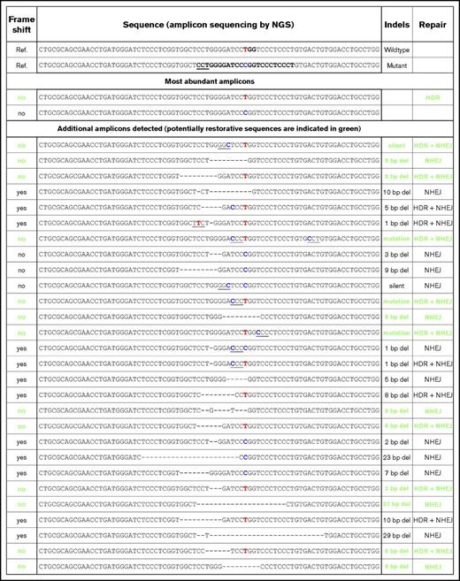
Our gene editing approach was next applied to primary HSCs isolated from the umbilical CB of patient II.4. After thawing, 60 000 CD34+ cells homozygous for the double K39N/W272R Mpl mutation were obtained from 104 × 106 CB mononuclear cells (0.058%), consistent with a CAMT type I diagnostic. Twenty-four hours after isolation, two-thirds of the cells were subjected to gene editing using 40 pmol of gRNA#1WR/Cas9 RNPs and the WR>WT repair template. Parental and edited cells were then maintained in CD34+ expansion media, containing (among other cytokines) 100 ng/mL of Tpo for 5 days before being used in functional assays to assess editing efficiency. Figure 4D shows the sequencing results of unedited (top panels) and edited (lower panels) cells at day 5 after editing. The homozygous K39N mutation could be found in both populations, as expected. However, in the edited cell population, the W272R mutation seemed to be mostly heterozygous. We concluded that most of the edited cells contained only 1 modified copy of MPL, reverting the sequences from R272 to W272. It is also interesting to note the presence of additional sequencing traces in the lower right panel of Figure 4D compared with the upper right panel (magenta rectangle), indicating off-target effects of gRNA#1WR being able to cut a previously edited sequence. This likely reflects the use of nonhomologous end joining (NHEJ) instead of HDR editing as a repair mechanism by the cell machinery. To determine the extent to which other repair mechanisms could generate restorative sequences in the MPL locus, we performed amplicon sequencing by next-generation sequencing. Data in Figure 5 indicate that several alternatively edited alleles, obtained by NHEJ only or a combination of HDR editing and NHEJ, are capable of encoding functional Mpl proteins. These alternate alleles are composed mostly of in-frame deletions of the mutant residue.
Next-generation sequencing of MPL exon 5 PCR amplicons after gene editing. Amplicons generated from gDNA obtained from patient II.4 edited CD34+ cells were subjected to next-generation sequencing. Sequences, other than properly HDR edited or unedited, that can potentially yield functional Mpl proteins are indicated in green.
Next-generation sequencing of MPL exon 5 PCR amplicons after gene editing. Amplicons generated from gDNA obtained from patient II.4 edited CD34+ cells were subjected to next-generation sequencing. Sequences, other than properly HDR edited or unedited, that can potentially yield functional Mpl proteins are indicated in green.
We used 2 different assays to show that only 1 functional allele was required for edited cells to deliver functional Mpl to the cell surface and proliferate in the presence of Tpo, regardless of whether the repair was obtained through the HDR editing or the NHEJ mechanism. Receptors on the surface of edited cells were detected by using a fluorescently labeled anti-Mpl antibody and were quantified by flow cytometry. As shown in Figure 4E, control CD34+ and II.4 edited CD34+ cells displayed similar amounts of surface Mpl proteins, whereas unedited II.4 CD34+ cells did not bind Mpl antibodies. We specifically selected for cells with surface expression of Mpl by maintaining cultures before and after editing in high Tpo conditions (100 ng/mL) as the sole growth factor. Finally, single-colony assay results (Figure 4F) demonstrated that edited CD34+ cells from patient II.4 were able to generate a significantly higher number of megakaryocytic colonies in the presence of Tpo than unedited cells from this patient and compared with control CB CD34+ cells. Results observed in this experiment reflect the presence of both efficiently edited cells and cells bearing alternate MPL exon 5 sequences that restored functionality. We speculate that alternative exon 5 rearrangements may couple in cis with the activating K39N mutation, which provides edited cells with a proliferation advantage superior to that of unedited CD34+ cells. Growth curves of cell lines that express Mpl K39N support this hypothesis (Figure 3B-C).
Discussion
To the best of our knowledge, this is the first report of a double mutation in MPL (G117T/T814C) that results in intracellular retention of Mpl receptors in the ER and the first study to fully characterize the pathogenic mechanism behind the association of this mutation with CAMT. In this CAMT type I family, the Mpl W272R mutation was present in cis with the activated K39N variant associated with hereditary thrombocytosis. We show that the trafficking defect of the Mpl W272R receptor effectively prevents surface expression and thus Tpo-mediated signaling. Although receptors bearing only the K39N mutation are also partially retained in the ER, a fraction of this hyperactive form of Mpl does reach the cell surface. Hence the absence of thrombocytosis for the 3 family members bearing the Mpl K39N polymorphism in a heterozygous fashion can be explained by the in cis loss-of-function W272R mutation and 1 normal copy of the MPL gene. Other combinations of the W272R mutation with another in cis genetic alteration in a heterozygous setting would likely give rise to a similar phenotype because the W272R mutation is responsible for the Mpl trafficking defects. For the 3 children who were homozygous for Mpl K39N/W272R, the W272R mutation overrides the activating K39N mutation because the Mpl receptors are retained in the ER and cannot respond to extracellular ligand. Consistent with reports of defective feedback regulation of circulating Tpo in CAMT,25 family members who are homozygous for the MPL K39N/W272R mutant presented with highly elevated Tpo levels in blood and a deficit in 2 hematopoietic lineages. The single point mutation T814C of MPL has recently been identified in a database generated by whole-exome sequencing of gastric cancer patients,26 indicating that additional cases may occur.
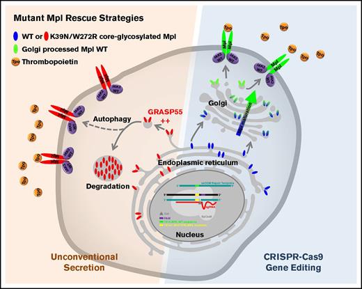
Importantly, we were able to design and test several strategies to rescue Mpl receptor trafficking and function (summarized in Figure 6). Like the most common CFTR mutation (DeltaF508) in cystic fibrosis,27 the doubly mutated Mpl protein retains partial functionality. GRASP55 overexpression enabled Mpl K39N/W272R to reach the surface by the autophagy secretory pathway22 where it could bind Tpo and trigger JAK-STAT–mediated signaling (Figure 3A). This approach may eventually be translated into patient treatment now that recent drug screening studies have identified compounds that activate autophagy.28 We also demonstrated that CRISPR-Cas9 gene-editing methods can be used to repair a disease-causing mutation in the Mpl coding sequence. We showed that transfected cell lines expressing mutant Mpl were efficiently edited to restore Mpl WT sequence trafficking to the cell surface and response to Tpo. Because CAMT patients typically have high levels of circulating Tpo,25 even a partial recovery of Mpl surface expression might normalize hematopoiesis or, at a minimum, provide additional time to match donors for a bone marrow transplantation.
Functional rescue strategies for Mpl mutants. Schematic summary of the 2 rescue approaches used to restore Mpl function: (1) overexpression of GRASP55 to force immature Mpl receptor expression at the cell surface using unconventional autophagy-dependent secretion and (2) CRISPR-Cas9 gene editing to convert mutated Mpl DNA sequence to WT sequence. sgRNA, single-guide RNA.
Functional rescue strategies for Mpl mutants. Schematic summary of the 2 rescue approaches used to restore Mpl function: (1) overexpression of GRASP55 to force immature Mpl receptor expression at the cell surface using unconventional autophagy-dependent secretion and (2) CRISPR-Cas9 gene editing to convert mutated Mpl DNA sequence to WT sequence. sgRNA, single-guide RNA.
We also applied CRISPR-Cas9 to primary CB-derived hematopoietic cells from study patient II.4 and were able to partially restore WT MPL sequence. Edited cells displayed a normal surface expression of the receptor and could generate in vitro megakaryocytic colonies. Despite a mutation-specific design of gRNA#1WR, we could see significant amounts of off-target effect in edited cells. A silent mutation of the PAM sequence from NGG to NTG,29 carried by the ssODN repair template, would likely circumvent this issue, although without allowing a truly scarless editing strategy. In addition, we found that alternate genomic sequences, potentially restorative of Mpl function, were also induced by our gene editing strategy. Some of these sequences resulted from a combination of HDR editing and NHEJ, whereas others were the result of NHEJ alone. This is an important finding, which indicates that alternate editing strategies can be developed and refined to restore Mpl function. Hence, our study provides in vitro proof‐of‐principle that MPL mutations detected in CAMT patients may be corrected by modern gene engineering methods and/or by autophagy-activating drugs. The promise of gene therapy for CAMT and other hereditary hematologic disorders awaits successes in the rapidly advancing field of genome editing of patient-derived stem cells.17,30 Because only a subset of hematopoietic progenitor cells may need to be edited to achieve long-term polyclonal hematopoiesis, this approach could represent a cure for the majority of CAMT patients.
The full‐text version of this article contains a data supplement.
Acknowledgments
The authors thank Matthew L. Fero for critical reading of the manuscript, Shayna R. Lucero for expert cell culture support, Isabelle Guiraud for excellent technical work on cDNA cloning, and Sara Girault for sequencing the patient’s genomic DNA.
This work was supported in part by grants from the Department of Defense, Congressionally Directed Medical Research Program (CA140409) (C.C. and B.S.W.), the American Cancer Society (126768-IRG-14-187-19) (C.C.), and Ligue contre le Cancer, comité du Gard (S.C.). Images in this paper were generated in the University of New Mexico (UNM) Cancer Center Fluorescence Microscopy Shared Resource supported by National Institutes of Health, National Cancer Institute (NCI) grant P30CA118110 and National Institutes of Health, National Institute of General Medical Science grant P50GM085273. Data were generated in the UNM Shared Flow Cytometry and High Throughput Screening Resource Center supported by the UNM Health Sciences Center and the UNM Cancer Center with support from NCI grant 2P30CA118100-11.
Authorship
Contribution: E.J. and T.L.-B. enrolled study patients; C.C., R.G., E.H.C., and S.C. performed experiments; C.C., R.G., S.C., and B.S.W. analyzed results and created the figures; C.C. and B.S.W. designed the research; and C.C., S.H., and B.S.W. wrote the paper with contributions from all authors.
Conflict-of-interest disclosure: The authors declare no competing financial interests.
Correspondence: Cédric Cleyrat, Department of Pathology and Comprehensive Cancer Center, University of New Mexico Health Sciences Center, MSC08-4640, Cancer Research Facility, Room 207A, Albuquerque, NM 87131-0001; e-mail: ccleyrat@salud.unm.edu.
References
Author notes
S.C. and B.S.W. contributed equally to this study.

