Key Points
R-BAC is associated with high rate of sustained remissions in older patients with MCL.
After 7 years of follow-up, the median OS and PFS were not reached, with no signal of late toxicity.
Abstract
The combination of rituximab, bendamustine, and low-dose cytarabine (R-BAC) has been studied in a phase 2 prospective multicenter study from Fondazione Italiana Linfomi (RBAC500). In 57 previously untreated elderly patients with mantle cell lymphoma (MCL), R-BAC was associated with a complete remission rate of 91% and 2-year progression-free survival (PFS) of 81% (95% confidence interval [CI], 68-89). Here, we report the long-term survival outcomes, late toxicities, and results of minimal residual disease (MRD) evaluation. After a median follow-up of 86 months (range, 57-107 months), the median overall survival (OS) and PFS were not reached. The 7-year PFS and OS rates were 55% (95% CI, 41-67), and 63% (95% CI, 49-74), respectively. Patients who responded (n = 53) had a 7-year PFS of 59% (95% CI, 44-71), with no relapse or progression registered after the sixth year. In the multivariate analysis, blastoid/pleomorphic morphology was the strongest adverse predictive factor for PFS (P = .04). Patients with an end of treatment negative MRD had better, but not significant, outcomes for both PFS and OS than patients with MRD-positive (P = 0.148 and P = 0.162, respectively). There was no signal of late toxicity or an increase in secondary malignancies during the prolonged follow-up. In conclusion, R-BAC, which was not followed by maintenance therapy, showed sustained efficacy over time in older patients with MCL. Survival outcomes compare favorably with those of other immunochemotherapy regimens (with or without maintenance), including combinations of BTK inhibitors upfront. This study was registered with EudraCT as 2011-005739-23 and at www.clinicaltrials.gov as #NCT01662050.
Introduction
Mantle cell lymphoma (MCL) is an aggressive histotype of non-Hodgkin lymphoma characterized by continuous relapses over time, with no standard frontline therapy. Therapeutic choices for patients who are ineligible for transplants include R-CHOP (rituximab, cyclophosphamide, doxorubicin, vincristine, and prednisolone) or bendamustine and rituximab (BR),1-3 both followed by rituximab maintenance.4,5
Compared with R-CHOP, BR is currently recommended as the preferred first-line regimen in contemporary clinical practice guidelines.6 The R-BAC regimen, which is based on the addition of an intermediate dose of cytarabine to BR,7 has also been included in clinical guidelines.6,8 This combination has been supported by preclinical studies showing that bendamustine and cytarabine have distinct and synergistic mechanisms of action, especially when administered sequentially.9 Between 2012 and 2014, the Fondazione Italiana Linfomi (FIL) conducted a phase 2 multicenter trial (RBAC500),7 analyzing the efficacy and safety of the R-BAC regimen (rituximab, bendamustine, and intermediate-dose cytarabine) in patients with MCL not eligible for autologous transplants. R-BAC was not followed by rituximab maintenance. The primary analysis of the study, with a median follow-up of 35 months, showed a high CR rate (91%), with a 2-year overall survival (OS) of 86% (95% confidence interval [CI], 74-93), and progression-free survival (PFS) of 81% (95% confidence interval [CI], 68-89). Despite this relevant antitumoral activity, R-BAC was more toxic than BR, particularly in terms of the hematotoxicity between cycles. A recent real-life report confirmed that R-BAC was significantly more effective than BR, but more toxic, with doses that were frequently reduced to spare hematotoxicity.10 Finally, the phase 3 randomized SHINE study showed that the addition of ibrutinib to BR, as compared with placebo, followed by double maintenance, significantly improved the PFS in older patients with MCL.11 With this in mind, we performed a long-term analysis on the efficacy and toxicity end points of the RBAC500 trial.
Methods
Study design and participants
The RBAC500 was a multicenter, single-arm, phase 2 study that recruited patients who were previously untreated and had a confirmed histological diagnosis of MCL. The study involved 29 FIL centers. To be included, patients had to be older than 65 years and fit per the comprehensive geriatric assessment, or aged from 60 to 65 years if they were ineligible for high-dose chemotherapy plus autologous stem-cell transplant, fit or unfit per the comprehensive geriatric assessment (for the modified comprehensive geriatric assessment, see Appendix 2 of the original report).7 The diagnostic criteria of MCL included positivity for cyclin D1 and SOX11 expression (mandatory in patients who were cyclin D1 expression or t [11;14] negative). Patients with in-situ MCL or non-nodal leukemic disease were excluded; for the complete list of inclusion and exclusion criteria see the original report.7
This study was conducted in accordance with the principles of the Declaration of Helsinki and Good Clinical Practice. Ethics approval was granted by the institutional review board of each participating institution, and all patients provided written informed consent.
Procedures
Baseline assessment included bone marrow (BM) biopsy, tumor staging with contrast-enhanced computed tomography (CT), and positron emission tomography. Paraffin blocks of the diagnostic specimens were collected for central pathological review, which was performed by an expert hematopathologist (S.A.P.), in accordance with the criteria of the World Health Organization classification.12 All patients received RBAC500 (rituximab 375 mg/m2 on day 1; bendamustine 70 mg/m2 over 30-60 minutes on days 2 and 3; cytarabine 500 mg/m2 over a 2 hour infusion starting 2 hours after bendamustine from days 2 to 4; all administered IV) every 4 weeks for up to 6 cycles. None of the patients received rituximab maintenance. Prophylaxis and toxicity management were described in the original paper7 with granulocyte colony-stimulating factor, which was mandatory following each cycle. Patients who did not respond to the first 2 cycles were excluded from the study. Response assessment was performed per the Lugano criteria.13 Follow-up, including clinical evaluation, laboratory tests, and CT, was performed every 3 months for the first year after the end of treatment and then every 6 months for 2 more years. After the third year, the patients were followed up with visits per the local hematologists. Patients were required to be examined at least 2 times per year for clinical examination and blood tests. A CT was mandatory for any clinical suspicion of relapse, appearance of adenopathies, organomegaly, or alterations in the blood exams. Queries were sent to the centers twice a year by FIL offices to update the state of the disease and inform them of late toxicities or secondary malignancies (SMs).
Centralized assessment of minimal residual disease (MRD) was performed in the EuroMRD standardized laboratory of the University of Torino (Torino, Italy) at diagnosis, before cycle 3, and at 1 month, 6 months, 12 months, and 24 months after the end of treatment. We used allele-specific oligonucleotide-droplet digital polymerase chain reaction (ddPCR) analysis14 to assess MRD in BM and peripheral blood (PB) samples, using patient-specific primers and consensus probes to detect immunoglobulin heavy-chain (IGH) variable region gene rearrangement or the IGH::BCL1 product of the t (11;14) translocation. The sensitivity of MRD detection via ddPCR was 2E-05, allowing for the detection of a single event among 75 000 cells analyzed. For survival analysis, a positive sample in at least 1 of the tissues analyzed (either BM or PB) was defined as MRD positivity.
The primary efficacy and safety outcomes were reported in the original article.7 For this study, we estimated the long-term OS and PFS as primary efficacy outcomes. The toxicity was analyzed in terms of SMs or any other toxicity reported by the investigators during the follow-up period.
Statistical analysis
Patient clinical characteristics and demographics were summarized using descriptive statistics. PFS and OS were estimated using the Kaplan–Meier method, and the groups were compared using the log-rank test. The OS from the progression of disease (POD; OS-2) was defined as the time from POD to death due to any cause. Multivariable analysis was performed using Cox regression models. Toxicity, completed treatment rates, and treatment response rates were compared using χ2 or Fisher test. The cumulative incidence of SM was estimated using the method proposed by Gooley et al, 115 considering death from any cause as a competing event.
Results
Demographic features, cycle delivery, and dose reductions
Between May 2012 and February 2014, 57 patients from 29 centers were consecutively enrolled in the RBAC500 trial. The main characteristics of these patients, whose median age was 71 years (range, 61-79 years), were described in the original report.7 Briefly, 25 patients (44%) had high-risk MCL International Prognostic Index (MIPI), 16 (31%) had ki67 ≥30%, and 14 (25%) had blastoid/pleomorphic morphology. None of the patients had non-nodal leukemic disease per the inclusion criteria. Overall, 54 (95%) patients received at least 4 cycles of RBAC500, and 38 (67%) recieved 6 cycles, with a median of 6.0 (range, 2-6) cycles per patient (for distribution of patients and treatment see Figure 1, “Trial Profile” from the original report7). The majority of the patients (75%) had dose reductions of the R-BAC regimen as per the protocol, which mostly consisted of avoiding the third day of cytarabine.
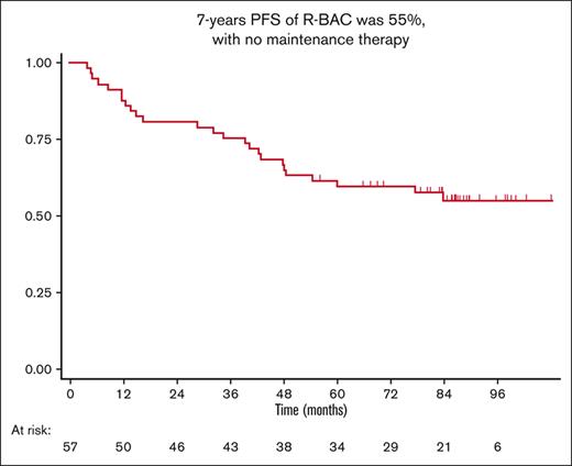
Survival curves at a median follow-up of 86 months, and MRD evaluation. (A) PFS of all patients (7-year PFS, 55% [95% CI, 41-67]); (B) OS of all patients (7-year OS, 63% [95% CI, 49-74]); (C) Duration of response for the 52 responding patients (7-year duration of response, 59% [95% CI, 44-71]); (D) PFS (7-year PFS, 65% vs 40%; P = .14) and (E) OS (7-year OS, 65% vs 45%; P = .16) based on the MRD results at the end of the treatment (blue curve, positive; red curve, negative).
Survival curves at a median follow-up of 86 months, and MRD evaluation. (A) PFS of all patients (7-year PFS, 55% [95% CI, 41-67]); (B) OS of all patients (7-year OS, 63% [95% CI, 49-74]); (C) Duration of response for the 52 responding patients (7-year duration of response, 59% [95% CI, 44-71]); (D) PFS (7-year PFS, 65% vs 40%; P = .14) and (E) OS (7-year OS, 65% vs 45%; P = .16) based on the MRD results at the end of the treatment (blue curve, positive; red curve, negative).
Efficacy outcomes
As previously described, all responding patients achieved CR (52; 91%). Two patients (4%) had progressive disease during induction, whereas 3 patients (5%) were considered nonresponders because of early treatment interruption due to toxicity.
At the time of this report, after a median follow-up of 86 months (range, 57-107 months), 35 patients (61%) were still alive, whereas 22 patients died (39%). The median OS and PFS were not reached (Figure 1A-B). The 7-year PFS and OS rates were 55% (95% CI, 41-67) and 63% (95% CI, 49-74), respectively. The 7-year duration of response of the 52 responding patients was 59% (95% CI, 44-71; Figure 1C).
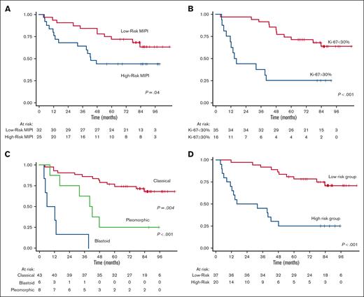
Upon univariate analysis, adverse predictive factors affecting PFS were high-risk MIPI (P = .04), Ki67≥30% (P < .001), blastoid/pleomorphic morphology (P < .001; Figure 2A,B,C, respectively), and failure to achieve CR at the end of treatment (P < .001). Patients who had 4 cycles had similar PFS rates to those who completed full treatment (6 cycles), because no difference was observed between patients with dose reductions as per protocol along cycles (P = .2 for both).
Survival curves for PFS. (A) MIPI score, (B) Ki67 value, (C) morphological variant, (D) or risk group defined as follows: low-risk (Ki67 < 30% and classical morphological variant); high-risk group (Ki67 ≥ 30% and/or blastoid/pleomorphic morphological variant).
Survival curves for PFS. (A) MIPI score, (B) Ki67 value, (C) morphological variant, (D) or risk group defined as follows: low-risk (Ki67 < 30% and classical morphological variant); high-risk group (Ki67 ≥ 30% and/or blastoid/pleomorphic morphological variant).
In the multivariate analysis, after computing variables that were significant in the univariate analysis, morphology (blastoid/pleomorphic variant) was the strongest risk factor for PFS (hazard ratio [HR], 3.12; P = .04; 95% CI, 1.05-9.28; Table 1).
Multivariate analysis
| Variable . | HR . | P > |z| . | [95% CI] . |
|---|---|---|---|
| Morphology (classic vs others) | 3.12 | .041 | 1.05-9.28 |
| High-risk MIPI | 2.14 | .096 | 0.87-5.24 |
| Ki-67 ≥30% | 2.53 | .085 | 0.88-7.25 |
| Variable . | HR . | P > |z| . | [95% CI] . |
|---|---|---|---|
| Morphology (classic vs others) | 3.12 | .041 | 1.05-9.28 |
| High-risk MIPI | 2.14 | .096 | 0.87-5.24 |
| Ki-67 ≥30% | 2.53 | .085 | 0.88-7.25 |
Long-term side effects, SMs, and causes of death
There were 22 registered deaths during the study period. The majority of them (17; 77%) were due to lymphoma progression, whereas the remaining 5 (23%) were due to other causes (2 because of SM, 2 because of sepsis, and 1 because of a septic shock in secondary acute myeloid leukemia). No other late toxicity was addressed as possibly related to the induction treatment.
During follow-up, 6 patients developed SMs: 1 with prostate cancer, 1 with bladder cancer, 2 with head and neck cancer (1 with larynx and 1 with thyroid cancer), 1 with lung cancer, and 1 with secondary acute myeloid leukemia. The cumulative incidence of SMs at 7 years was 11.2% (95% CI, 4.5-21.3), and the median time to SM was 68 months (range, 55-91 months; supplemental Figure 1).
Salvage therapy
Of the 25 patients who were refractory or relapsed, 3 (12%) did not receive any further treatment because of rapid deterioration of clinical status. Six (24%) patients received ibrutinib monotherapy as second line, of whom 4 responded (3 still in CR). The remaining 16 patients received miscellaneous treatment (7 received R-CHOP or R-CHOP-like, 3 received BR, 1 received high-dose methotrexate for central nervous system relapse, 2 received lenalidomide, and 3 received radiation therapy).
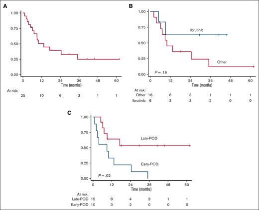
Overall, the OS from the time of first relapse (OS-2) was 8.9 months (range, 0-61.9 months; Figure 3A). The median OS-2 for patients treated with ibrutinib was longer, albeit not significantly, than for patients treated with other approaches (2 year OS-2, 63% [95% CI, 14-89] vs 36% [95% CI, 11-63], P = .16; Figure 3B).
OS at the time of first POD (OS-2). (A) OS-2 in all 25 patients with relapsed/refractory disease, (B) OS-2 in patients who received ibrutinib as second line (n = 6) vs other patients with relapsed and treated patients (n = 16), and (C) POD-24 in 25 patients who had relapsed/refractory disease: early- vs late-POD.
OS at the time of first POD (OS-2). (A) OS-2 in all 25 patients with relapsed/refractory disease, (B) OS-2 in patients who received ibrutinib as second line (n = 6) vs other patients with relapsed and treated patients (n = 16), and (C) POD-24 in 25 patients who had relapsed/refractory disease: early- vs late-POD.
Furthermore, patients who were refractory to induction therapy or experienced POD within 24 months (POD-24, n = 10) had a significantly inferior OS-2 than patients with late-POD (22% [95% CI, 3-51] vs 53% [95% CI, 21-78]; P = .02; Figure 3C).
MRD
Of the 57 patients, 45 (79%) had a molecular marker (29 had IGH rearrangements, 8 had IGH::BCL1 targets, and 8 had both). Follow-up DNA samples from the BM and/or PB of 31 patients (28 IGH and 3 BCL1) were available for MRD analysis. For this report, samples were reanalyzed via highly sensitive, standardized ASO-ddPCR, in addition to previously reported nested PCR results.14
No significant differences in baseline characteristics were observed between analyzed and nonanalyzed patients, except for BM involvement (P = .003; supplemental Table 1), because no differences were noted for PFS and OS rates (7 year PFS and OS 40% [95% CI, 14-68] vs 59% [95% CI, 42-72; P = .38] and 67% [95%CI, 34-86] vs 62% [95%CI 46-74; P = .95], respectively).
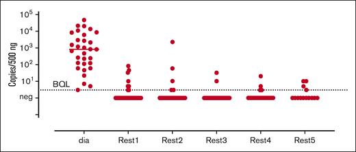
After 2 cycles of RBAC500 (REST1), 20 of 28 patients (71%) tested as MRD-negative in the BM, and 23 of 28 (82%) in the PB. At the end of treatment (REST2), 22 of 27 (81.5%) patients tested as MRD-negative in BM, and 22 of 26 (85%) in the PB. Of the 20 patients with available follow-up samples at one-year after the end of treatment (REST4), 16 (80%) continued testing as MRD-negative in the BM, and 18 of 20 (90%) in the PB. In the following time points, the number of negative patients slightly decreased over time, as shown in Figure 4.
Molecular responses evaluated in the BM and PB during the subsequent time points.
Molecular responses evaluated in the BM and PB during the subsequent time points.
In a landmark analysis starting from the response at the end of treatment, patients who tested as MRD-negative had better, but not significant, outcomes for both PFS and OS than patients who tested as MRD-positive (7-year PFS 65% [95% CI, 42-81] vs 40% [95% CI, 5-75; P = .148]; 7-year OS, 65% [95%CI, 42-81] vs 40% [95% CI, 5-75; P = .162]; Figure 1D,E).
Further details on patients behavior based on the MRD analysis at different time points are shown in supplemental Figures 2A-B and 3A-B.
Discussion
We report on the long-term follow-up of the RBAC500 phase 2 trial, showing that this regimen achieved its goal of maintaining activity over time, with median PFS and OS figures that were not yet reached after prolonged follow-up. These results, which were achieved without maintenance therapy, support the use of R-BAC in older patients with MCL, even when judicious dose reductions are implemented to avoid hematotoxicity.
As for previous observations, blastoid/pleomorphic morphology is the most relevant independent predictor of adverse survival. Unfortunately, at the time of the study conception, no study of TP53 function was planned, which limits our knowledge on the activity of R-BAC in TP53 mutated patients, and represents a limitation of this study. Indeed, the FIL VR-BAC trial (NCT03567876), which had completed recruitment ∼1 year ago,16 will fill this gap.
The results obtained with R-BAC compare favorably with those of other immunochemotherapy regimens (with or without maintenance) in similar populations. The combination of bortezomib, rituximab, cyclophosphamide, doxorubicin, and prednisone (VR-CAP) frontline therapy was tested against R-CHOP in a randomized trial,17 showing a median PFS of 24.7 months but prolonged median OS of 90.7 months. In 2020, Kluin-Nelemans et al reported a long-term update of the phase 3 European MCL Network Elderly trial, in which the median OS was 6.4 years. Maintenance therapy with rituximab following response to R-CHOP was associated with a median PFS of 5.4 years.18 In the STiL study, patients treated with BR had a median PFS of 3 years. Our trial had apparently similar population to that included in the STiL study,2 with a median age of 70 (64.5-74) and 71 (67-75), respectively, but was associated with a 3-year PFS of 76%.7 Similarly, in the bright study, the median PFS after long-term follow-up for the BR arm was ∼48 months.19 A direct comparison between BR and R-BAC has been performed in a real-life study using a propensity score to match patients for characteristics at presentation and reduce selection bias. This study10 showed that patients treated with R-BAC had a 2-year PFS of 87% ± 3% compared with 64% ± 7% for BR (P = .001). In terms of toxicity, R-BAC was associated with significantly more pronounced grade 3 to 4 thrombocytopenia than BR (50% vs 17%); however, the efficacy of R-BAC was preserved when the doses were reduced. Overall, the present trial and real-life experience in patients treated with R-BAC consistently reported similar efficacy results but significantly reduced toxicity when the regimen was administered at attenuated doses, in a 2-day fashion, or with flat doses of bendamustine and cytarabine (ie, 100-500 mg total dose, respectively, on 2 consecutive days, skipping the third day of cytarabine).7,10 In clinical practice, after the first cycle we recommend adopting dose reductions in patients experencing grade 3-4 hemotooxicity, while patients who are unfit, or older than 75, may take advantage of the 100-500 regimen.
The SHINE trial11 is a large randomized international trial reporting the association of ibrutinib, a Bruton tyrosine kinase inhibitor, in combination with BR, followed by rituximab maintenance, in older patients with previously untreated MCL. This study established the superiority of the ibrutinib-containing arm, together with BR and maintenance rituximab, in terms of PFS with respect to BR plus maintenance but with placebo. With a median follow-up of 84.7 months (very similar to that of this study), the median PFS in the ibrutinib-containing winner arm was 80.6 months, which resembles our observation with the R-BAC regimen, in which cytarabine is added to BR instead of ibrutinib, but no maintenance was administered. Overall, the demographic characteristics of the population analyzed in the SHINE trial were comparable with those described in this study (median age, 71 years for both; advanced Ann Arbor Stage in 89.3% and 91%, high-risk MIPI in 35.6% and 31%, respectively; however, much higher prevalence of blastoid/pleomorphic variants in the R-BAC 500 trial was 25% and 7.3%, respectively). The 7-year OS was 55% in the ibrutinib group of SHINE and 63% in the RBAC500 group. We acknowledge that comparisons between different studies usually hide pitfalls; however, we still describe a PFS curve for R-BAC that is largely similar to the PFS achieved by BR + ibrutinib + maintenance rituximab and ibrutinib in the SHINE trial. Furthermore, avoidance of maintenance may have been of importance in the era of COVID-19, in which concerns have been raised regarding infectious complications for patients who are long-term maintained with anti-CD20 antibodies.20
R-BAC was associated with high MRD negativity at the end of induction, with 81.5% of the analyzed patients scoring negative in the BM and 85% scoring negative in the PB. This percentages are in line with that of the MRD results of intensive chemoimmunotherapy regimens usually offered to younger patients.21,22
The cumulative incidence of SM (including skin cancers) at 7 years in our cohort was 11.2% (95% CI, 4.5-21.3), and the median time to SM was 68 months (range, 55-91), with 3 patients (5%) who died during follow-up due to SM. One patient developed secondary leukemia. This is in line with the SHINE study,11 where SM (also including skin cancers) was observed in 20.8% and 18.8%, respectively, in the 2 arms, and 3.8% of deaths were related to SM. In the VR-CAP vs R-CHOP study,17 the reported rate of SM was lower (4.1%, 10 patients in each group), and mortality was 1.6%. In younger patients treated with rituximab-hyper fractionated cyclophosphamide, vincristine, doxorubicin and dexamethasone, a 5% rate of secondary myelodysplasia has been reported23 It is difficult to draw conclusions on the risk of developing SM according to different regimens; however, we acknowledge that this topic deserves specific studies in MCL.
In this study, the OS from the time of POD (OS-2) was 8.9 months. Patients who were refractory to induction therapy or who experienced POD-24, n = 25, had significantly inferior 2 year OS-2 than patients with late-POD (22% [95% CI, 3-51] vs 53% [95% CI, 21-78]; P = .02; Figure 3C), confirming observations from others both in younger and older patients.24-26 Indeed, ibrutinib was not available in Italy at the time of relapse for most patients included in this study, which explains why only a minority of patients were treated at relapse with this compound.
In conclusion, we reported the long-term outcomes among older patients with MCL treated upfront with R-BAC in a prospective RBAC500 FIL trial. The responses were durable without maintenance therapy and compared favorably with those of modern examples of combination therapies. This regimen was not associated with any unexpected long-term toxicity. With a median PFS and OS exceeding 50% after 7 years, this regimen has significantly affected the life expectancy of older patients with MCL, a disease that, a couple of decades ago, was associated with a median OS <3 years in similar populations.
Acknowledgments
The authors thank all patients who participated in this trial. The authors also thank the Fondazione Italiana Linfomi secretary and particularly Elisa Masiera, Claudia Peracchio, and Antonella Ferranti. The authors also thank Emilia Elizbieta Florea and Anna Bordin for their scientific and administrative contributions to the work and the AIL section of Verona. The authors thank Valentina Tabanelli for contribution to the central pathological review. This research was financially supported by Progetto di Ricerca Sanitaria Finalizzata 2009 grant (RF-2009-1469205) and 2010 grant (RF-2010-2307262); AO S Maurizio, Bolzano/Bozen, Italy; Fondi di Ricerca Locale, Università degli Studi di Torino, Italy; Fondazione Neoplasie Del Sangue, Torino, Italy; and Fondazione Cassa di Risparmio di Torino (project code 2015.1044), Torino, Italy.
Authorship
Contribution: C.V. and M.C.T. conceived and wrote the manuscript; C.V., M.C.T., S.F., and A.E. analyzed and interpreted the data; S.F. and M.F. analyzed the MRD; S.A.P. performed the central pathological review; and all authors were involved in patient recruitment, data collection, and database assembly and approved the final version of the manuscript.
Conflict-of-interest disclosure: M.C.T. serves on the advisory board for Incyte, Bristol Myers Squibb (BMS), Gilead Science, and Novartis, and serves on the speakers’ bureau for Incyte, Gilead Science, Novartis, and Janssen. C.P. serves on the advisory board for Takeda, Incyte, AbbVie, AstraZeneca, and Janssen. S.F. receives research funding from Janssen, MorphoSys, Gilead, and BeiGene; provides consultancy for EusaPharma, Janssen, Sandoz, and AbbVie; serves on the advisory board for EusaPharma, Janssen, Clinigen, Incyte, and Italfarmaco; and receives speakers honoraria from Janssen, EusaPharma, and Servier, Gentili. A.D.R. serves on the advisory board for Takeda and Jannsen, and is a consultant for Incyte and Gilead. S.A.P. serves on the advisory board for Celgene, NanoString, Roche, and BeiGene. F.M.Q. has an advisory role with AstraZeneca and Janssen; is a speaker for Janssen; and is a consultant for Sandoz. A.R. serves on the advisory board for Takeda, Incyte, and Italfarmaco, and is a consultant for Takeda and Servier. S.V. serves on the advisory board for AbbVie. V.R.Z. serves on the advisory board of Gentili, Gilead, MSD, Servier, and Takeda; gives presentations at Janssen and Takeda; is a consultant for Roche; and receives travel expenses and accomodations from Janssen and Takeda. A.A. serves on the advisory board and speaker’s bureau for and receives travel expenses from Janssen, AbbVie, Gilead, Novartis, Gentili, and Servier. F.M. serves on the advisory board for Roche, Gilead, Incyte, MSD, and Takeda. C.V. provides consultancy for AbbVie, BMS, Incyte, Roche, Pfizer, Janssen, Kyowa Kirin, Gentili, and BeiGene; serves on the speakers bureau of AbbVie, BMS, AstraZeneca, Servier, Incyte, Roche, Pfizer, Novartis, Gentili, Janssen, Kite-Gilead, and BeiGene; and receives research funding from Janssen. The remaining authors declare no competing financial interests.
Correspondence: Carlo Visco, Department of Engineering for Innovation Medicine, Section of Hematology, University of Verona, Piazzale L. Scuro 10A, 37129 Verona, Italy; e-mail: carlo.visco@univr.it.
References
Author notes
Data are available upon request from the corresponding author, Carlo Visco (carlo.visco@univr.it).
The full-text version of this article contains a data supplement.


![Survival curves at a median follow-up of 86 months, and MRD evaluation. (A) PFS of all patients (7-year PFS, 55% [95% CI, 41-67]); (B) OS of all patients (7-year OS, 63% [95% CI, 49-74]); (C) Duration of response for the 52 responding patients (7-year duration of response, 59% [95% CI, 44-71]); (D) PFS (7-year PFS, 65% vs 40%; P = .14) and (E) OS (7-year OS, 65% vs 45%; P = .16) based on the MRD results at the end of the treatment (blue curve, positive; red curve, negative).](https://ash.silverchair-cdn.com/ash/content_public/journal/bloodadvances/7/15/10.1182_bloodadvances.2023009744/2/m_blooda_adv-2023-009744-gr1.jpeg?Expires=1769082976&Signature=EhFRkR0SQviimsKPhRnk447sAb82ysqejKKX3-BPHANNKljbB-9yPlycZbMOlYaRosq8Arkmd3DotAXOH8FMmqSLdVf1aK3hCuR~~4fcGtdHe0P1V9WoKdwPijF2KvfF~~kDLn4iOGTvq63sB3A-ieiag0Fac6N4Kytl12B0YUxvEV4a5WxuzzCkf-ZgB1~4zSeuEZCemRrHsb4ngk3OTYkZEo8s1ANlvxaCFoVloB9Gb-brb56XiPPkuuIRlY4NZRm-IOZLyjwUD0V3yd8v-fw0Gz8RKE78kIaG05x67EJYckztxBnt295kj0rgDlQwkIllbvPm99pYAy9nt08f-A__&Key-Pair-Id=APKAIE5G5CRDK6RD3PGA)


