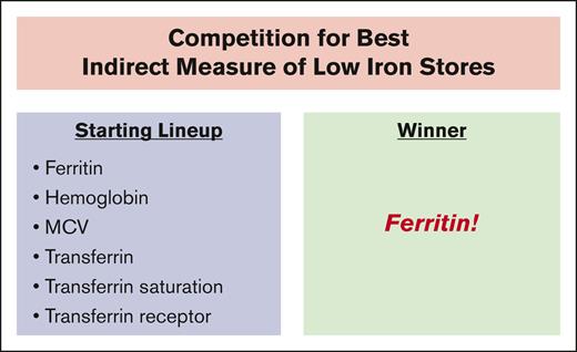
In this issue of Blood Advances, Lahtiharju et al1 report a retrospective analysis of 6610 patients from Helsinki University Hospital with predominantly hematologic diagnoses who had results available for both indirect measures of iron status and bone marrow aspirates, stained with Prussian blue for iron. Bone marrow iron stain is the gold standard for evaluating iron stores. Defining iron deficiency as either absent or low-stainable bone marrow iron, the prevalence of iron deficiency was 22.5%. Of the 6 indirect measures of iron status measured—ferritin, hemoglobin, mean corpuscular volume (MCV), transferrin, transferrin saturation, and transferrin receptor—serum ferritin had the greatest accuracy in identifying iron deficiency (see figure). Using receiver operating characteristics analysis, ferritin had an area under curve of 88% for females and 89% for males for predicting low or absent bone marrow iron.
Of the 6 indirect measures of iron status measured—ferritin, hemoglobin, MCV, transferrin, transferrin saturation, and transferrin receptor—serum ferritin had the greatest accuracy in identifying iron deficiency.
Of the 6 indirect measures of iron status measured—ferritin, hemoglobin, MCV, transferrin, transferrin saturation, and transferrin receptor—serum ferritin had the greatest accuracy in identifying iron deficiency.
Iron is an essential nutrient that is vital for hemoglobin production and many other physiological processes. Iron deficiency is a common clinical problem, often due to menstrual blood loss, increased physiologic requirements of pregnancy and lactation, and the iron demand of growth spurts in childhood and adolescence.2 Decreased iron absorption can also be a factor in conditions such as gluten enteropathy, autoimmune gastritis, status-post gastrectomy or proximal bowel resection, and TMPRSS6 mutations.3 Iron deficiency is encountered almost daily in clinical practice, and it is critical to not miss the diagnosis because iron deficiency may be the early sign of a potentially fatal condition such as a gastrointestinal or genitourinary malignancy.4,5 Diagnosing iron deficiency, identifying the cause, and providing curative therapy has saved the lives of many patients and is an essential responsibility of every health care provider.
Although making the correct diagnosis of iron deficiency is critical for patient care, the use of indirect markers obtained through routine phlebotomy presents a challenge. Low serum iron, transferrin saturation, hemoglobin, and MCV are characteristic of iron deficiency but can also result from inflammation. In addition, hemoglobin and MCV may be low due to genetic causes and, in the case of hemoglobin, to diurnal variation rather than iron deficiency. Transferrin concentrations tend to increase with iron deficiency and decrease with inflammation, whereas transferrin receptor levels rise with both iron deficiency and inflammation.6,7 Serum ferritin is an attractive marker because a very low level (<10 μg/L) is diagnostic of iron deficiency as documented by absent stainable iron in a bone marrow aspirate. However, serum ferritin can be normal or even elevated when iron deficiency is documented by absent bone marrow stainable iron.8 A recent Cochrane review of 512 adults presenting for medical care at various institutions found that a serum ferritin <30 μg/L had 98% specificity and 79% sensitivity for diagnosing iron deficiency as documented by absent bone marrow iron.9
Lahtiharju et al make a remarkable contribution by evaluating the relationship of indirect measures of iron status to bone marrow iron stain in 6610 patients and providing evidence that ferritin is the most reliable single biomarker for predicting reduced iron stores. This finding is consistent with studies dated decades ago that established ferritin as a key standalone indicator of iron status.8 This study also underscores the limitations of ferritin, particularly its low sensitivity. Using a ferritin cutoff of 30 μg/L, the specificity rates were impressively high at 97% for females and 99% for males, but the sensitivity rates were only 54% and 35%, respectively. This indicates that while a ferritin of 30 μg/L or lower is highly specific for low or absent bone marrow iron, it is not sensitive enough to detect all cases of low iron stores, especially in patients with comorbid conditions that can increase ferritin levels. It is important to recognize that in this study, there were females with serum ferritin >1750 μg/L and males with serum ferritin > 4967 μg/L who had low or absent bone marrow iron stores.
The other biomarkers studied by the authors demonstrated inferior diagnostic performance compared to ferritin except for transferrin in females. Multivariate logistic regression models including these biomarkers did not improve the prediction of low or absent bone marrow iron stores over ferritin alone.
The clinical implications of the current study are important. The high specificity of ferritin at the 30 μg/L cutoff suggests that it can be a reliable marker for ruling in low or absent iron stores in patients with a high pretest probability of the condition. However, the low sensitivity indicates that clinicians should be cautious in ruling out low or absent iron stores based solely on ferritin levels, particularly in patients with conditions that can elevate ferritin, such as inflammation or liver disease. In such cases, additional diagnostic tests or clinical judgment may be necessary to accurately diagnose low or absent iron stores. We cannot emphasize too strongly that bone marrow aspirate with iron staining is often necessary, as the finding of absent iron necessitates an evaluation that may uncover a gastrointestinal or genitourinary malignance at an early, curable stage.
The reliability of the bone marrow iron determination depends on the adequacy of the specimen including the presence of spicules in the aspirate, and it would have been good to have specific comments about these factors in this study. In addition, it would have been good to provide their analysis based on the classical definition of iron deficiency, which is absence of stainable iron in an adequate bone marrow aspirate. The retrospective design and single institution character of the study are offset by the very impressive sample size.
In conclusion, this study reinforces the importance of ferritin as the primary biomarker for diagnosing iron deficiency, while also highlighting its limitations in terms of sensitivity. Clinicians should consider these findings when interpreting laboratory measures of iron status, particularly in patients with comorbid conditions. Importantly, the presence of a normal or elevated serum ferritin does not negate the potential need for a bone marrow aspirate to assess potential metabolic or malignant conditions that may present with iron deficiency.
Conflict-of-interest disclosure: The authors declare no competing financial interests.

