The CXCR4 chemokine receptor is a Gi protein–coupled receptor that triggers multiple intracellular signals in response to stromal cell-derived factor 1 (SDF-1), including calcium mobilization and p44/42 extracellular signal-regulated kinases (ERK1/2). Transduced signals lead to cell chemotaxis and are terminated through receptor internalization depending on phosphorylation of the C terminus part of CXCR4. Receptor endocytosis is also required for some receptors to stimulate ERK1/2 and to migrate through a chemokine gradient. In this study, we explored the role played by the 3 intracellular loops (ICL1-3) and the C terminus domain of CXCR4 in SDF-1–mediated signaling by using human embryonic kidney (HEK)–293 cells stably expressing wild-type or mutated forms of CXCR4. ICL3 of CXCR4 is specifically involved in Gi-dependent signals such as calcium mobilization and ERK activation, but does not trigger CXCR4 internalization after SDF-1 binding, indicating that ERK phosphorylation is independent of CXCR4 endocytosis. Surprisingly, ICL2, with or without the aspartic acid, arginine, and tyrosine (DRY) motif, is dispensable for Gi signaling. However, ICL2 and ICL3, as well as the C terminus part of CXCR4, are needed to transduce SDF-1–mediated chemotaxis, suggesting that this event involves multiple activation pathways and/or cooperation of several cytoplasmic domains of CXCR4.
Introduction
The chemokine receptor CXCR4 is a member of the large family of 7-transmembrane domain receptors coupled to heterotrimeric Gi proteins.1,2 This receptor is also a fusion coreceptor for T-cell tropic and dual-tropic HIV-1 strains.3 Its ligand, the CXC chemokine stromal cell-derived factor 1 (SDF-1), also named CXCL12, activates multiple signal transduction pathways. This chemokine was first described as a powerful chemoattractant for peripheral blood lymphocytes,4 CD34+ progenitor cells,5 and pre– and pro–B-cell lines.6SDF-1 stimulation of cells expressing CXCR4 results in the increased phosphorylation of focal adhesion components, such as proline-rich tyrosine kinase 2 (Pyk-2), p130Cas, focal adhesion kinase (FAK), paxillin, Crk and Crk-L,7,8 extracellular-signal regulated kinases 1 and 2 (ERK-1 and -2), phospholipase C-γ (PLC-γ), protein kinase C (PKC), and phosphatidylinositol 3-kinase (PI3-K), and in activation of the Janus kinase signal transducers and activation of transcription (JAK/STAT) pathway9 and nuclear factor κ-B (NFκB).7
SDF-1 also triggers CXCR4 internalization, involving G protein–coupled receptor kinases (GRKs), followed by binding of β-arrestin.2,10,11 CXCR4 regulation by β-arrestin is mediated by the C terminal tail and the third intracellular loop (ICL3).10 Beside the role of receptor internalization in desensitization that terminates receptor signaling, G protein–coupled receptor (GPCR) endocytosis may be necessary to activate several pathways and functions such as chemotaxis12-14 and ERK/mitogen-activated protein kinase (MAPK)15-17 cascade activation. However, conflicting results have been obtained, in which chemotaxis18-20 and MAPK21,22 signaling have been shown to be independent of GPCR endocytosis. Furthermore, signals regulating chemotaxis are largely unknown. Gi activation, presumably involving βγ20,23,24 and PI3-K phosphorylation,25,26 are not sufficient, although necessary.27
Despite the wide range of molecules activated after SDF-1 binding to CXCR4, there is scant information on the link between the role of the intracellular domains of CXCR4 and the activation of signal transduction pathways. For this purpose, we constructed stable clones by using the well-established human embryonic kidney 293 (HEK-293) cell system already expressing a truncated form of CD4 incapable of transducing a signal on its own. This cell line, which is widely used for GPCR signaling studies, has also been useful for analysis of chemotaxis14,20,24,28,29 and β-arrestin–dependent internalization because of its high endogenous levels of β-arrestin.11,30 31 Transfected cells express either a mutated form of CXCR4 in which each intracellular loop (ICL) was replaced by a scrambled amino acid sequence of ICL1 that does not contain serine (Ser), threonine (Thr), and tyrosine (Tyr), or a CXCR4 form truncated at position 308. In addition, we analyzed cells expressing CXCR4 in which the mutated ICL2 still contains the aspartic acid, arginine, and tyrosine (DRY) amino acid sequence. We demonstrated that ICL3 is the only intracellular domain involved in transducing Gi-dependent signals after SDF-1 binding. MAPK activation, one of the Gi-mediated signals, is independent of CXCR4 internalization, whereas the truncated CXCR4 form, unable to internalize after SDF-1 binding, still phosphorylates ERK. Interestingly, ICL2 is not involved in Gi signaling or in CXCR4 internalization, indicating that in CXCR4, the DRY motif is not required for coupling to Gi proteins. In contrast, chemotaxis needs multiple transduction pathways, including Gi signaling through ICL3 and signaling via ICL2 and the C terminal part of CXCR4, and/or cooperation of these domains to be efficient.
Materials and methods
Materials
SDF-1α, polyclonal rabbit anti–SDF-1α antibody, and mouse monoclonal (mAb) anti-CXCR4 (MAB173) antibody were purchased from R & D Systems (R & D Systems Europe, Abington, United Kingdom). Anti-ERK1/2 and antiphosphorylated ERK1/2 antibodies were purchased from Santa Cruz Biotechnology (Tebu, Le Perray en Yvelines, France) and Promega (Promega France, Lyon, France), respectively. Fluorescein isothiocyanate (FITC)–labeled Fab′2 antimouse and antirabbit immunoglobulins were from Sigma-Aldrich (L'Isle d'Abeau Chesnes, France). Bordetella pertussis toxin (PTX) was purchased from Calbiochem (France Biochem, Meudon, France). Geneticin (G418) and Zeocin were from Invitrogen (Abington, United Kingdom).
Construction of the CXCR4 mutants
The mutant receptor cDNAs were prepared by polymerase chain reaction (PCR) by using the pBABE.Fusin vector obtained through the National Institutes of Health AIDS Research and Reference Reagent Program from N. Landau (Aaron Diamond AIDS Research Center, The Rockefeller University, New York, NY) as template. The primers used at each extremity of the CXCR4 gene were 5′Fus (5′-GTG CAC GAA TTC GGC TTA AGT GAC GCC GAG-3′) and 3′Fus (5′-CCG ATG CTC GAG TTA CTT GTC ATC GTG GTC C-3′). The primers used for the construction of the mutated loops were 5′ICL1m (5′-CAA GAT GCA GAA GCT GAA GGC AAG GCT GCA CCT GTC AGT G-3′) and 3′ICL1m (5′-CCT TCA GCT TCT GCA TCT TGG CGT CTC TTG CAC CCA TGA CCA GGA TGA C-3′); 5′ICL2m (5′-CAA GAT GCA GAA GCT GAA GGC AGT GGT CTA TGT TGG CGT-3′) and 3′ICL2m (5′-CCT TCA GCT TCT GCA TCT TGG CGT CTC TTG CCA GAC TGA TGA AGG CCA-3′) or 3′ICL2mDRY (5′-CCT TCA GCT TCT GCA TCT TGG CGT CTC TTG CGT AGC GGT CCA GAC TGA TG-3′); 5′ICL3m (5′-CAA GAT GCA GAA GCT GAA GGC AGT CAT CCT CAT CCT GGC T-3′) and 3′ICL3m (5′-CCT TCA GCT TCT GCA TCT TGG CGT CTC TTG CGA TGA TAA TGC AAT AGC A-3′). The PCR fragments obtained with the primer pairs 5′Fus/3′ICLm and 5′ICLm/3′Fus were used as template for an overlapping PCR with 5′Fus and 3′Fus as primers. Each fragment was cloned in the pcDNA3 Zeo expression vector (Invitrogen) and sequenced on an Applied Biosystems (Courtaboeuf, France) Model 373A automated sequencer, using Taq polymerase and dye terminator. The CXCR4.7TM mutant was previously described.32
Cell culture and transfection
HEK-293 cells expressing a truncated form of CD4 lacking the cytoplasmic domain (CD4.403)32 were cultured in Dulbecco modified Eagle medium (DMEM) supplemented with 1% penicillin-streptomycin, 1 mg/mL G418, 1% glutamax, and 10% fetal calf serum (Gibco-Life Technologies, Cergy Pontoise, France), and 107 cells were transfected with 10 μg pcDNA3 Zeo containing the CXCR4 mutants (CXCR4.ICL1m, CXCR4.ICL2m, CXCR4.ICL2mDRY, and CXCR4.ICL3m) by using the FuGENE 6 transfection reagent (Roche Diagnostics, Meylan, France) according to the manufacturer's instructions. The presence of the external part of CD4 at the cell surface allows us to verify the capability of the CXCR4 mutants to support HIV-1 infection. Clones able to grow in the presence of 250 μg/mL Zeocin were selected by flow cytometry for receptor surface expression.
HIV-1 infection
Cells (1 × 10⁵) were infected for 4 days with 100 μL suspension of R7-GFP HIV-1 in which Nef-coding sequences were replaced by a modified form of green fluorescent protein (GFP),33 and GFP+ cells were analyzed by flow cytometry. The culture supernatant used to infect the different transfected HEK cell lines was collected after infection for 7 days of the CEM lymphoblastoid cell line. At this time, 21% of CEM cells were GFP+ and reverse transcriptase (RT) activity was 170 000 cpm/mL. GFP+ cells were analyzed by flow cytometry.
Flow cytometry
Cells (1 × 10⁵) were incubated for 1 hour at 4°C with 50 μL phosphate-buffered saline containing 0.2% bovine serum albumin (PBS-BSA) or PBS-BSA supplemented with the appropriate mAb. After 3 washes with PBS-BSA, bound mAb was revealed by addition of 50 μL of a 1/50 dilution of fluorescein-conjugated (FITC) secondary immunoglobulin. After 30 minutes' staining, cells were washed with PBS-BSA, and fluorescence intensity at 543 nm was measured on an EPICS XL4-C cytofluorometer (Beckman-Coulter France, Villepinte, France). To study the direct binding of SDF-1α to CXCR4 expressed on transfected HEK/CD4.403 cells, 1 × 10⁶cells were incubated in 30 μL PBS for 1 hour at 4°C to prevent subsequent internalization, and 30 μL SDF-1α solution (100, 200, or 400 nM) was then incubated for 20 minutes at 4°C after cell centrifugation. After washing with PBS-BSA, 30 μL anti–SDF-1α antibody at 10 μg/mL was added for 30 minutes, and bound antibody was revealed as described previously.
To study CXCR4 internalization on transfected HEK/CD4.403 cells, 1 × 10⁶ cells were incubated at 37°C for 30 minutes with SDF-1α at 200 nM. After washes with PBS-BSA, 30 μL anti-CXCR4 monoclonal antibody (MAB173) was added at 10 μg/mL for 45 minutes, and bound antibody was revealed as previously described.
Chemotaxis assay in adherent cells
The technique used was adapted from Goya et al.34Briefly, tissue culture inserts (Nunc) pore size 8 μm, 10 mm in diameter, were coated with 20 μg/mL collagen type I (Sigma) for 2 hours at 37°C. Cells were starved of serum overnight, trypsinized, washed 3 times in migration buffer (DMEM, 0.25% BSA), counted, and diluted to 5 × 10⁵ cells/mL. A volume of 100 μL of cells was placed in the upper chamber, whereas a solution of SDF-1α (75, 125, or 250 nM) in migration buffer or migration buffer alone was in the lower chamber. Migration was allowed to proceed for 6 hours at 37°C, 5% CO2. After this time, the cells were removed from the upper chamber by washing in PBS and scraping. The migrated cells were visualized by fixing and staining in 0.5% crystal violet in 20% methanol followed by repeated washing in H2O. The number of migrating cells in 5 fields (×20) was counted by 2 investigators. Results are presented as a migration index calculated as follows: number of migrating cells with SDF-1α/number of migrating cells in migration buffer alone.
Calcium signaling
Cells (1 × 10⁷) were resuspended in Hanks solution (Gibco-Life Technologies) and loaded with Fluo-3-AM at 2 μM for 20 minutes at room temperature. After 3 washes with Earle balanced salt solution (EBSS) containing 0.1% BSA (EBSS-BSA), 106 cells/mL were stimulated with buffer alone (EBSS-BSA) or SDF-1α at 250 nM. Ionomycine at 10−6 M was then added to verify the capability of the cells to induce a calcium influx. Ratio of fluorescence of bound to free Fluo-3 was analyzed each 10 seconds on an EPICS XL4-C cytofluorometer.
ERK phosphorylation induced after SDF-1α binding on CXCR4
Cells were starved of serum overnight at 37°C, 5% CO2. After 3 washings in PBS, 5 × 10⁶cells/mL were resuspended in 100 μL PBS and incubated at 37°C for 15 minutes. Cells were then stimulated with SDF-1α at 125 nM for 2 minutes, lysed in Tris (tris(hydroxymethyl)aminomethane)–HCl 50 mM (pH 8), Triton X-100 1%, NaCl 100 mM, MgCl2 1 mM, benzamidine 2 mM, leupeptine 2 μg/mL, phenylmethylsulfonyl fluoride (PMSF) 150 μM containing NaF, Na3VO4, β glycerophosphate, andpara-nitrophenylphosphate (PNPP).35 Cell lysates were subjected to electrophoresis through 10% sodium dodecyl sulfate–polyacrylamide gel electrophoresis (SDS-PAGE) and electrotransferred to polyvinylidene difluoride (PVDF) membranes (Millipore). Membranes were then blocked in Tris-buffered saline (TBS), 0.05% Tween 20, and 10% milk for 1 hour at 20°C. Blots were incubated overnight at 4°C with the primary antibody diluted 1/2500 in TBS-Tween-5% milk. After 30 minutes of washing in 6 changes of TBS-Tween, the blots were incubated for 1 hour at 20°C with peroxidase-coupled antiserum diluted 1/2000 in TBS-Tween-5% milk. After further washing, the immune complexes were revealed by enhanced chemiluminescence (ECL; NEN) and subjected to autoradiography. Quantification of ERK1/2 phosphorylation was performed by using the Scion program (Frederick, MD) after autoradiography scanning.
Statistical analysis
Variance analysis was performed after arc sine transformation of the data36: *P < .05; **P < .01; ***P < .001.
Results
Construction and expression of the CXCR4 mutants
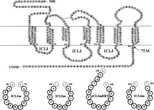
To investigate the role of the intracellular domains of CXCR4 in signal transduction after SDF-1α binding, CXCR4 mutants were generated and stably transfected in HEK cells expressing the CD4 molecule deleted of its cytoplasmic tail. The amino acids of each loop were replaced by those of ICL1, and the amino acids Ser, Thr, and Tyr (putative sites of phosphorylation) were changed to alanine (Ala). Furthermore, the sequence of ICL1 was completely modified by interchanging the amino acid order. In this way, the global charge of the loop was respected, but the putative binding sites to cytoplasmic molecules were destroyed. The mutated ICL sequences are described in Figure 1.
Schematic representation of CXCR4 receptor and mutants.
The 7 transmembrane domain structure is depicted. The truncated CXCR4.7TM mutant was generated by introducing a stop codon after the residue 308. The intracellular loops of CXCR4 subjected to mutagenesis are represented in bold. CXCR4.ICL1m, CXCR4.ICL2m, CXCR4.ICL2mDRY, and CXCR4.ICL3m were constructed by substituting the native loop sequences at the positions indicated with a modified ICL1 loop unable to transduce a signal.
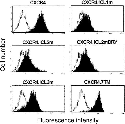
Cell surface expression of the CXCR4.ICL mutants was measured by flow cytometry and compared with expression of the CXCR4 wild-type molecule and the CXCR4.7TM mutant on HEK/CD4.403 cells (Figure2). CXCR4 and CXCR4.7TM are very well expressed, at an almost identical level, at the cell surface. CXCR4.ICL3m is well expressed on HEK/CD4.403 cells, but at a lower level to that of CXCR4 and CXCR4.7TM. CXCR4.ICL1m and the 2 CXCR4.ICL2 mutants, with or without the DRY sequence, are weakly expressed at the cell surface.
Expression of wild-type and mutated CXCR4 molecules at the surface of the HEK/CD4.403 cell line.
Cells were incubated with medium containing the anti-CXCR4 mAb at 10 μg/mL. Bound mAb was detected with a FITC-labeled antimouse immunoglobulin. The white histogram represents binding of anti-CXCR4 mAb to HEK/CD4.403 cells, and black histograms to the different CXCR4 molecules analyzed. The fluorescence intensity was recorded in log mode on an EPICS XL4 cytofluorometer. Data representative of 1 to 5 independent experiments are shown.
CXCR4 mutants expressed on HEK/CD4.403 cells are functional
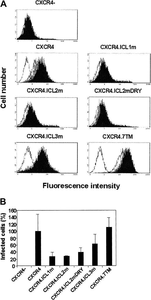
To confirm that the CXCR4.ICL1-3m and CXCR4.7TM molecules are still able to bind SDF-1α when expressed on HEK/CD4.403 cells, SDF-1α at 100, 200, or 400 nM was incubated with the 4 HEK/CD4.403/CXCR4.ICL mutants, HEK/CD4.403/CXCR4.7TM, HEK/CD4.403/CXCR4, and HEK/CD4.403 cells. SDF-1α binding to these cell lines was revealed by an anti–SDF-1α antibody. As shown in Figure 3A, SDF-1α binds to CXCR4.ICL mutants and CXCR4.7TM, and the level of SDF-1 binding is parallel with that of CXCR4 expression. These CXCR4 mutants are also capable of serving as a coreceptor for HIV-1 entry (Figure 3B). As for SDF-1 binding, the degree of infection is strictly dependent on the level of cell surface expression.
Functional cell surface expression of wild-type and mutated CXCR4 receptors after stable transfection of the HEK/CD4.403 cell line.
(A) Cells were incubated with PBS alone (white histograms) or a solution of SDF-1 at 100 nM (light gray histograms), 200 nM (dark gray histograms), or 400 nM (black histograms) in PBS at 4°C. After incubation of the anti–SDF-1 antibody at 10 μg/mL, bound antibody was detected with a FITC-labeled antirabbit immunoglobulin. Data representative of 1 to 5 experiments are shown. (B) Cells expressing CD4.403 and wild-type or mutated CXCR4 molecules were infected by the HIV–R7-GFP33 for 4 days as described in “Materials and methods,” and GFP+ cells were analyzed by flow cytometry. The percentage of R7-GFP–infected cells expressing wild-type CXCR4 is defined as 100% and corresponds to the ratio of GFP+ cells expressing wild-type CXCR4 to GFP+ cells that do not express CXCR4. The histograms represent the percentage of infected cells. Data are expressed as means ± SDs of 4 different experiments.
SDF-1α triggers internalization of the CXCR4.ICL mutants but does not induce downmodulation of CXCR4.7TM
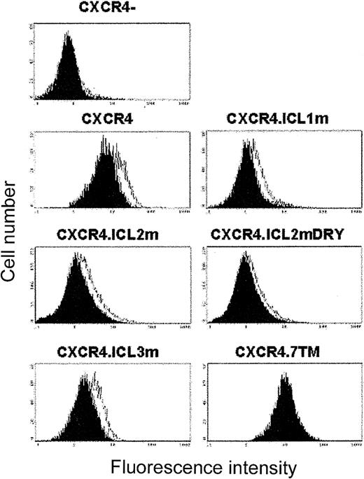
β-Arrestin plays an essential role in GPCR desensitization and has been described to bind in vitro to both the third intracellular loop and the C terminus part of CXCR4. Downmodulation of all the CXCR4 mutants expressed on HEK/CD4.403 cells was analyzed after incubation of SDF-1α at 200nM. On stimulation with SDF-1, surface expression of CXCR4.ICL mutants is reduced at a level comparable to that obtained with wild-type CXCR4 (Figure4). Conversely, SDF-1α binding to the CXCR4 molecule with C terminal truncation does not trigger endocytosis. CXCR4 expression at the cell surface is thus differentially regulated after SDF-1α binding, depending on the intracellular interaction sites involved in receptor internalization. To verify that no competition occurs in the binding of SDF-1α and the anti-CXCR4 antibody (MAB173) to CXCR4, sequential incubation of these 2 molecules was performed with HEK/CD4.403/CXCR4 cells. The binding sites of SDF-1α and MAB173 on CXCR4 are different because binding of one of these molecules does not hamper binding of the other (data not shown).
SDF-1–induced internalization of wild-type and mutated CXCR4 molecules stably expressed at the surface of the HEK/CD4.403 cell line.
Cells were incubated with PBS alone (white histograms) or a solution of SDF-1 at 200 nM (black histograms) in PBS at 37°C. After incubation of the anti-CXCR4 mAb at 10 μg/mL, bound Ab was detected with a FITC-labeled antimouse immunoglobulin. Results of data from 1 experiment representative of 4 are shown.
Gi-dependent signals only depend on the third intracellular loop of CXCR4
SDF-1α binding to CXCR4, CXCR4.7TM, CXCR4.ICL1m, CXCR4.ICL2m, and CXCR4.ICL2mDRY expressed on HEK/CD4.403 cells induced calcium mobilization from ionomycin-sensitive intracellular stores but failed to trigger calcium response in HEK/CD4.403 and HEK/CD4.403/CXCR4.ICL3m cells. Pretreatment with PTX completely blocked SDF-1α–induced calcium mobilization, indicating that ICL3 is involved in Gi-dependent calcium influx (Figure5). Increase in SDF-1 concentration (500 nM) did not enhance the level of calcium mobilization induced (data not shown).
SDF-1–induced calcium mobilization.
Stably transfected HEK/CD4.403 cells expressing wild-type and mutated CXCR4 receptors were loaded with Fluo-3-AM. SDF-1–induced calcium flux was measured as described in “Materials and methods” in the presence (○) or absence (●) of PTX preincubated overnight at 100 ng/mL. Representative curves of [Ca2+]i changes after stimulation with SDF-1α and ionomycin (I) from 1 to 4 experiments are shown.
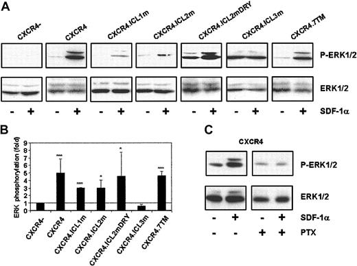
Next, we analyzed the activation of ERK, another Gi-dependent event, triggered after SDF-1α binding to CXCR4. After SDF-1α stimulation, ERK phosphorylation occurred in the HEK/CD4.403/CXCR4, HEK/CD4.403/CXCR4.ICL1m, HEK/CD4.403/CXCR4.ICL2m, HEK/CD4.403/CXCR4.ICL2mDRY, and HEK/CD4.403/CXCR4.7TM cells, whereas no activation of this kinase was found in HEK/CD4.403 and HEK/CD4.403/CXCR4.ICL3m cells (Figure6A). This signal was inhibited by PTX (Figure 6B), confirming that this signal is dependent on Gi proteins.
SDF-1–mediated ERK activation of HEK/CD4.403 cells expressing wild-type and mutated CXCR4 molecules.
(A) Serum-starved transfected cells were stimulated for 2 minutes with SDF-1 at 125 nM and lysed as described in “Materials and methods.” Protein samples were run on an SDS-polyacrylamide gel and Western blotted with an antiphosphoERK1/2 antibody. Protein loading was controlled by using a total anti–ERK1/2 antibody. (B) Densitometric analysis of phosphorylated ERK expression. Data are presented as fold increases of ERK phosphorylation, in which the amount of ERK phosphorylation in cells that do not express CXCR4 is assigned a value of 1.0 after SDF-1 activation. All the relative intensities were calculated by normalizing the intensity of phosphorylated ERK protein to its ERK protein loading control. Results shown are from 2 to 5 independent experiments; error bars reflect SDs. Statistical analysis was performed as described in “Materials and methods” (*P < .05, **P < .01, ***P < .001). (C) ERK phosphorylation induced by SDF-1 in HEK/CD4.403/CXCR4 cells in the presence or absence of PTX.
SDF-1α–induced chemotaxis requires several intracellular domains of CXCR4
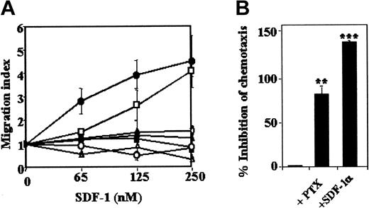
Chemotaxis of HEK/CD4.403, HEK/CD4.403/CXCR4, HEK/CD4.403/CXCR4.ICL1-3m, and HEK/CD4.403/CXCR4.7TM cell lines to SDF-1α was first studied. Only HEK/CD4.403/CXCR4 and HEK/CD4.403/CXCR4.ICL1m cells underwent SDF-1α–induced chemotaxis in a dose-dependent manner (Figure 7A), indicating that chemotaxis needs the presence of the second and the third intracellular loops as well as the C terminus part of CXCR4. PTX strongly, but not totally, inhibits chemotaxis induced by SDF-1α (Figure 7B). Elimination of the gradient by adding the same concentration of the chemokine in the upper and lower chamber at the same time completely inhibits chemotaxis, indicating that the migration observed was not due to chemokinesis (Figure 7B).
SDF-1–induced chemotaxis of HEK/CD4.403 cells stably transfected with wild-type or mutated CXCR4 molecules.
Cells were subjected to chemotaxis by using SDF-1α at 65, 125, or 250 nM, fixed, stained, and counted under a high-power field microscope. (A) Migration index obtained with the different clones (⋄, CXCR4−; ●, CXCR4+; ■, CXCR4.ICL1m; ○, CXCR4.ICL2m; ▴, CXCR4.ICL2mDRY; ▪, CXCR4.ICL3m; ▵, CXCR4.7TM). Results are expressed as means of at least 5 independent experiments; error bars reflect SDs. (B) Inhibition of SDF-1–induced chemotaxis in HEK/CD4.403/CXCR4 cells. Cells expressing wild-type CXCR4 were preincubated overnight with PTX (100 ng/mL). SDF-1 was also added to the upper and lower chemotaxis chambers (250 nM) to eliminate the chemokine gradient. Data are expressed as means ± SDs of 3 independent experiments. Statistical analysis was performed as described in “Materials and methods” (*P < .05, **P < .01, ***P < .001).
These results highlight the complexity of SDF-1α–induced chemotaxis in which several intracellular domains are involved.
Discussion
Multiple PTX-sensitive and -insensitive signaling pathways are activated through CXCR4, but little is known of the components of CXCR4 necessary to transduce those signals. The present study was aimed at determining the role of the cytoplasmic domains of CXCR4 in different signaling events after SDF-1 stimulation.
The ubiquitous structure of the GPCRs led to the assumption that the 7 transmembrane (TM) domains that confer 3 intracellular and 3 extracellular loops in addition to the N and C terminal segments at opposite membrane surfaces are the minimum to achieve structural stability and to trigger activation of signal molecules. The 3 intracellular loops of CXCR4 are highly basic, with 4, 6, and 8 basic amino acids in the ICL1, ICL2, and ICL3, respectively. The replacement of each loop by the mutated ICL1 still containing its own basic residues allowed preservation of the general basic properties of these loops. ICL1 is the shortest loop, composed of 11 residues, which are sufficient to link the TM1 to TM2 and induce a functional conformation of the entire CXCR4 molecule. We thus postulated that replacement of each loop by the ICL1 modified to destroy the putative sites involved in binding cytoplasmic signaling proteins but keeping its characteristics of charge and length could be a novel and interesting way to analyze the role of the intracellular loops of GPCRs. First, our data demonstrate that this approach allows cell lines to be derived that stably express CXCR4 mutants at the cell surface. Additionally, these mutants are able to bind SDF-1α and to support HIV-1 infection. As SDF-1 also binds to glycosaminoglycans, present at the cell surface,37 we verified that they did not interfere with our binding assay by using heparin and chondroitin sulfate as competitors (data not shown). Thus, the original amino acid sequence of each ICL is not necessary for a functional configuration of CXCR4. In particular, the DRY motif in the N terminus part of ICL2, which is highly conserved in the GPCRs and was shown to be essential for insertion into the cell-surface membrane,8 is not important for CXCR4 folding and presentation at the cell surface. Indeed, CXCR4.ICL2m and ICL2mDRY are expressed at the same level and are capable of binding to SDF-1α. However, ICL1 and ICL2, but not the DRY sequence, play a role in CXCR4 conformation and/or stability because they are less expressed at the HEK cell surface than the wild-type CXCR4 molecule. Similar data were observed in COS cells, a transformed kidney cell line from African green monkey, expressing transient CXCR4 mutants38,39 and different CXCR4 chimeras.39 However, several CXCR4 chimeras that presented reduced expression levels were still able to induce calcium mobilization.39
Numerous studies have shown that in most cases the second and the third ICLs of GPCRs are the major sites for the receptor to interact with G proteins, even if ICL1 of some receptors played a role in transducing G protein–dependent signals.40,41 The current study indicates that ICL1 is not essential for CXCR4 activation even if this loop is needed for CXCR4 to be well expressed at the cell surface. This result agrees with those found by Ling et al42 who demonstrated that a mutated form of CXCR4 in which the N-terminal segment is directly connected to TM3 was still able to transduce SDF-1–induced chemotaxis, calcium influx, and activation of Gi proteins.
The DRY box in ICL2 is highly conserved among GPCRs, and its mutation in different receptors such as rhodopsin, the α- and β-adrenergic receptors,43-45 and CCR546,47 eliminates signaling. However, mutation of this motif did not always trigger abolition of receptor activation.8,48 It was previously shown that mutation of the DRY motif of CXCR4 to asparagine, alanine, alanine (NAA) largely eliminated the ability to signal. However, this mutant retained partial G protein-coupling activity, as a small calcium mobilization was noted. Furthermore, this mutant was very weakly expressed at the surface of transitory transfected cells.39 Stably transfected cells that express functional CXCR4 molecules containing a mutated form of ICL2 with or without DRY are still able to transduce Gi-dependent signals, indicating that this loop and the DRY box are dispensable for Gi-mediated signaling, although this loop participates to SDF-1–induced chemotaxis. Indeed, CXCR4 in which the ICL2 loop is entirely replaced by a mutated form of ICL1 which is unable to transduce a signal yet binds SDF-1 and triggers calcium mobilization and ERK activation. The ability of the CXCR4.ICL2m receptors to signal in response to SDF-1 in spite of their low cell surface expression indicate that these mutants are expressed at a functional level. It is worth noting that SDF-1–mediated signal is not strictly related to mutant expression level, whereas SDF-1 binding and HIV-1 infection are dependent on the quantity of cell surface receptor.
Extensive work has shown that the third cytoplasmic loop is a major determinant of G protein coupling for several receptors.49-52 Furthermore, the CXCR4 ICL3 contains the BBXXB motif (RKALK) in which B is a basic and X a nonbasic residue, described as a structural determinant for Gi-stimulating function.53 To analyze the role of ICL3 of CXCR4 in transduction of Gi-dependent signaling, we studied several Gi-dependent activation signals after SDF-1 binding to CXCR4 in which ICL3 was replaced by a mutated form of ICL1. This mutant, which is well expressed at the cell surface and capable of binding efficiently to SDF-1, is unable to induce calcium mobilization and ERK phosphorylation, Gi-dependent events, after SDF-1 stimulation. This result emphasizes that the mutated ICL1 used to replace the other loops of CXCR4 and designed to destroy all the putative activation sites is unable by itself to transduce a signal. Calcium mobilization was already shown to be abolished after SDF-1 binding to CXCR4 that contained deletions in ICL3.38Gi-dependent signaling through CXCR4 is thus supported by only one loop, without cooperation of other cytoplasmic domains.
The third intracellular loop, as well as the C terminus part of CXCR4, has also been involved in GPCR interaction with arrestin.54-57 It was previously demonstrated that ICL3 and the C terminus of CXCR4 directly interact with β-arrestin, producing differential regulation of receptor functions, such as internalization and ERK activation.10 In this study, SDF-1–mediated internalization of cells expressing the mutated form of ICL3 occurs, at a level identical to that observed with wild-type CXCR4, indicating that this loop is dispensable for receptor endocytosis. It is worth noting that this mutant possesses the entire C terminus domain that was shown to bind efficiently to GRK and β-arrestin. When this part of the CXCR4 molecule (CXCR4.7TM) is withdrawn, internalization of this mutant expressed on HEK cells following SDF-1 binding is totally abolished. Similar data were obtained by using a CXCR4 form deleted of the last 41 amino acids,58 whereas truncation of 3410 or 352 amino acids leads to only a decrease in SDF-1–induced internalization. Length of deletion in the C terminal part of CXCR4 thus determines the level of agonist-dependent internalization. This observation is in contradiction with the fact that truncation of the last 34 amino acids in the C terminal part of CXCR4 is sufficient to induce a complete inhibition of phosphorylation of this receptor.2,59 One hypothesis could be that major sites for GRK phosphorylation, easily detectable by phosphorylation studies, should correspond to Ser/Thr rich part of the cytoplasmic tail at position 318 to 352 and especially the dileucine motif (Ile328 and Leu329) and the serines 324, 325, 338, and 339,11 but Ser and Thr present at the very beginning of the tail (Thr311, Ser312, and possibly Thr318) may also be phosphorylated and participate to the endocytotic process. Another possibility is that β-arrestins need the complete C terminal sequence to bind efficiently to CXCR4 and induce complete internalization after phosphorylation of the 318 to 352 amino acid region. The fact that CXCR4.ICL3m, which contains the C terminus part but lacks the 3 serines as well as potential sites in ICL3 able to activate cytoplasmic signaling molecules, undergoes complete endocytosis after SDF-1 binding emphasizes the importance of the C terminus domain of CXCR4 in internalization. This suggests that the binding of β-arrestin to ICL3 is not necessary for CXCR4 clathrin-mediated endocytic pathway but may be implicated in specific signaling after SDF-1 binding to CXCR4.
Interestingly, ERK is phosphorylated after SDF-1 binding to CXCR4.7TM, in absence of receptor internalization. The role of clathrin-induced endocytosis in GPCR-mediated ERK activation has yielded, until now, conflicting results. Although the present work does not provide insight into the mechanism of ERK phosphorylation by SDF-1 binding to CXCR4, this result underlines the fact that CXCR4 endocytosis is not necessary for ERK activation. In several cell types, including HEK-293 cells, GPCR-stimulated ERK involves the ligand-independent transactivation of receptor tyrosine kinases, such as the EGF receptor.60-62In our cellular model, SDF-1–mediated phosphorylation of ERK through CXCR4 is independent of the transactivation of the EGF receptor (data not shown).
Chemotaxis through CXCR4 needs several distinct signaling pathways. Neither CXCR4.ICL2m and CXCR4.ICL2mDRY, able to transduce both calcium and ERK signaling and to undergo internalization, nor CXCR4.ICL3m, unable to transduce Gi-dependent signaling but undergoing internalization and CXCR4.7TM transducing ERK activation and calcium mobilization without endocytosis, can induce this signaling event. Indeed, only CXCR4.ICL1m can still transduce signals necessary for chemotaxis. This mutant shows the same cell surface expression level as CXCR4.ICL2m and CXCR4.ICL2mDRY, and these 3 mutants transduce comparable levels of Gi-dependent signals. However, we cannot totally exclude the fact that the weak expression of CXCR4.ICL2m and CXCR4.ICL2mDRY may be responsible for the absence of chemotaxis signaling. The specific signaling functions required for GPCRs to mediate chemotaxis are complex and poorly understood. Giactivation and release of free βγ subunits was shown necessary but probably not sufficient for chemotaxis.20 PI3-K is required for SDF-1–induced migration of hematopoietic progenitor cells8 and T lymphocytes.25,26 PKC is also required for SDF-1–induced tyrosine phosphorylation of focal adhesion proteins and for migration of hematopoietic progenitor cells.8 Migration through a chemotactic gradient could also need chemokine receptor internalization that engages receptor phosphorylation by GRK that may explain the fact that the C terminus domain of CXCR4 could be involved in this process. Further investigation on the biologic processes required for cell mobilization induced after SDF-1 binding to CXCR4 is needed.
In summary, this study is the first that can dissociate the role of each intracellular domain of CXCR4 in transducing Gi-dependent signaling such as calcium influx and ERK activation, Gi-independent event (internalization), and cell migration. Our results indicate that the ICL3 of CXCR4 alone supports binding to Gi proteins, whereas the second, third, and C terminal intracellular domains are involved in chemotaxis. We have also dissociated ERK activation from receptor internalization.
Prepublished online as Blood First Edition Paper, September 5, 2002; DOI 10.1182/blood-2002-03-0978.
Supported by institutional funds from the Centre National de la Recherche Scientifique (CNRS) and grants from the Agence Nationale de Recherches sur le syndrome de l'immunodeficience acquise (SIDA) (ANRS) and Ensemble contre le SIDA, by an EMBO short-term fellowship (ASTF no. 9468) (B.J.M.), and by financial support from Professor A. J. Pinching (B.J.M. and K.E.N.).
J.R. and B.J.M. contributed equally to this work.
The publication costs of this article were defrayed in part by page charge payment. Therefore, and solely to indicate this fact, this article is hereby marked “advertisement” in accordance with 18 U.S.C. section 1734.
References
Author notes
Martine Biard-Piechaczyk, Laboratoire Infections Rétrovirales et Signalisation Cellulaire CNRS UMR 5121, Institut de Biologie, 4 Boulevard Henri IV, CS89508, 34060 Montpellier Cedex 34960, France; e-mail:piechacz@xerxes.crbm.cnrs-mop.fr.

![Fig. 5. SDF-1–induced calcium mobilization. / Stably transfected HEK/CD4.403 cells expressing wild-type and mutated CXCR4 receptors were loaded with Fluo-3-AM. SDF-1–induced calcium flux was measured as described in “Materials and methods” in the presence (○) or absence (●) of PTX preincubated overnight at 100 ng/mL. Representative curves of [Ca2+]i changes after stimulation with SDF-1α and ionomycin (I) from 1 to 4 experiments are shown.](https://ash.silverchair-cdn.com/ash/content_public/journal/blood/101/2/10.1182_blood-2002-03-0978/4/m_h80233648005.jpeg?Expires=1763747312&Signature=fAAGt74WdNTWQqs9mh6lApALrb9hw~c3hd3VCBOWkOZlHbLNbFAwYuDsd2HB16eJ34nS9fq8ZXM1Y4hLnwPA7BSrLNWam4ingb1-bZszvElzekawFhvLDwdGXv4pAVO38hNvXfsaJ5JtuzF0Go2RwVz6h7ZxX5HjhAaP7x59empwBWNhsKqYi7ckzXvyP6Y1do4a-Z5SU-eKyvlUIK~eqAZSPfgfXfcJsJBTL--csrmUk7yG8chJXmDwHiie7xIw83OyJkxYMInqRo46T-0OAKfsd9VJts4hTtjaC06gjCm7OTnfMasAWrHfGwGZeiu-bnILCtOvvrSI1a2CJNQIkQ__&Key-Pair-Id=APKAIE5G5CRDK6RD3PGA)
