In light of the emerging concept of a protective function of the mitogen-activated protein kinase (MAPK) pathway under stress conditions, we investigated the influence of the anthracycline daunorubicin (DNR) on MAPK signaling and its possible contribution to DNR-induced cytotoxicity. We show that DNR increased phosphorylation of extracellular-regulated kinases (ERKs) and stimulated activities of both Raf-1 and extracellular-regulated kinase 1 (ERK1) within 10 to 30 minutes in U937 cells. ERK1 stimulation was completely blocked by either the mitogen-induced extracellular kinase (MEK) inhibitor PD98059 or the Raf-1 inhibitor 8-bromo-cAMP (cyclic adenosine monophosphate). However, only partial inhibition of Raf-1 and ERK1 stimulation was observed with the antioxidant N-acetylcysteine (N-Ac). Moreover, the xanthogenate compound D609 that inhibits DNR-induced phosphatidylcholine (PC) hydrolysis and subsequent diacylglycerol (DAG) production, as well as wortmannin that blocks phosphoinositide-3 kinase (PI3K) stimulation, only partially inhibited Raf-1 and ERK1 stimulation. We also observed that DNR stimulated protein kinase C ζ (PKCζ), an atypical PKC isoform, and that both D609 and wortmannin significantly inhibited DNR-triggered PKCζ activation. Finally, we found that the expression of PKCζ kinase-defective mutant resulted in the abrogation of DNR-induced ERK phosphorylation. Altogether, these results demonstrate that DNR activates the classical Raf-1/MEK/ERK pathway and that Raf-1 activation is mediated through complex signaling pathways that involve at least 2 contributors: PC-derived DAG and PI3K products that converge toward PKCζ. Moreover, we show that both Raf-1 and MEK inhibitors, as well as PKCζ inhibition, sensitized cells to DNR-induced cytotoxicity.
Introduction
The anthracycline daunorubicin (DNR) represents one of the major antitumor agents widely used in the treatment of acute myeloid leukemias. Despite intense efforts, its mechanism of action is still not fully understood. However, it is generally believed that DNR-induced cytotoxicity is related to DNA damage because of the intercalation of the drug and its interaction with nuclear topoisomerase II.1 It has been shown that DNR induced apoptosis in myeloid leukemia cell lines.2 However, present knowledge does not allow us to determine whether apoptosis simply reflects DNA lesions or represents an independent cytotoxic mechanism triggered by a specific signaling pathway.3
The characterization of DNR-activated signaling pathways leading to apoptosis is made still more complex by the emerging evidence that DNR may also activate intracellular signals, which have been shown to contribute to cell survival in other stress conditions. Indeed, we have shown that DNR induced sustained extracellular-regulated kinase 1 (ERK1) tyrosine phosphorylation in U937 cells.4 ERKs are members of the mitogen-activated protein kinase (MAPK) family. Each MAPK cascade consists of a module of 3 kinases: a MAPK kinase, which phosphorylates and activates a MAPK kinase, which in turn phosphorylates and activates a MAPK. The classical MAPK module consists of a Raf-1 kinase, mitogen-induced extracellular kinase 1 (MEK-1), and extracellular-regulated kinase (ERK). MAPKs are activated in response to a variety of growth factors, cytokines, and oncogenes and regulate a wide array of cellular processes such as gene expression, differentiation, and proliferation.5 More recently, it has been reported that ERK exerts a protective function against apoptosis induced by growth factor deprivation and that the dynamic balance between ERK and Jun kinase (JNK) pathways may be important in determining cell survival.6 The MAPK signaling pathway can also be activated at various extents by stress conditions such as heat shock,7 UV radiation, and ionizing radiation,8,9 as well as exposure to a variety of cytotoxic molecules, including tumor necrosis factor-α (TNFα),10 hydrogen peroxide,11 and cell permeant ceramides.4 Moreover, Raf-1 or MEK inhibitors were found to sensitize cells to oxidative stress11 and to ionizing radiation.12 Following these studies, it has been proposed that ERK activation by cytotoxic agents may contribute to cell survival. However, as far as DNR is concerned, the mechanism by which this drug activates ERK1 in leukemic cells, and whether or not ERK1 activation influences DNR cytotoxicity, remained unknown.
Raf-1 activation can be mediated through phosphorylation on either serine or tyrosine residues, depending on the stimuli and mediators that are involved in signal propagation, such as reactive oxygen species (ROS)11 and diacylglycerol (DAG). Indeed, studies have shown that, on phosphatidylcholine (PC) hydrolysis, elevated levels of PC-derived DAG caused the activation of Raf-1 through protein kinase C ζ (PKCζ), a phorbol ester–insensitive atypical PKC isoform.13 Interestingly, PKCζ may also be stimulated by phosphoinositide 3 kinase (PI3K) lipid products14; therefore, PKCζ has emerged as a common downstream effector of distinct signaling pathways converging toward MAPK.15 Altogether, these findings suggest that ROS, PC-derived DAG, and PI3K products are important mediators for Raf-1 activation.
On the basis of these findings and considerations, we speculated that these mediators could be involved via Raf-1 in DNR-induced ERK1 activation. Three lines of evidence argued for this hypothesis: (1) DNR generates ROS through reduction of its semiquinone radical by nicotinamide adenine dinucleotide phosphate (NADPH)–dependent flavin reductase,16 (2) DNR triggers PC hydrolysis through the stimulation of PC-specific phospholipase C (PC-PLC) activity with subsequent production of both DAG and phosphocholine,17and (3) DNR stimulates PI3K activity and subsequent production of phosphoinositide 3-phosphate (PI-3P).18
In light of those observations in which DNR may induce not only proapoptotic but also equally antiapoptotic pathways, we investigated whether it was possible to increase DNR cytotoxicity by negatively regulating the cell survival pathways. In this study, we evaluated the effects of DNR (0.1 μM and 1 μM) on PKCζ, ERK1, and Raf-1. In addition, we have used the antioxidant N-acetylcysteine (N-Ac) as well as different kinase (PI3K inhibitor wortmannin, MEK inhibitor PD98059, and Raf-1 inhibitor 8-bromo-cyclic adenosine monophosphate [cAMP]) or phospholipase inhibitors (PC-PLC inhibitor D609) and PKCζ kinase–defective cells to evaluate the possible role of ROS, PC-derived DAG, and PI3K products in the DNR-induced PKCζ and Raf-1/MEK/ERK–mediated antiapoptotic pathway and their contribution to cell sensitivity to DNR.
Materials and methods
Drugs and reagents
DNR (Cerubidine) was supplied from Laboratoire Roger Bellon (Neuilly-sur-Seine, France). PD98059 was purchased from Alexis (San Diego, CA); wortmaninn and N-Ac were supplied from Sigma Chemical (St Louis, MO). D609 (xanthogenate tricyclodecan-9-yl) was purchased from Calbiochem (San Diego, CA). All other drugs and reagents were purchased from Sigma, Carlo Erba (Rueil-Malmaison, France), or Prolabo (Paris, France). The kinase-defective PKCζ cDNA, generously provided by Jorge Moscat (Molecular Biology Center, Madrid, Spain) was inserted in the PRcCMV plasmid (Invitrogen, Groningen, The Netherlands). The Leu275Thr substitution suppresses a critical amino acid for adenosine triphosphate (ATP) binding in the catalytic domain, thereby inactivating the enzyme that behaves as a dominant-negative mutant.19
Cell culture
The human leukemic cell line U937 (monocytic) was purchased from American Type Culture Collection (ATCC; Rockville, MD). U937 cells were transfected with either an insertless vector (pRcCMV) or a kinase-defective PKCζ-containing vector (pRcCMV-ζ mut). Stable transfection of U937 cells was performed by electroporation at the capacitance of 960 μF and 300 V/0.4 cm followed by selection in the presence of 1 mg/mL G418 (Life Technology, Cergy-Pontoise, France) and limiting dilution cloning as previously described.20 Cells were cultured in RPMI 1640 medium supplemented with 10% heat-inactivated fetal calf serum, 2 mM L-glutamine, 100 μg/mL streptomycin, and 100 μg/mL penicillin (all from Eurobio, les Ulis, France) at 37°C and 5% CO2. In all experiments, exponentially growing cells (5 × 10⁵ cells/mL) were preincubated with or without various inhibitors before addition of DNR.
Fresh leukemic cells were obtained from unselected patients with acute myeloid leukemia (AML) at diagnosis. Bone marrow (BM) aspirates were collected in heparinized syringes, and mononuclear cells were separated by centrifugation through a Ficoll-Hypaque density gradient. Cells were washed twice in Iscoves modified Dulbecco medium (IMDM), resuspended at a final concentration of 2 × 10⁷cells/mL, and cryopreserved in IMDM containing 10% dimethyl sulfoxide and 50% fetal calf serum (FCS). Percentages of blast cells on BM smears at diagnosis varied from 50% to 99%. After processing (Ficoll separation, freezing, and thawing), the percentage of leukemic cells was higher than 90% in all cases.
Determination of ROS
Production of ROS was detected by using a C2938 fluorescent probe as previously described.21 Briefly, exponentially growing cells were labeled with 0.5 μM C2938 for 1 hour and then incubated in the absence or presence of DNR at 37°C for various periods of time. The cells were washed in phosphate-buffered saline (PBS), and cell fluorescence was determined by using flow cytometry on a fluorescence-activated cell sorter scan (FACScan; Becton Dickinson, Paris, France).
Immunoprecipitation and Western blot
For Western blot, 5 × 10⁶ cells were lysed in RIPA buffer (50 mM Tris (tris(hydroxymethyl)aminomethane) pH8, 150 mM NaCl, 1% Triton X100, 1% sodium deoxycholate, 1% sodium dodecylsulfate [SDS], 5 mM EDTA [ethylenediaminetetraacetic acid], 1 mM dithiothreitol [DTT], 1 mM sodium vanadate, 10 mM β-glycerophosphate, 50 mM sodium fluoride, 5 μg/mL leupeptin, 0.1 mM phenylmethylsulfonyl fluoride [PMSF]) for 5 minutes on ice, then sonicated. Protein cell extracts (40 μg; determined by using the Bradford method) were resolved by electrophoresis in 10% SDS-polyacrylamide, transferred onto nitrocellulose membrane (Hybond-C; Amersham, Les Ullis, France), and probed with anti-P–MAPK (New England Biolabs, Saint Quentin en Yvelines, France), anti-ERK1 (Santa Cruz, Le Perray-en Yvelines, France). Bound proteins were detected by enhanced chemiluminescence (ECL) detection system (Amersham).
Immunoprecipitation was performed essentially as described previously.22 Briefly, approximately 10 × 106 cells were pelleted and then lysed with 1 mL RIPA. Protein cell extracts (200 μg) were incubated with 3 μg antiphosphotyrosine PY20 antibody (Transduction Laboratories, Lexington, KY) overnight at 4°C. Immune complexes were collected by incubation with 20 μg protein G Sepharose beads (Sigma-Aldrich, Saint Quentin Fallavier, France) for 2 hours at 4°C and eluted by boiling for 5 minutes in denaturation solution. Western blot was then performed on extracts as described.
Protein kinase activity assays
After treatment of cells (3 × 10⁶/mL) with DNR or DiC8 for the indicated times, cell extracts were prepared by lysing cells in buffer containing 20 mM HEPES (N-2-hydroxyethylpiperazine-N′-2-ethanesulfonic acid) pH 7.4, 12 mM EDTA, 250 mM NaCl, 1% NP-40, 2 μg/mL leupeptin, 2 μg/mL aprotinin, 1 mM PMSF, 0.5 μg/mL benzamidine, and 1 mM DTT. Cell extracts (150-250 μg/sample) were immunoprecipitated with 0.3 μg anti-ERK1 (C-16) or anti–Raf-1 (C-12) (Santa Cruz), or anti-PKCζ antibodies (Gibco BRL, Paris, France) for 60 minutes at 4°C. Immune complexes were collected by incubation with protein A/G Sepharose beads (Pierce, Rockford, IL) for 60 minutes at 4 °C. The beads were extensively washed with lysis buffer and kinase buffer (20 mM HEPES, pH 7.4, 1 mM DTT, 25 mM NaCl). Kinase assays were performed for 15 minutes at 30°C using myelin basic protein (MBP; Sigma) as a substrate for ERK1, Raf-1, or PKCζ activities in 20 mM HEPES, pH 7.4, 10 mM MgCl2, 1 mM DTT, and 0.37 MBq (10 μCi) (γ32P) ATP (ICN, Orsay, France). Reactions were stopped with the addition of 15 μL 2 × SDS sample buffer, boiled for 5 minutes, and subjected to SDS-polyacrylamide gel electrophoresis (PAGE) (9%). Phosphorylated MBP was visualized by staining with Coomassie blue, the dried gel was analyzed by autoradiography, and the corresponding bands were scrapped and quantitated by scintillation counting.
Analysis of phosphatidylcholine
PC quantitation was performed by labeling cells to isotopic equilibrium with [methyl-3H] choline. Quantitation of PC was determined in the aqueous phase of [3H] choline-labeled cell extracts by thin-layer chromatography as previously described.17
In vitro lipid kinase assay
The phosphorylated phosphoinositides were evaluated as previously described.23 Exponentially growing cells were incubated in RPMI containing 10% FCS overnight. After various times of incubation with DNR, the cells were washed once with ice-cold PBS, then once with buffer 1 (50 mM HEPES, 150 mM NaCl, 10 mM EDTA, 10 mM Na4P2O7, 2 mM orthovanadate, 100 mM NaF, pH 7.4) and lysed in 300 μL buffer 2 (buffer 1 containing 1 μg/mL aprotinin, 1 mM PMSF, 1 μg/mL leupeptin, 1% NP-40) for 15 minutes. After centrifugation, the supernatants were incubated at 4°C for 90 minutes with protein A-Sepharose (Pharmacia Biotechnology, Les Ullis, France) previously incubated with the polyclonal anti-p85 antibody (Upstate Biotechnology, Lake Placid, NY). The beads were washed twice with each of the following buffers: (1) PBS with 1% NP-40; (2) 0.5 M LiCl, 0.1 M Tris, pH 7.6; and (3) 10 mM Tris, pH 7.5, 100 mM NaCl, 1 mM EDTA. The beads were then incubated with a buffer containing 20 mM HEPES, 0.4 mM EGTA (ethyleneglycoltetraacetic acid), and 0.4 mM Na2HPO4 (30 μL). Then 10 μL of 10 mg/mL phosphatidylinositol (previously sonicated in 5 mM HEPES) and 10 μL reaction buffer (50 mM MgCl2, 100 mM HEPES, 50 μM ATP containing 0.0111 MBq (0.3 μCi)/sample of γ[32P]ATP) (ICN) were added to the beads that were further incubated for 10 minutes at room temperature. The reaction was stopped by the addition of 15 μL of 4 N HCL. Lipids were extracted with methanol/chloroform (1/2, vol/vol). Lipids contained in the organic phase were separated by thin-layer chromatography. After autoradiography, the PI-3P fractions were scraped off, and the incorporated radioactivity was assayed by scintillation counting.
Drug cytotoxicity studies
Cells were treated with DNR. Cell viability was measured by methyl thiazolyl tetrazolium (MTT) assay.
Cytochemical staining
Changes in cellular chromatin of U937 cells were evaluated by fluorescence microscopy by DAPI (4′, 6′-diamino 2-phenylindol) staining as previously described.21 Cell samples from patients with AML were stained with May-Grünwald-Giemsa (MGG). Results are expressed as percentage of apoptotic cells.
Results
Effects of DNR on ERK phosphorylation and activity
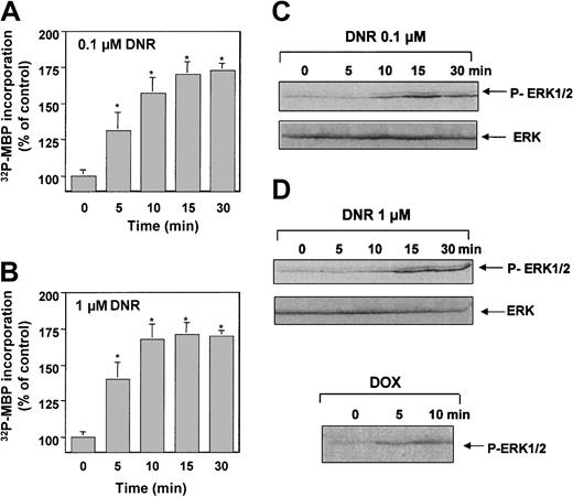
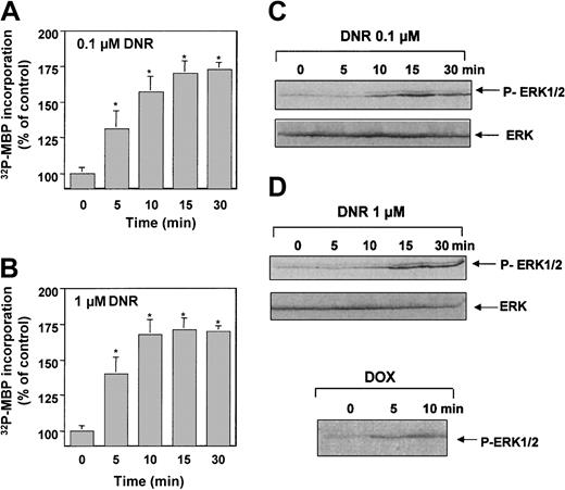
As we have previously described,4 incubation of U937 cells with either 0.1 μM or 1 μM DNR induced ERK1 activation. p44 ERK1 tyrosine phosphorylation was observed within 4 minutes and remained relatively stable for at least 30 minutes (data not shown). The magnitude of ERK1 phosphorylation (∼3-fold increase) was similar to that induced by 16 nmol/L phorbol 12-myristate 13-acetate (PMA) used at 15 minutes as a control (data not shown). The phosphorylation of ERK1 correlated with a time-dependent increase in kinase activity, with a maximum increase of about 170% within 10 to 15 minutes (Figure 1A-B). In addition, we evaluated the influence of DNR on the phosphorylation of ERK proteins by using an antibody directed against phosphorylated forms of both ERK1 and ERK2 (antiphospho-MAPK antibody). As shown in Figure 1C, treatment with either 0.1 μM or 1 μM DNR resulted in increased phosphorylation of not only p44 ERK1 but also p42 ERK2. ERK tyrosine phosphorylation was not limited to DNR, but was also observed to a similar extent with another anthracycline doxorubicin at either 0.1 μM (data not shown) or 1 μM doxorubicin (Figure 1D). These experiments were conducted in U937 cells incubated overnight in serum-free media. Similar experiments were performed in the presence of serum, and the magnitude of DNR-induced ERK stimulation was found to be similar (∼2-fold increase), although basal ERK activity was moderately increased, compared with serum-starved conditions.
Anthracycline-triggered activation of ERK1 in U937 cells.
ERK1 kinase activity was evaluated in U937 cells treated for 5 to 30 minutes with 0.1 μM (A) or 1 μM DNR (B) by MBP phosphorylation as described in “Materials and methods.” Results are mean ± SEM of 3 independent determinations. *P < .01. (C) ERK activation was assayed in U937 cells treated with either 0.1 μM or 1 μM DNR with anti-P–MAPK as described in “ Materials and methods.” Results are representative of 3 independent experiments. (D) ERK activation was also determined in 1 μM doxorubicin (DOX)–treated cells.
Effects of pharmacologic inhibitors on DNR-induced ERK1 activity
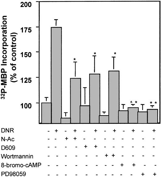
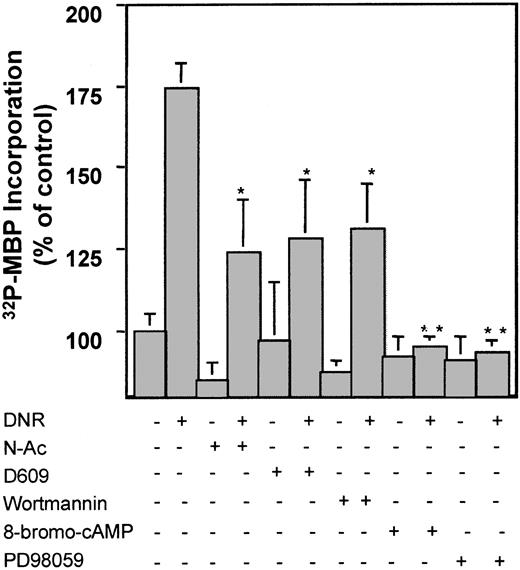
To decipher the signaling components that influenced ERK activity, we used a series of pharmacologic inhibitors that have been documented to interfere with DNR signaling, including the antioxydant N-Ac, the PC-PLC inhibitor D609, or the PI3K inhibitor wortmannin.24The influence of these compounds on the MAPK cascade was evaluated by an immunokinase assay after anti-ERK1 immunoprecipitation. We found that N-Ac, D609, and wortmannin resulted in a significant (but incomplete) inhibition of DNR-induced ERK1 kinase activity (Figure2); whereas ERK1 activity was completely blocked by Raf-1 inhibitor 8-bromo-cAMP and MEK inhibitor PD98059. These results suggested that ROS, DAG, and PI3K products partially contributed in ERK1 activation triggered by DNR, whereas both Raf-1 and MEK acted directly upstream of ERK1.
Effect of pharmacologic inhibitors on DNR-induced ERK1 stimulation.
U937 cells were either untreated or preincubated in the absence or presence of various pharmacologic inhibitors: 25 mM N-Ac for 2 hours, 10 μg/mL D609 for 1 hour, 25 nmol/L wortmannin for 30 minutes, 250 μM 8-bromo-cAMP for 30 minutes, or 50 μM PD98059 for 30 minutes. ERK1 kinase activity was evaluated, following a further incubation of 15 minutes with 1 μM DNR, as described in “ Materials and methods.” Results are mean ± SEM of 4 independent determinations. *P < .05; **P < .01 compared with cells treated with DNR alone.
Role of the ROS-dependent PI3K pathway in DNR-induced ERK1 activation
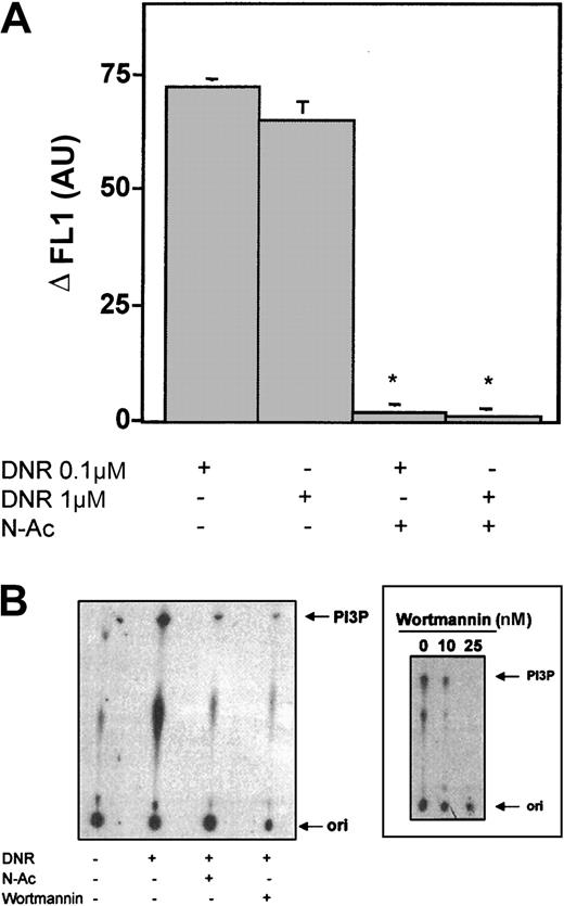
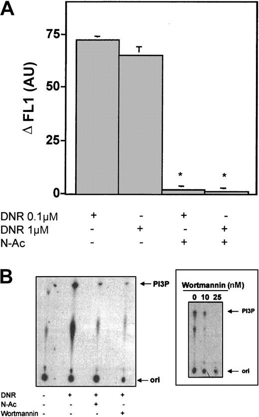
In an attempt to characterize the signaling pathways leading to ERK1 activation by DNR, we first studied the role of N-Ac and wortmannin in DNR signaling. Incubation of U937 cells with DNR led to H2O2 generation that was not dose dependent (Figure 3A). H2O2generation was observed within the first 5 minutes of incubation and was inhibited by N-Ac (Figure 3A). DNR (0.1 and 1 μM) also triggered within 5 minutes PI3K activation and subsequent generation of PI-3P, which was inhibited by wortmannin, at the optimal concentration of 25 nmol/L (Figure 3B). Interestingly, PI3K stimulation was also totally inhibited by N-Ac, demonstrating an essential role for ROS upstream of PI3K. Because both N-Ac and wortmannin significantly (but incompletely) inhibited DNR-induced ERK1 kinase stimulation (Figure 2), these results suggested that DNR triggered a ROS-dependent PI3K pathway that contributed to ERK1 activation.
Role of the ROS-dependent–PI3K pathway on DNR-induced ERK1 stimulation.
(A) U937 cells were incubated with C2938 fluorescent probe for 1 hour followed by incubation in the absence or presence of 25 mM N-Ac for 2 hours followed by 0.1 μM or 1 μM DNR for 5 minutes. The cells were washed, and cell fluorescence was determined by using flow cytometry as described in “ Materials and methods.” Results are mean ± SEM of 3 independent determinations; *P < .01. (B) U937 cells were preincubated in the absence or in the presence of 25 mM N-Ac for 2 hours or 25 nM wortmannin for 30 minutes followed by 1 μM DNR for 5 minutes. PI3K kinase activity was assayed by thin-layer chromatography (TLC) as described in “ Materials and methods.” (Inset) Dose-effect inhibition of basal PI3K activity by wortmannin after 30 minutes of incubation. Results are representative of 3 independent experiments. ori indicates origin; PI3P, phosphatidylinositol-3-phosphate.
Role of PC-derived DAG pathway in DNR-induced ERK1 activation
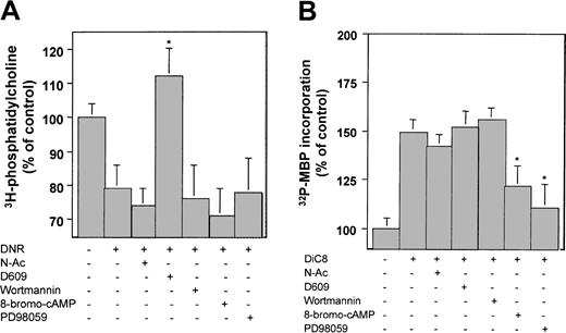
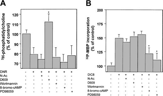
In a previous study, we have also shown that DNR (0.1-1 μM) triggered within 4 minutes PC-PLC–dependent PC hydrolysis and subsequent generation of DAG. Preincubation with D609 resulted in the expected inhibition of DNR-induced PC hydrolysis,17whereas N-Ac, wortmaninn, 8-bromo-cAMP, and PD98059 presented no effect (Figure 4A). Because D609 was found to decrease DNR-induced ERK1 activation (Figure 2), this suggested that PC-derived DAG played a role in ERK1 stimulation in DNR-treated cells. To further investigate this hypothesis, we used the cell permeant DAG analog DiC8 to mimic DNR-induced endogenous DAG production. As shown in Figure 4B, treatment of U937 cells with DiC8 resulted in partial ERK1 activation, which was not affected by N-Ac, D609, or wortmannin but was significantly inhibited by 8-bromo-cAMP and PD98059 (Figure 4B). These observations suggested that DNR also elicited a ROS-PI3K independent pathway that consists in PC-PLC–mediated PC hydrolysis, DAG production, and DAG-induced ERK1 stimulation. Overall, one can conclude that DNR triggered 2 independent pathways that both converge to activate ERK1: a PC-PLC/DAG (ROS independent) pathway and a ROS-dependent PI3K pathway. Furthermore, these 2 signaling pathways appeared to equally contribute to ERK1 stimulation.
Role of the PC-PLC–derived DAG pathway on DNR-induced ERK1 stimulation.
U937 cells were either untreated or preincubated in the absence or presence of various pharmacologic inhibitors: 25 mM N-Ac for 2 hours, 10 μg/mL D609 for 1 hour, 25 nmol/L wortmannin for 30 minutes, 250 μM 8-bromo-cAMP for 30 minutes, or 50 μM PD98059 for 30 minutes. (A) Phosphatidylcholine levels were evaluated, following a further incubation of 5 minutes with 1 μM DNR, as described in “Materials and methods.” (B) ERK1 kinase activity was evaluated as described in “Materials and methods” following a further incubation of 5 minutes with 25 μM DiC8. Results are mean ± SEM of 3 independent experiments. *P < .01 compared with cells treated with DNR alone.
Activation of Raf-1 by DNR
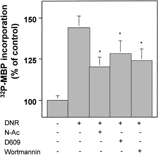
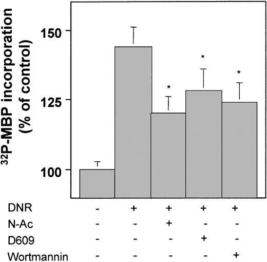
Because of the known function of Raf-1 as a MEK regulator, which in turn can phosphorylate ERK, we investigated the role of this serine-threonine kinase in DNR-induced ERK1 activation. DNR (1 μM) induced an increase in Raf-1 activity that became detectable at 10 minutes and remained stable between 10 to 30 minutes (data not shown) (1.5-fold at 15 minutes, Figure5). Similar results were observed with 0.1 μM DNR (data not shown). To further investigate the role of DNR-induced ROS, DAG, and PI3P production in DNR-triggered Raf-1 kinase activation, we evaluated the influence of N-Ac, D609, and wortmannin on Raf-1 activity in cells treated by DNR. These pharmacologic inhibitors presented essentially similar results as those observed on ERK1 activity. Indeed, preincubation with N-Ac, D609, or wortmannin only partially inhibited DNR-induced Raf-1 kinase activity (Figure 5). Therefore, both PC-derived DAG and PI3K products pathway contributed independently to Raf-1 activation. Previous studies have shown that both PC-derived DAG and PI3P may activate the atypical phorbol ester–insensitive PKCζ isoform,13,14,25 and that on activation PKCζ may phosphorylate and activate Raf-1.26 Therefore, we speculated that PKCζ could play a critical role in DNR-induced ERK activation as a common target of DAG and PI3P.
DNR triggered activation of Raf-1 in U937 cells.
U937 cells were either untreated or preincubated in the absence or presence of various pharmacologic inhibitors: 25 mM N-Ac for 2 hours, 10 μg/mL D609 for 1 hour, or 25 nM wortmannin for 30 minutes. U937 cells were further incubated for 15 minutes with 1 μM DNR, and Raf-1 kinase activity was measured as described in “Materials and methods.” Results are mean ± SEM of 3 independent determinations. *P < .01 compared with cells treated with DNR alone.
Role of PKCζ on DNR-induced MAPK activation
As shown in Figure 6A, DNR induced an increase in PKCζ activity, which peaked at 4 minutes (2-fold stimulation). Furthermore, although preincubation with either D609 or wortmannin significantly (but incompletely) inhibited DNR-induced PKCζ stimulation (53% and 67% decrease, respectively) (data not shown), cotreatment with both inhibitors abrogated DNR-induced stimulation of PKCζ. Moreover, 8-bromo-cAMP did not influence DNR-induced PKCζ stimulation. These results suggested the central role of PKCζ as the converging element of both DAG and PI3K-mediated signaling pathways and situated PKCζ upstream of Raf-1 in DNR signaling. Therefore, we speculated that PKCζ may play a critical role in DNR-induced ERK activation. To investigate such a hypothesis, we used U937 cells expressing a kinase-defective PKCζ variant (Z4 cells). As shown in Figure 6B, DNR (1 μM) induced a 2.5-fold increase in PKCζ activity in U937 cells transfected with empty vectors (C10 cells) but not in Z4 cells. The stimulation observed in C10 cells was detectable as soon as 5 minutes and remained stable up to 30 minutes (data not shown). Furthermore, we found that DNR induced ERK1 and ERK2 phosphorylation in C10 control cells but not in Z4 cells as evaluated by immunoblotting with antiphospho-MAPK antibody (Figure 6C). These findings confirmed that PKCζ function is critical for DNR-induced MAPK activation.
Role of PKCζ on DNR-induced MAPK activation.
(A) U937 cells were either untreated or preincubated in the absence or in the presence of both 10 μg/mL D609 for 30 minutes and 25 nmol/L wortmannin for 1 hour, 250 μM 8-bromo-cAMP for 30 minutes followed by 1 μM DNR for 4 minutes, and PKCζ kinase activity was measured as described in “ Materials and methods.” Results are mean ± SEM of 3 independent determinations. *P < .01 compared with cells treated with DNR alone. (B) C10 and Z4 cells were incubated with 1 μM DNR, and PKCζ kinase activity was measured as described in “ Materials and methods.” Results are mean ± SEM of 3 independent determinations. *P < .01. (C) Control cells (C10) and kinase-defective PKCζ cells (Z4) were incubated with 1 μM DNR, and ERK activation was evaluated by anti-P–MAPK as described in “Materials and methods.” Results are representative of 3 independent experiments.
Influence of PKCζ and MAPK activation on DNR-induced cytotoxicity
To evaluate the influence of the PKCζ/Raf-1/MAPK pathway on DNR-induced cytotoxicity, cells were pretreated 30 minutes with either 8-bromo-cAMP or PD98059 and then exposed to 0.1 μM or 1 μM DNR for 1 hour, and cell viability was measured by MTT assay at 48 hours. Neither PD98059 nor 8-bromo-cAMP alone influenced cell growth (Figure 7A), and neither significantly influenced DNR-induced apoptosis when the drug was used at 1 μM (data not shown). However, PD98059 or 8-bromo-cAMP significantly decreased cell viability when DNR was used at 0.1 μM (Figure 7A,B). Furthermore, on the basis of the role of PKCζ in DNR-induced MAPK activation, we speculated that the abrogation of PKCζ activation might also result in enhanced DNR-induced cytotoxicity. For this reason, we compared the sensitivity of C10 cells and Z4 cells to DNR. As shown in Figure 7C, DNR used at 0.1 μM was found to be 2-fold more toxic in Z4 cells than in C10 cells, whereas DNR used at 1 μM was found to be equitoxic (data not shown). Moreover, we found that DNR-induced apoptosis was enhanced in Z4 cells, compared with C10 cells, when the drug was used at the concentration of 0.1 μM (Figure7D). Collectively, these results showed that PKCζ/Raf-1/ERK pathway inhibition facilitated DNR-induced apoptosis and cytotoxicity.
Effect of PD98059 and 8-bromo-cAMP, a PKCζ kinase–defective mutant, on DNR-induced cytotoxicity.
(A) U937 cells were left untreated or pretreated in the absence or presence of 250 μM 8-bromo-cAMP for 30 minutes or 50 μM PD98059 for 30 minutes followed by 0.1 μM DNR for 1 hour. Cell viability was determined at 48 hours by MTT assay. (B) C10 and Z4 cells were treated by 0.1 μM DNR for 1 hour. Cell viability was determined at 48 hours by MTT assay. (C) C10 and Z4 cells were treated by 0.1 μM DNR for 1 hour and apoptosis by DAPI staining as described in “ Materials and methods.” All results are mean ± SEM of 3 independent determinations. *P < .01. (D) Cell samples from patients with AML were left untreated or pretreated in the presence of 50 μM PD98059 for 30 minutes followed by 0.1 μM DNR for 1 hour. Apoptotic cells were determined by MGG.
Finally, to evaluate the clinical relevance of ERK activation in DNR-induced cytotoxicity, we examined apoptosis in 4 samples from patient with AML treated with 0.1 μM DNR in the presence or not of 50 μM PD98059. As shown in Figure 7E, DNR alone induced variable levels of apoptosis, ranging from 3% to 40%. In DNR-sensitive samples, DNR and PD98059 displayed simple additive effect (sample 1 and 2), whereas in DNR-resistant cells, PD98059 significantly increased DNR-induced apoptosis (sample 3 and 4). This finding confirmed that ERK down-regulation can increase DNR cytotoxicity in drug-resistant AML cells.
Discussion
Compared with ultraviolet (UV) or ionizing radiation, the implication of the MAPK signaling pathway in the cellular response to antitumor drugs has received little attention. This could be explained by the fact that it appeared from previous studies that most drugs, except for cisplatinum,27 were unable to activate ERK proteins. Thus, it has been reported that paclitaxel,28aracytine, vinblastine, or VP-167 did not stimulate the classical MAPK module, whereas most of them are strong inducers of the JNK/SAPK (stress-activated protein kinase) pathway. Similarly, it has been claimed that the anthracycline doxorubicin was unable to activate the MAPK signaling pathway in different cellular models, including tumor epithelial cells7 or T-cell leukemia cells.29 Conflicting with these findings, our study shows that the anthracycline DNR, as well as doxorubicin, increased phosphorylation of ERK proteins, as well as it induced a modest but significant stimulation of ERK1 kinase activity in U937 cells. Moreover, we show that DNR stimulated the serine/threonine kinase Raf-1 in U937 cells treated with either 0.1 μM or 1 μM and that DNR-induced ERK1 activation was inhibited by 8-bromo-cAMP, a Raf-1 kinase inhibitor. Collectively, these results show that DNR activates the classical Raf-1/ERK MAPK module. Our observation that both doxorubicin and DNR stimulate ERK1 in a myeloid leukemia cell model suggests that this effect is cell type specific.
Our study indicates that the magnitude of ERK1 stimulation was not significantly different whether the cells were treated with either 1 μM or 0.1 μM DNR. In a previous study, we showed that a minimal DNR dose of 0.5 μM was required for stimulating ceramide production.30 Therefore, ceramide does not appear to be a critical mediator in DNR-induced MAPK activation. In fact, although still debated, the role of ceramide in Raf-1 activation has been questioned.31 Moreover, we also found that the MEK inhibitor PD98059 had no influence on JNK1 activation and apoptosis induced by DNR used at 1 μM (data not shown). Our findings suggest that, in cells treated with the dose of 1 μM DNR, there is no cross-interaction between the sphingomyelin/JNK and MAPK pathways as it has been suggested for other stimuli.32 This could explain why PD98059 had no effect on DNR-induced cytotoxicity and apoptosis when the drug was used at a concentration sufficient for activating the sphingomyelin-ceramide pathway (ie, 1 μM).
We observed that D609 inhibited DNR-induced ERK1 activation. In a previous study, we showed for the first time that DNR stimulated within 4 to 10 minutes a PC-PLC responsible for transient PC hydrolysis with subsequent generation of both DAG and phosphocholine in U937 cells that were inhibited by D609, a potent PC-PLC inhibitor33; dose-effect studies showed significant PC hydrolysis starting at 0.1 μM DNR ( = 6%) and maximal at 1 μM ( = 15%).17On the basis of these previous findings, we speculated that DAG originated from the PC breakdown could play a role in the activation of MAPK by DNR. Indeed, it has been shown that a dominant-negative mutant of Raf-1 blocked the transduction of the mitogenic signal generated by PLC-mediated hydrolysis of PC.13 Moreover, previous studies have suggested that DAG may directly interact with Raf-134 or interfere with Raf-1 function through a PKC-dependent mechanism.35 In fact, D609 inhibited Raf-1 kinase activation in cells treated either with 0.1 μM or 1 μM DNR. Furthermore, DiC8 stimulated both Raf-1 activity (data not shown) and ERK1 (Figure 4). These results suggested that ERK1 activation was, at least in part, mediated by DAG originated from PC breakdown through Raf-1 kinase stimulation.
In this study, we show that wortmannin inhibited DNR-induced ERK1 stimulation. We have previously shown that DNR induced within 2 to 10 minutes a wortmannin-inhibitable transient PI3K stimulation followed by the production of PI3K enzyme lipid products in a dose range between 0.1 μM and 1 μM DNR.18 Thus, the inhibitory effect of wortmannin suggested that PI3P mediated, at least in part, DNR-induced ERK1 activation. To investigate at which level of the MAPK module PI3K products operated, we evaluated the effect of wortmannin on DNR-induced Raf-1 stimulation. In fact, as D609 did, wortmannin partially inhibited Raf-1 activation in cells treated either with 0.1 μM or 1 μM DNR. This result confirms that ERK1 activation is not only mediated by DAG that originated from PC breakdown but also by PI3K lipid products.
The observation that D609 and wortmannin were equally efficient for blocking both Raf-1 and ERK1 kinase activities suggested that PC-derived DAG and PI3K products stimulated common downstream effectors capable of directly or indirectly stimulating Raf-1. Therefore, we speculated that these 2 mediators could target PKCζ, an atypical PKC isoform, that has been found to be activated by both PC-derived DAG13 and PI3K products through PDK-114,36and, at least in some circumstances, operates in a signaling complex as a direct activator of Raf-1.25,26 We demonstrate that the anthracycline DNR, at the doses of 0.1 μM and 1 μM, activated PKCζ and that both wortmannin and D609 inhibited DNR-triggered PKCζ activation. Although we have not shown in this study the direct interaction of PKCζ and Raf-1 in DNR-treated cells, these results strongly suggest that DNR-induced Raf-1 kinase stimulation was mediated by both PC-derived DAG and PI3K products through PKCζ. The central role of PKCζ in this signaling is supported by the fact that the expression of PKCζ kinase-defective mutant resulted in the abrogation of DNR-induced ERK protein phosphorylation. It is possible that this enzyme mediates other drug-activated signaling pathways that may interfere with drug-induced cytotoxicity. For example, it has been recently described that VP-16, another topoisomerase II inhibitor, activated PKCζ and that PKCζ inhibition resulted in enhanced drug-induced cytotoxicy.20 In the latter study, it has been proposed that PKCζ's protective effect could be mediated by nuclear factor κB (NF-κB).20 Whether DNR-induced PKCζ stimulation leads to NF-κB activation, in parallel to MAP kinase stimulation, should be investigated.
The present study shows that pretreatment with 8-bromo-cAMP and PD98059 resulted in an increase in DNR cytotoxicity. Moreover, in cells expressing PKCζ-defective kinase variant, DNR-induced apoptosis and cytotoxicity were also facilitated. These results suggest that the PKCζ/Raf-1/ERK signaling pathway contributes to cell survival. As a matter of fact, Raf-1 has been recently involved in resistance to doxorubicin in epithelial cells.37 However, when PKCζ, Raf-1, or MEK were inhibited, enhanced drug cytotoxicity was essentially observed when DNR was used at a low dose. This result suggests that, in the DNR-sensitive U937 cells, MAPK stimulation represents a futile cellular response to high-dose chemotherapy. However, Raf-1/ERK activation may contribute to cell survival when drug concentrations are reduced because of low-dose schedules, pharmacokinetic alterations, or altered biodistribution. Moreover, on the basis of these findings, one can also speculate that constitutive MAPK activation, as evidenced in fresh AML cells,38perhaps in response to hematopoietic growth factor paracrine or autocrine production39 may significantly alter DNR cytotoxicity in clinical settings.24 For this reason, it should be of interest to evaluate the chemosensitizing effect of newly described Raf-1 or MEK inhibitors.40 41 In this perspective, it is interesting to note that PD98059-mediated ERK inhibition sensitized DNR-resistant fresh AML cells.
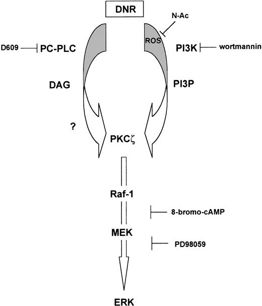
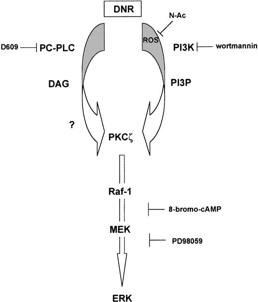
To conclude, our results suggest that DNR activates the classical Raf-1/ERK pathway and that Raf-1 activation is mediated through complex signaling that involves at least 2 contributors: PC-derived DAG and PI3K lipid products that converge toward PKCζ (Figure8). This study provides the first evidence of the implication of PKCζ in the cellular response to anthracyclines and points out this enzyme as a possible target for pharmacologic manipulation to improve DNR-induced cytotoxicity.
Hypothetical schema of DNR-induced pathway leading to activation of ERK.
PC-derived DAG and ROS-dependent PI3-K products generated by DNR activate distinct pathways, which converge toward PKCζ. Activated PKCζ is then responsible for the activation of Raf-1/MEK/ERK through a still-unknown mediator. DNR, daunorubicin; PC-PLC, phosphatidylcholine-phospholipase C; DAG, diacylglycerol; ROS, radical oxygen species; PI3K, phosphoinositide-3 kinase; PI3P, phosphatidylinositol-3-phosphate; PKCζ, protein kinase C ζ; MEK, mitogen extracellular kinase; ERK, extracellular-regulated kinase; ?, directly or indirectly.
Prepublished online as Blood First Edition Paper, October 24, 2002; DOI 10.1182/blood-2002-05-1585.
Supported by la Ligue Nationale Contre le Cancer and les Comités Départementaux du Gers, de l'Aveyron, du Lot, et de la Haute-Garonne (J.-P.J.), and La Faculté de Médecine Toulouse-Rangueil (G.L.). I.P. and N.M. are recipients of fellowships from la Ligue Nationale Contre le Cancer and la Ligue Departementale Contre le Cancer du Tarn et Garonne, respectively.
The publication costs of this article were defrayed in part by page charge payment. Therefore, and solely to indicate this fact, this article is hereby marked “advertisement” in accordance with 18 U.S.C. section 1734.
References
Author notes
Véronique Mansat-De Mas, Laboratoire d'Hématologie, Pavillon Caubet, Hôpital Purpan, 31059 Toulouse, France; e-mail:demas.v@chu-toulouse.fr.

