Abstract
Hepatitis C virus (HCV) may be associated with the mixed cryoglobulinemia syndrome and other B-cell lymphoproliferative disorders (LPDs). The t(14;18) translocation may play a pathogenetic role. Limited data are available regarding the effects of antiviral therapy on rearranged B-cell clones. We evaluated the effects of interferon and ribavirin on serum, B-lymphocyte HCV RNA, and t(14; 18) in 30 HCV+, t(14;18)+ patients without either mixed cryoglobulinemia syndrome or other LPDs. The t(14;18) translocation was analyzed by both bcl-2/JH polymerase chain reaction and bcl-2/JH junction sequencing in peripheral blood mononuclear cells in all patients. Fifteen untreated patients with comparable characteristics served as controls. Throughout the study, the presence or absence of both t(14;18) and HCV RNA sequences were, in most cases, associated in the same cell samples. At the end of treatment, t(14;18) was no longer detected in 15 patients (50%) with complete or partial virologic response, whereas it was persistently detected in nonresponders (P < .05), as well as in 14 of 15 control patients. In 4 responder patients, t(14;18) and HCV RNA sequences were no longer detected in blood cells after treatment, but were again detected after viral relapse; the same B-cell clones were involved in the pretreatment and posttreatment periods. In conclusion, this study suggests that antiviral therapy may induce regression of t(14;18)–bearing B-cell clones in HCV+ patients and that this phenomenon may be related, at least in part, to the antiviral effect of therapy. This in turn suggests that antiviral treatment may help prevent or treat HCV-related LPDs.
Introduction
Infection with hepatitis C virus (HCV) could be associated with lymphoproliferative disorders (LPDs), including the mixed cryoglobulinemia syndrome (MCS) and B-cell non-Hodgkin lymphoma (NHL).1-5 The pathogenetic mechanisms involved are still unknown.
We previously showed a significantly higher prevalence of bcl-2 recombination [t(14;18) (q32;q21)]—a B-lymphocyte (BL)–specific chromosomal rearrangement—in peripheral blood mononuclear cell (PBMC) samples from HCV-infected patients, especially in case of HCV-related MCS, as well as clonal expansion of translocated BLs, suggesting that inhibition of BL apoptosis by bcl-2 overexpression plays a key role in LPD pathogenesis.6-8 Other reports dealt with the detection of bcl-2 rearrangement in HCV-related type II mixed cryoglobulinemia (MCII).9-11 KitayCohen and coworkers detected bcl-2 rearrangement by fluorescence in situ hybridization in 13 of 15 (86%) patients with MCII. This prevalence was significantly higher than in HCV+ patients without mixed cryoglobulinemia (16%; P < .001).9 A significantly higher rate of bcl-2 rearrangement was also found by Zuckerman and coworkers using a polymerase chain reaction (PCR)–based approach10 in MCII patients than in controls, although to a lesser degree (39%, P < .001).
The pathogenetic importance of t(14;18) in LPDs has been supported by animal studies; mice transgenic for t(14;18) develop an indolent polyclonal follicular lymphoproliferation that progresses to a monoclonal high-grade malignancy. Interestingly, in transgenic mice, the penetrance of B-lymphoid tumors was low and latency long, suggesting that prolongation of B-cell life span increases the risk of transformation to lymphoma.12,13 The t(14;18) translocation is not sufficient to induce clinical neoplasm and may be found in apparently healthy people, with the prevalence increasing with age.14-19 However, in some individuals, t(14;18) may represent an early stage in a complex, multistep process that leads to malignant lymphoma. If our interpretation of the pathogenic process of MCS is correct, inhibition of abnormal B-cell expansions at earlier stages of the pathogenic process may prevent the development of some neoplasias.
Limited data are available with respect to the effects of antiviral treatment on clonal HCV-related B-cell expansion. These data generally refer to patients with frank LPDs, especially mixed cryoglobulinemia with or without NHL.8,11,20 In addition, it is unclear whether such effects are mediated only by the antiproliferative properties of interferon (IFN). In this study, to better investigate whether a correlation exists between the presence of t(14;18) translocation bearing BLs and viral infection, a population of chronically HCV-infected patients harboring the t(14;18) translocation, despite the absence of MCS or other HCV-associated LPDs, was followed during antiviral treatment for modifications in detection of circulating translocated BLs, viral load, and HCV RNA sequences in peripheral BLs.
Patients and methods
Patients
Ninety men and 60 women (mean age, 55 ± 7 years; range, 48-62 years) with HCV-related chronic liver disease (CLD), consecutively referred to the outpatient clinic at the Department of Internal Medicine, University of Florence School of Medicine, between September 1998 and February 2001, were considered for this study. Ninety-five patients had chronic hepatitis (CH), 46 cirrhosis (C), and 9 C and superimposed hepatocellular carcinoma (HCC). All patients had circulating anti-HCV antibodies, serum HCV RNA, no previous antiviral or immunosuppressive treatment, no evidence of alcohol abuse or other causes of liver disease, nor evidence of either MCS or other HCV-associated LPDs according to previously described criteria.5,7,21,22 Patients were negative for hepatitis B surface antigen (HBsAg), hepatitis B virus DNA (HBV DNA), anti-HIV, IgM anti-δ, anti–hepatitis B core (HBc), anti–Epstein-Barr virus (EBV), anti-cytomegalovirus (CMV), and anti–herpes simplex virus (HSV). Determinations of serum cryoglobulins, complement fraction levels, rheumatoid factor (RF), and autoantibodies and thoracoabdominal computed tomography scanning were performed in all patients.
Blood samples were tested for t(14;18) translocation. Fifty-seven of the 150 patients (38%) scored t(14;18)+ in PBMCs. Twenty-five of these patients had already been included in a previous study aimed at assessing the prevalence of t(14;18) translocation in HCV+ patients with CLD in comparison with HCV+ patients with MCS or other control populations.7
The 57 patients positive for t(14;18) were tested for eligibility to undergo antiviral treatment, according to standard criteria, as previously described.23 Thirty-seven patients (22 men, 15 women; mean age 50 ± 8 years) were treated for 6- or 12-month cycles of interferon α (IFNα) at 3 to 5 million IU given 3 times/wk plus ribavirin (RBV; 1-1.2 g/d). At the end of treatment, the virologic response to therapy was defined as follows: (1) complete response (CR), characterized by disappearance of serum HCV RNA; (2) partial response (PR), characterized by persistently detectable HCV viremia, but with more than one half reduction of initial load; and (3) nonresponse (NR), characterized by no appreciable modifications in viral titers. After treatment, patients underwent a 12-month follow-up. At the end of the follow-up period, patients were defined as sustained responders (SRs), in case of maintenance of serum HCV RNA negativity or relapsers, in case of viremia recurrence. Blood samples were taken from treated patients every 6 months.
The remaining 20 patients were not treated because they either refused treatment or had contraindications. Fifteen of these patients gave their consent to be evaluated for 24 months and served as controls. These patients did not significantly differ from the treated patients for sex, age, viral load, or severity of liver disease (9 men and 6 women; mean age 50 ± 11 years, 10 CH and 5 C, 10 with genotype 1, 4 with genotype 2, and 1 with genotype 3; mean serum HCV RNA levels 1.6 × 10⁶ and mean alanine aminotransaminase [ALT] values 3.2 [as multiple of upper limit of normal values]; 13 scoring positive for HCV RNA sequences in peripheral B cells). In addition, to evaluate the prevalence of t(14;18) translocation in a healthy, age- and sex-matched population, using our methodology, an additional group of 85 healthy subjects (43 men and 42 women; mean age, 51.4 ± 8 years) was analyzed.
All patients gave their written informed consent to participate in the study, which was performed in accordance to the principles of the Declaration of Helsinki and approved by the local ethics committee.
Determination of t(14;18)
The t(14;18) translocation in PBMCs was detected by a “nested” PCR method (MBR bcl-2/JH PCR) on total DNA, as previously shown.7,8 This technique was characterized by a limit of sensitivity of approximately one rearranged cell in 105 to 106 normal cells. Amplification products were analyzed by ethidium bromide staining and Southern hybridization using a specific digoxygenin (DIG)–labeled probe. Each sample was analyzed at least twice and all samples that were negative on PCR were analyzed on at least 4 occasions. When possible, different cell samples taken at the same time (synchronous) or at different times (metachronous) were also analyzed. Between 2.5 to 3 × 10⁵ mononuclear cells were tested in each reaction. Positive and negative control samples were included in each experiment.7 To avoid false-positive PCR results by PCR product carryover, we also used further precautions, as already described.5 To ensure that DNA could be amplified in all samples, PCR was also performed by using primers for the human HLA gene (exon 2 of HLA-DRB gene), as previously shown.7 Finally, both strands of the PCR products were directly sequenced in part by cycle sequencing and solid-phase sequencing techniques7,24 and in part by automated sequencing (Abi Prism, Perkin Elmer, Norwalk CT).
HCV RNA determination
Viral RNA was extracted from 150 μL serum and amplified by one-tube “nested” reverse transcriptase (RT)–PCR. To avoid false-positive results by PCR product carryover, we adopted precautions similar to those used for bcl-2/JH determination.7
HCV RNA sequences were detected in BLs as previously shown.25 Briefly, total RNA from approximately 2.5 × 10⁵ cells was heated for 10 minutes at 65°C with 0.4 mM of the outer antisense primer, and then incubated for 45 minutes at 42°C in the RT reaction mixture (10 mM dithiothreitol, 1 mM deoxyribonucleoside triphosphate (dNTP), 10 U RNAsin; Invitrogen, Karlsruhe, Germany), and 200 U Moloney murine leukemia virus RT (Invitrogen). Samples were then heated at 100°C for 10 minutes. Negative controls were represented by sera and PBMCs from healthy subjects, distilled water, and extraction buffer. Serial dilutions of HCV RNA+ sera, liver, and PBMCs from chronically HCV-infected patients were used as positive controls. The synthesized cDNA was then 2 times amplified (nested PCR) in a 100-μL reaction mixture containing Taq polymerase buffer, 1 mM dNTP, 1.5 mM MgCl2, 1 mM of each outer primer (first reaction) or inner primers (second reaction), and 1 U Taq polymerase (Invitrogen). In the second PCR reaction 5 μL of the first PCR products were used. The primers are derived from the highly conserved 5′-untranslated region (outer primers: 5′ GCC ATG GCG TTA GTA TGA GT 3′ and 5′ TGC AGG GTC TAC GAG ACC T 3′; inner primers: 5′ GTG CAG CCT CCA GGA CCC CC 3′ and 5′ GGG CAC TCG CAA GCA CCC TAT 3′). Amplification products were analyzed by ethidium bromide staining and hybridization with specific DIG-labeled probe (Southern blot analysis). Each sample was analyzed at least twice.
PBMC isolation and B-cell separation
Total PBMCs were isolated from peripheral blood by Percoll (Amersham, Freiberg, Germany) gradient fractionation. Separation of CD19+ B cells from peripheral blood was achieved through immunomagnetic isolation using Dynabeads M-450 Pan-B (Dynal, Oslo, Norway), according to the manufacturer's instructions. Briefly, total PBMCs were incubated with an appropriate volume of anti-CD19–coated magnetic bead solution (30 μL/7 mL initial peripheral blood) for 30 minutes at 4°C under agitation. After extensive wash, isolated BLs were collected by magnetic capture, then resuspended in 2% fetal bovine serum (FBS) in phosphate-buffered saline (PBS) and counted.
Statistical analysis
Data are expressed as the mean ± SD and were analyzed by the Fisher exact test, using True Epistat 4.0 statistic software (Epistat Service, Richardson, TX).
Results
Among the 37 patients who began antiviral treatment, 7 dropped out because of side effects. All the 15 untreated (control) patients completed the 24-month follow-up period.
Characteristics of the 30 treated HCV+, t(14;18)+ patients (18 men, 12 women; mean age, 49 ± 8 years) who completed the study are outlined in Table 1. Despite absence of MCS, trace amounts of mixed cryoglobulins were detected in sera from 3 patients; in 2 cases type II cryoglobulins (IgM/k) were identified (patient nos. 15 and 29), in the remaining patient, cryoglobulin typing was not possible (patient no. 3). HCV RNA sequences were detected in BLs from 28 of 29 patients (96.5%; Table 1).
At the end of treatment (Table 1), BLs bearing the t(14;18) were no longer detected in 15 of 30 cases (50%). Disappearance of t(14;18)+ BLs was significantly correlated with complete (P < .05) or partial response (P < .05) to antiviral treatment. In particular, translocated BLs disappeared in 9 of 14 CR patients, in 6 of 10 PR patients, and in none of the 6 NR patients.
Considering HCV RNA detection in BLs, HCV RNA sequences disappeared in 13 of the 28 tested patients (46%; Table 1).
Throughout the study, the presence or absence of both t(14;18) and HCV RNA sequences were in most cases associated in BL samples from the same patients (Table 1).
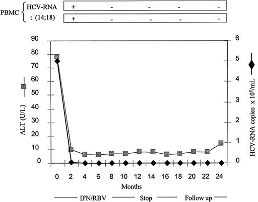
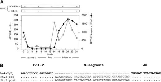
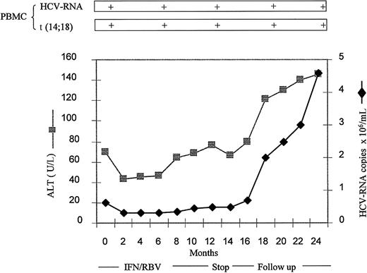
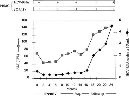
At the end of follow-up, complete data were available for 17 patients. These data are outlined in Table 2. In particular, in one SR patient (no. 5) who maintained CR in the follow-up period, both t(14;18) and HCV RNA sequences remained undetectable in BLs (Figure 1). In 2 patients who had relapse (nos. 3 and 15), both t(14;18) and HCV RNA sequences were again detected in BLs after viral and clinical relapse (Figure 2). Among the 6 PR patients (nos. 1, 2, 13, 17, 19, 25) where t(14;18) was no longer detected at the end of treatment, 4 remained t(14;18)– (nos. 13, 17, 19, 25) and 2 (nos. 1 and 2) scored again t(14;18)+ during the follow-up. All NR patients with t(14;18) translocation at the end of treatment maintained the translocated BL clone also at the end of follow-up (Table 2; Figure 3).
Results in one patient with SR. Analysis of serum ALT values, serum HCV RNA titer, BL HCV RNA, and t(14;18) translocation in a representative SR patient (no. 5) who maintained negativity of both BL HCV sequences and t(14;18) during follow-up.
Results in one patient with SR. Analysis of serum ALT values, serum HCV RNA titer, BL HCV RNA, and t(14;18) translocation in a representative SR patient (no. 5) who maintained negativity of both BL HCV sequences and t(14;18) during follow-up.
Results after viral and clinical relapse. (A) ALT values, serum HCV RNA titers, BL HCV RNA detection, and t(14;18) translocation in a representative patient (no. 3) who had a relapse during follow-up after a transient CR. (B) DNA sequence of bcl-2/JH junctions detected in total DNA extracted from peripheral BLs of the same patient, taken at time 0 (pretreatment) and at 9 months of follow-up (posttreatment); bcl-2, N-segment, JH: sequences of the bcl-2/JH junction corresponding to bcl-2 gene (chromosome 18), N-segment, and JH region of the IgH locus (chromosome 14), respectively. Line bcl-2/JH: alignment was done on bcl-2 (GenBank accession no. M14745) and IgH locus J-regions (accession no. M25625; line bcl-2/JH). Pt indicates patient number (Tables 1 and 2); pre and post, sequences corresponding to peripheral blood samples taken before and after IFNα treatment, respectively. Dots indicate identical nucleotides; gaps in sequences (–) were introduced for clarity.
Results after viral and clinical relapse. (A) ALT values, serum HCV RNA titers, BL HCV RNA detection, and t(14;18) translocation in a representative patient (no. 3) who had a relapse during follow-up after a transient CR. (B) DNA sequence of bcl-2/JH junctions detected in total DNA extracted from peripheral BLs of the same patient, taken at time 0 (pretreatment) and at 9 months of follow-up (posttreatment); bcl-2, N-segment, JH: sequences of the bcl-2/JH junction corresponding to bcl-2 gene (chromosome 18), N-segment, and JH region of the IgH locus (chromosome 14), respectively. Line bcl-2/JH: alignment was done on bcl-2 (GenBank accession no. M14745) and IgH locus J-regions (accession no. M25625; line bcl-2/JH). Pt indicates patient number (Tables 1 and 2); pre and post, sequences corresponding to peripheral blood samples taken before and after IFNα treatment, respectively. Dots indicate identical nucleotides; gaps in sequences (–) were introduced for clarity.
Results in NR patient. Analysis of serum ALT values, serum HCV RNA titer, BL HCV RNA, and BL t(14;18) translocation in a representative NR patient who maintained positive BL HCVRNA and t(14;18)+, during follow-up.
Results in NR patient. Analysis of serum ALT values, serum HCV RNA titer, BL HCV RNA, and BL t(14;18) translocation in a representative NR patient who maintained positive BL HCVRNA and t(14;18)+, during follow-up.
Sequence analysis performed in all positive cases carefully confirmed that the detected translocations were not derived from contaminant amplicons because of the presence of unique nucleotides at the junction of bcl-2 and JH (N segment). In 4 cases (2 PR patients [nos. 1 and 2] and 2 CR patients [nos. 3 and 15]) where t(14;18) temporarily disappeared, but was again detectable in PBMCs at the end of follow-up, sequence analysis showed that the same B-cell clones were involved in bcl-2 rearrangement either in the pretreatment or posttreatment periods (Figure 2).
Among the 15 untreated (control) patients, nobody cleared the virus from either serum or BLs. No modification of initial t(14;18) translocation status was observed in 14 of these control patients in all samples tested during the study. In the remaining control (genotype 2), starting from month 12, BLs bearing the t(14;18) translocation became undetectable. Finally, t(14;18) translocation was detected in 5 of 85 healthy subjects (5.88%; prevalence in HCV+ patients versus healthy subjects, P < .001).
Discussion
This study shows a striking association between the virologic response to treatment and detection of t(14;18) translocation (a BL-specific event) in peripheral blood cells.
Previous studies showed disappearance of BLs bearing t(14;18) following effective antiviral treatment in populations of patients with evidence of LPDs, and in particular MCS.8,11 It is presumed that HCV-infected patients without evidence of LPDs may represent a more sensitive model to test the possible influence of therapy on the expansion of translocated BL clones. Accordingly, in this study, the effect of antiviral therapy was assessed in a population of HCV+ patients with peripheral BLs harboring the t(14;18) translocation despite the absence of MCS or other HCV-related LPDs. In addition, the virologic status of patients was assessed not only by monitoring viremia levels, but also through determination of HCV RNA sequences in peripheral BLs.26 A positive correlation was observed between virologic response to treatment and regression of translocated BL clones.
In this study the prevalence of patients harboring HCV sequences in PBMCs was high when compared with previous studies performed in HCV+ patients.27 Also, because only qualitative methods were used for the detection of t(14;18) and HCV RNA and no studies were performed on isolated B cells with t(14;18), this study could not delineate the role of HCV in inducing t(14;18) translocation. Further studies are needed to determine the role of HCV, if any, in t(14;18) translocation and the development of B-cell malignancies in patients infected with HCV.
In addition, in the present study, genomic HCV sequences were detected in the PBMC population, which may be affected by t(14;18) translocation (B cells). Due to the fact that HCV RNA has been demonstrated in mononuclear and T cells in addition to B cells,26-28 further studies will ascertain whether, in case of bcl-2 rearrangement, the HCV infection of peripheral lymphocytes is or is not confined to the B-cell subset.
Detection of HCV sequences does not constitute sufficient argument to prove replication into the cell. The detection of HCV RNA could simply reflect absorption of viral particles on the cellular surface membrane. Such absorption could be due to the recently shown selective binding between HCV-E2 protein and CD81 molecule, abundantly present on the BL surface,29 as well as Fc receptor–mediated binding of antibody-coated viral particles.30 It is also impossible to exclude that the associated detection in BLs between HCV RNA sequences and t(14;18) may simply be mediated by the initial HCV particles binding to the BL surface with stimulation of HCV receptors. In fact, as previously hypothesized, HCV interaction with CD81 may contribute to the high frequency of this rearrangement in patients with HCV infection.7 However, the possible involvement of other unknown mechanisms that may be more directly related to viral infection should be taken into account as well.
Due to the antiproliferative effect of IFN, previous studies showing regression of BL clones following antiviral treatment in HCV+ subjects could not determine whether such an effect is or is not entirely related to this activity.8,11,20 With respect to more specific studies investigating t(14;18)+ patients, disappearance of this rearrangement following negativization of serum HCV RNA by antiviral therapy was previously observed in HCV+ patients, generally showing an MCS.8,11 In the present work, performed in a large population of treated HCV+ patients without MCS, virologic analysis also included detection of genomic HCV sequences in BLs and serum HCV RNA titers. We not only observed that clearance of HCV RNA from serum as well as from BLs was associated with disappearance of translocated BL clones, but, more interestingly, that despite a similar IFN regimen, none of the NR patients became t(14;18)–. According to these observations, the present study more strongly suggests that the antiviral value of IFN therapy, and not only its antiproliferative effect, is key in determining the observed effect on expanded BL clones. In addition, reappearance of translocated BL clones was observed in this study in patients with recurrent HCV replication after an initial response. In these cases, the bcl-2/JH junctions corresponding to the pretreatment and posttreatment periods showed identical N-segments. This indicates that translocated BL clones regressed and became undetectable following antiviral treatment and reappeared following virologic relapse, thus confirming the hypothesis that the expanded BL clone needs persistence of the infective stimulus to be maintained.8,11,31 However, no data at present exist indicating the specific mechanisms that may be involved. The observation that translocated BLs became undetectable also in patients with significant reduction of the viral load but without complete viral clearance (HCV RNA still detectable in serum or peripheral BLs) may suggest that BL expansion is sensitive to changes in viral replication. Mechanisms involved may include viral factors such as a reduction of antigenic stimulation or BL surface CD81 molecules activation or possibly other still unclear factors related to cell infection.32 As already observed, even if a collaborative role of the antiproliferative properties of IFN appears obvious, the fact that none of the 6 patients without significant modifications of HCV titers become t(14;18)– makes the hypothesis of an exclusive role of the latter mechanism unlikely.
Although not yet thoroughly demonstrated, t(14;18) determination in BLs may carry predictive value, especially for the development of BL LPDs. This appears deducible also by the intrinsic precarious condition of prolonged BL life induced in B cells and is indirectly suggested by the model of transgenic mice for bcl-2, in which overexpression of bcl-2 leads to diffuse lymphadenopathy followed by malignant lymphoproliferation, culminating in a diffuse aggressive lymphoma.12 Recently, in an HCV-infected, t(14;18)+ subject, we observed over several years an evolution from a condition of diffuse lymphadenopathy diagnosed as “reactive” at pathologic analysis to MCS, and then to frank BL malignancy (C.G. et al, unpublished data 2003). More recently, Zuckerman and colleagues observed that 2 of 8 patients with translocation who did not undergo antiviral treatment evolved to frank BL NHL during follow-up, whereas none of the treated patients did.11 These data further sustain the prognostic value of t(14;18) translocation with respect to development or evolution of BL LPDs. The fact that none of our untreated patients developed LPDs during the follow-up is probably related to the absence of MCS at the time of inclusion; in this respect, further studies based on long-lasting observation periods seem useful.
Although the bcl-2 gene was first identified at the chromosomal breakpoint of t(14;18) translocation-bearing human follicular B-cell lymphomas33-35 and is found in this NHL type in about 70% to 80% of cases, it may be also found in diffuse lymphoma (about 20% of cases) and in nonmalignant conditions, such as persistent polyclonal B-cell lymphocytosis (PPBL),7,36,37 which is an expansion of memory B cells without evidence of evolution to a frank lymphoma, as well as in HCV+ MCS, a situation which may evolve to malignant B-cell NHL types generally different from follicular lymphoma (lymphoplasmacytoid lymphoma/immuncytoma are the most common lymphomas reported in this subpopulation).31,38 Despite this, in a large Italian survey, the type of idiopathic NHL more frequently associated with HCV infection was the follicular center type,31,39 whereas different types were preferentially associated in other studies. This creates interest for a more accurate analysis of the varying pathogenetic pathways in which bcl-2 rearrangement may be involved, possibly culminating in very different situations.
The current study was performed in HCV+, t(14;18)+ patients without evidence of LPDs. The effect of antiviral therapy in HCV+ patients with overt HCV+ LPDs has been the object of several studies, including very recent ones. In one HCV+ patient presenting with an atypical picture composed of mixed cryoglobulinemia, leukemic-like proliferation of B cells bearing marginal zone B-cell phenotypic markers, splenomegaly, and partial trisomy of chromosome 3, but without t(14;18) translocation, Casato and coworkers40 observed that, during IFN therapy, the decline in monoclonal B-cell populations paralleled the decline in viremia. Although no data about bcl-2 expression (and bcl-2/Bax ratio) in BLs was provided, total PBMC samples showed bcl-2 overexpression.40 This interesting work supports the therapeutic role of antiviral therapy in HCV-related LPDs. In this patient, a decrease in IFN therapy from daily to 3 weekly administrations and interruption of therapy were associated with parallel increases in viremia and monoclonal BLs.
More recently, the importance of antiviral therapy in HCV-related LPDs has been confirmed in a larger study performed in 14 patients with splenic lymphoma with villous lymphocytes (SLVL), 9 of whom also had HCV infection. Only HCV+ patients had regression of SLVL, which paralleled disappearance of HCV RNA. In contrast, no HCV– patient had regression of the LPD, despite similar treatment with IFN.41 No data about t(14;18) translocation determination in peripheral BLs was provided. However t(14;18) translocation has been described in SLVL, suggesting the need for further studies investigating this aspect.
In conclusion, the close association between the modifications of HCV replication induced by antiviral treatment, and detection of BL clones bearing t(14;18) suggests that HCV-related BL clone expansion needs the persistence of the infective stimulus to be maintained; this in turn suggests that antiviral treatment may be effective in preventing or treating HCV-related LPDs. Moreover, the effects of IFN therapy seem to be related, at least in part, to its antiviral properties. Further studies will be necessary to clarify which mechanisms are involved.
Prepublished online as Blood First Edition Paper, April 10, 2003; DOI 10.1182/blood-2002-05-1537.
Supported by the Italian Liver Foundation, MURST, the Fondazione Istituto di Ricerca Virologica O. B. Corsi, European Community HepaC-resist grant, and AIRC.
The publication costs of this article were defrayed in part by page charge payment. Therefore, and solely to indicate this fact, this article is hereby marked “advertisement” in accordance with 18 U.S.C. section 1734.
We gratefully acknowledge Prof Giampiero Buzzelli for helpful discussion and Ms Mary Diamond for kind help in the preparation of the manuscript.


