Comment on Majstoravich et al, page 1396
Majstoravich and colleagues have determined that the short microvilli of lymphocytes, although uniform in length and density, are highly dynamic actin filament bundle–containing structures that do not depend upon WASp for their morphology.
Lymphocytes are known to have short, actin filament–rich, microvillus-like projections, and these structures have been implicated in processes of key importance, such as the initial phase of rolling during extravasation and virus recognition. But, primarily because these structures are so short (∼ 0.3 μm), relatively little is known about their internal organization and dynamics. An issue of central importance is the role of Wiskott-Aldrich syndrome protein (WASp), the archetype of a family of actin nucleation–promoting factors that stimulate actin-related protein 2/3 (Arp2/3) complex–mediated actin polymerization. WASp is expressed preferentially in hematopoietic cells and is mutated in patients with Wiskott-Aldrich syndrome (WAS), which is characterized by immunodeficiency, thrombocytopenia, and eczema.FIG1
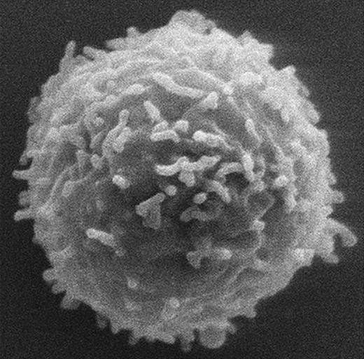
Lymphocytes from WASp knock-out mice have normal microvilli. See the complete figure in the article beginning on page 1396.
Lymphocytes from WASp knock-out mice have normal microvilli. See the complete figure in the article beginning on page 1396.
Of the classes of cell surface specialization assembled using an actin filament–based scaffold, 2 major ones, lamellipodia and filopodia, are believed to involve WASp family members directly or indirectly. The branched actin filament network of lamellipodia uses WASp family members to activate the Arp2/3 complex to form new branches.1 This lamellipodial actin network can, in turn, be rearranged, cross-linked, and elongated to form the parallel actin bundle scaffold of filopodia.2 In contrast, remarkably little is known about how the parallel actin bundle scaffolds of microvilli are established and maintained.3 Compared with lamellipodia and filopodia, microvilli may seem more stable, but recent studies indicate that the actin filaments in the parallel actin bundle at the core of epithelial cell microvilli turn over rapidly through actin treadmilling.3
Majstoravich and colleagues used scanning electron microscopy to examine lymphocytes isolated from mice and humans and the larger cells of a transformed pre-B-lymphoma line, and they found a remarkable conservation in microvillar length and density. In addition, using transmission electron microscopy, they observed what appeared to be a parallel actin bundle at the core. Importantly, when the authors exposed the lymphocytes to the actin monomer–sequestering drug Latrunculin A, they observed a rapid and reversible shrinkage of these projections. This behavior was consistent with the existence of actin treadmilling in the filaments of the core actin bundle at a rate of about 1 to 2 actin monomers per second, which is remarkably similar to the treadmilling rate measured in the brush-border microvilli of a kidney epithelial cell line.3 Finally, these authors found that lymphocytes isolated from WASp knockout mice or human WAS patients of 3 different genotypes showed minimal defects in microvillar length or density, suggesting that wild-type WASp was not required for expression of this baseline microvillar morphology. Although at first glance this result appears to contrast with earlier reports of microvillar abnormalities on lymphocytes from WAS patients4 and WASp-deficient mice,5 these earlier studies examined cells under conditions likely to favor lymphocyte activation. This raises the intriguing possibility that there are multiple mechanisms for microvillus formation in lymphocytes and suggests that a WASp-dependent pathway becomes dominant under conditions of activation.


