Subjects:
BloodWork
Topics:
chediak-higashi syndrome
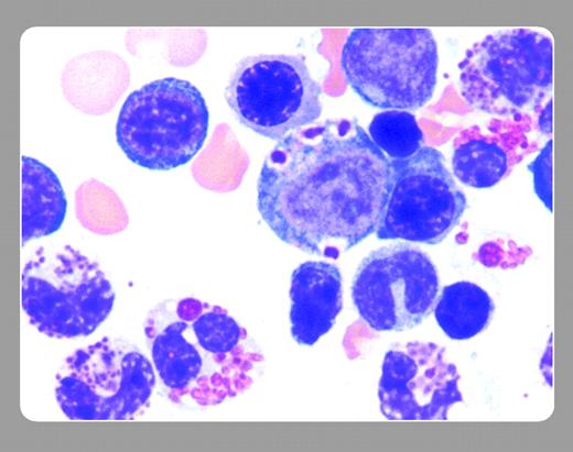
Bone marrow aspirate from a 17-year-old female with Chediak-Higashi syndrome is shown. Giant inclusions are present in the cytoplasm of the myeloid precursor cell (center of the image). Note also in both the granulocytes and eosinophils the multiple atypical large cytoplasmic granules that are characteristic of this disorder.FIG1
 John Lazarchick and Bart McRae, Medical University of South Carolina
 John Lazarchick and Bart McRae, Medical University of South Carolina
Many Blood Work images are provided by the ASH IMAGE BANK, a reference and teaching tool that is continually updated with new atlas images and images of case studies. For more information or to contribute to the Image Bank, visit www.ashimagebank.org.
Copyright © 2005 by The American Society of Hematology
2005


