Comment on Kerksiek et al, page 742
Dendritic cells (DCs) rely on the Rho guanosine triphosphatase (GTPase), Rac1, for their normal maturation, phagocytosis, and immunostimulatory capacity.
Dendritic cells (DCs) are unique among antigen-presenting cells for their critical role in stimulating naive T cells. They do this by continuously sampling their environment through constitutive antigen uptake and cross-presentation to T-cell populations. They require actin cytoskeletal remodeling to migrate through tissues and phagocytose antigen-bearing apoptotic cells. The small, monomeric Rho guanosine triphosphatases (GTPases) have been shown to be key signaling molecules required for regulating phagocytosis and actin cytoskeleton reorganization.1 Previous studies have suggested that the Rho GTPase, Rac1, may be critical for endocytosis and cross-presentation in DCs.2,3 These findings were based on 2 approaches: transfection of a phagocytic cell line with dominant-negative (DN) Rac1, Rac1 N17,2 and injection of large quantities of Rac1 N17 into in vitro–generated DCs.3 However, these studies were based on in vitro systems, and did not address the function of Rac1 in highly heterogeneous populations of DCs in vivo. The only way to address the physiologic function of Rac1 in DCs is to use a mouse model deficient in Rac1 expression. But the problem with knocking out Rac1 function in mice is that it is early embryonic lethal.
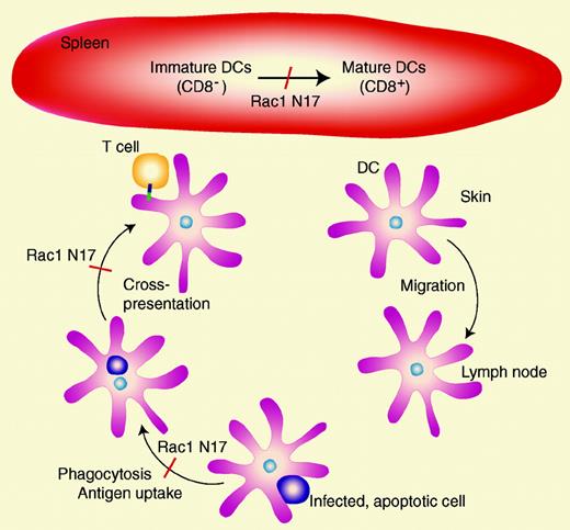
In this issue, Kerksiek and colleagues overcame this hurdle by using an elegant model of transgenic mice expressing DC-specific dominant negative Rac1 N17. Dominant-negative isoforms of Rho GTPases inhibit effector functions by binding to and arresting the function of upstream GDP/GTP exchange factors (GEFs). Transgenic C57Bl/6 mice were developed that expressed Rac1 N17 under the control of the DC-selective murine CD11c promoter. Unexpectedly, the expression of Rac1 N17 led to reduced maturation of DCs (measured as the appearance of CD8+ DCs in the spleen), but had no effect on DC migration from the skin to the lymph nodes. Rac1 N17 also significantly impaired apoptotic cell uptake by CD8+ DCs and subsequent cross-presentation to T cells as measured by proliferation assays. This observation was confirmed in animals infected with recombinant ovalbumin (OVA)–expressing Listeria monocytogenes. Since L monocytogenes replicates poorly in DCs, the DCs in wild-type animals are likely to endocytose infected, apoptotic cells and cross-present antigen to CD8+ and CD4+ T-cell populations specific for L monocytogenes. Recombinant OVA expression was required because immunodominant H2b-restricted cytotoxic T lymphocyte populations fail to expand upon infection of C57Bl/6 mice with wild-type L monocytogenes. OVA-expressing L monocytogenes stimulated an easily detectable expansion of CD8+ and CD4+ T cells from wild-type mice. However, substantially less expansion of these cells was detected in Rac1 N17–expressing mice. These findings suggest that in DCs, Rac1 is critical for maturation, uptake of apoptotic cells, and cross-presentation of exogenous antigens to T cells (see Figure). The lack of effect of Rac1 N17 on tissue migration of DCs is surprising, since Rac1 N17 would be predicted to inhibit actin cytoskeleton dynamics and reduce DC motility. This indicates that there may be other important regulatory molecules expressed in DCs that may compensate for the loss of Rac1 function. The generation of these transgenic mice is likely to lead to new findings on the role of Rho GTPases in DC function in a range of immunologic models. ▪
Rac1 is required for DC maturation, phagocytosis, antigen uptake, and cross-presentation to T cells in DC-specific expression of Rac1 N17 in transgenic mice. Migration of DCs from skin to lymph nodes was not impeded in these mice, suggesting that Rac1 is not required for DC migration through tissues.
Rac1 is required for DC maturation, phagocytosis, antigen uptake, and cross-presentation to T cells in DC-specific expression of Rac1 N17 in transgenic mice. Migration of DCs from skin to lymph nodes was not impeded in these mice, suggesting that Rac1 is not required for DC migration through tissues.


