Abstract
Although arsenic trioxide (As2O3) is an effective therapy in acute promyelocytic leukemia (APL), its use in other malignancies is limited by the toxicity of concentrations required to induce apoptosis in non-APL tumor cells. We looked for agents that would synergize with As2O3 to induce apoptosis in malignant cells, but not in normal cells. We found that trolox (6-hydroxy-2,5,7,8-tetramethylchroman-2-carboxylic acid), a widely known antioxidant, enhances As2O3-mediated apoptosis in APL, myeloma, and breast cancer cells. Treatment with As2O3 and trolox increased intracellular oxidative stress, as evidenced by heme oxygenase-1 (HO-1) protein levels, c-Jun terminal kinase (JNK) activation, and protein and lipid oxidation. The synergistic effects of trolox may be specific to As2O3, as trolox does not add to toxicity induced by other chemotherapeutic drugs. We explored the mechanism of this synergy using electron paramagnetic resonance and observed the formation of trolox radicals when trolox was combined with As2O3, but not with doxorubicin. Importantly, trolox protected nonmalignant cells from As2O3-mediated cytotoxicity. Our data provide the first evidence that trolox may extend the therapeutic spectrum of As2O3. Furthermore, the combination of As2O3 and trolox shows potential specificity for tumor cells, suggesting it may not increase the toxicity associated with As2O3 monotherapy in vivo.
Introduction
Arsenic has been used as a therapeutic agent for more than 2400 years. Until the 1930s, arsenic was used as a treatment for patients with chronic myelogenous leukemia. More recently, the use of arsenic in leukemia has resurfaced after reports from China that arsenic induced a high remission rate in acute promyelocytic leukemia (APL), including those who were resistant to therapy with all-trans retinoic acid.1,2
The activity of arsenic trioxide (As2O3) in APL is in part related to the disappearance of the promyelocytic leukemia–retinoic acid receptor α (PML-RARα) fusion protein, the gene product of the chromosomal translocation t(15,17) characteristic of APL, and the induction of differentiation.3,4 As2O3 can also induce apoptosis through a variety of mechanisms, which appear to be independent of PML-RARα degradation.5 In addition to causing mitochondrial toxicity,6 impairing microtubule polymerization,7 and deregulating a number of proteins and enzymes through binding to sulfhydryl groups,8-10 considerable evidence suggests that As2O3 induces the accumulation of reactive oxygen species (ROSs) and, subsequently, induces oxidative stress.11,12 Indeed, the intracellular redox status has been shown to be important in predicting whether a cell will respond to arsenic.11,13
Recently it has been shown that As2O3 stimulates apoptosis in additional malignant cells including acute myeloid leukemia, chronic myeloid leukemia, myeloma, and various solid tumor cells.14-17 However, higher concentrations of As2O3 are required to induce apoptosis in non-APL tumor cells, suggesting that higher, more toxic doses might be needed for clinical efficacy. Clinical trials are currently testing arsenic in the treatment of lymphoma and myeloma,18 but clear evidence of clinical benefit has, thus far, been largely restricted to patients with APL. Therefore, the sensitization of the resistant tumor cells to As2O3 could expand its therapeutic spectrum.
Different compounds have been reported to enhance As2O3-mediated apoptosis.19-21 Recently ascorbic acid (AA), a key antioxidant molecule, was reported to augment the toxicity of As2O3 in vitro.22,23 However, there is some evidence that the toxicity of ascorbate is due to ascorbic acid–mediated production of hydrogen peroxide, to an extent that varies with the medium used to culture the cells.24
Trolox (6-hydroxy-2,5,7,8-tetramethylchroman-2-carboxylic acid) is a hydrophilic vitamin E analog lacking the phytyl tail, with enhanced antioxidant capacity due to its increased cell permeability. It provides protection against oxidative reactions in aqueous solutions25 and against cisplatin-induced apoptosis in renal proximal tubular epithelial LLC-PK1 cells.26 There is also evidence that trolox inhibits DNA damage formation induced by singlet oxygen in human lymphoblast WTK-1 cells27 and protects red blood cells during photodynamic treatment.28
In spite of a large and consistent literature documenting antioxidant effects of trolox in several different experimental models, we report here that this compound can enhance As2O3-mediated cytotoxicity in APL, myeloma, and breast cancer cells. We show an increase in intracellular oxidative stress when trolox and As2O3 are combined, leading to caspase-3 activation and apoptosis in leukemic cells. We extend previous reports that trolox does not enhance the cytotoxicity of other chemotherapeutic agents to cancer cells and provide evidence that the enhancement of apoptosis by As2O3 may be limited to malignant cells.
Materials and methods
Cell lines
The arsenic trioxide–resistant APL cell line, NB4-M-AsR2 (AsR2), was generated by culturing NB4 cells in the presence of As2O3 at concentrations that were gradually increased over time.13 NB4 (provided by Dr M. Lanotte, Hospital Saint-Louis, Paris, France), AsR2, and multiple myeloma IM9 (American Type Culture Collection [ATCC], Manassas, VA) were maintained in RPMI 1640 media. MCF-7 and MDA-231 were obtained from ATCC and maintained in alpha modified essential medium (MEM). T47D (ATCC) was cultured in Dulbecco-MEM/F12 (D-MEM/F12). All media were purchased from Life Technologies (Bethesda, MD) and supplemented with 10% fetal bovine calf serum (FBS). AsR2 was routinely grown in RPMI containing 2 μM As2O3. In experiments examining the response of AsR2, the cells were first washed thoroughly to remove As2O3 and then cultured 24 hours in media alone prior to initiating the experiment. All cells were grown in a humidified chamber at 37°C with a 5% CO2 environment.
Growth assays
NB4, IM9, and AsR2 cells were seeded at 1 × 10⁵ cells/mL in 24-well plates. Cells were treated with various concentrations of As2O3 or doxorubicin, alone or in combination with 100 μM trolox for 6 days. Viable cells were counted by trypan blue exclusion on days 1, 3, and 6. All cells were maintained at a density lower than 1 × 10⁶ cells/mL through dilution as required, and media ± treatment was replaced every third day. MCF-7, MDA-231, and T47D were seeded in 24-well plates at a density of 4000 cells/well. The next day, fresh media containing As2O3 ± trolox was added. On the days indicated, cells were fixed in 10% trichloroacetic acid and subsequently stained with sulforhodamine B (SRB). Bound SRB was solubilized in 10 mM unbuffered Tris (tris(hydroxymethyl)aminomethane), and optical density was measured at 570 nm in a microplate reader.
Propidium iodide staining
Quantitation of apoptotic cells was performed as previously described.29 Cells were treated, washed in buffer (phosphate-buffered saline [PBS]/5% FBS/0.01 M NaN3) at 4°C, pelleted, and resuspended in 0.5 mL hypotonic fluorochrome solution containing 50 μg/mL propidium iodide (PI), 0.1% sodium citrate, and 0.1% Triton X-100. Fluorescence was measured on a Becton Dickinson fluorescence-activated cell sorter (FACS) Calibur (San Jose, CA). Cells undergoing DNA fragmentation and apoptosis (those in which PI fluorescence was weaker than the typical G0-G1 cell cycle peak) were quantified using CellQUEST software (BD Bioscience, Mississauga, ON, Canada).
Annexin V staining
Cells were stained with annexin V–fluorescein isothiocyanate (FITC) and propidium iodide in binding buffer according to the manufacturer's recommendations (BD Pharmingen, San Diego, CA). The fluorescent signals of FITC and PI were detected by FL1 at 518 nm and FL2 at 620 nm, respectively, on a FACSCalibur. Apoptotic cells (annexin V–positive/PI-negative) were quantified using the CellQUEST software.
Western blotting and immune kinase assays
Cell extracts were washed with cold PBS and resuspended in 0.1 mL lysis buffer (5 mM NaH2PO4, 1 mM dithiothreitol [DTT], 10% glycerol, 1 mM phenylmethylsulfonyl fluoride (PMSF), 10 μg/mL each aprotinin and leupeptin, pH 7.4) at 4°C. Extracts were centrifuged at 10 000 g at 4°C, and supernatants were transferred to fresh tubes. Protein concentration was determined with the Bio-Rad protein assay (Bio-Rad, Mississauga, ON). To detect heme oxygenase-1 (HO-1) or poly(adenosine diphosphate–ribose) polymerase (PARP), 50 μg protein was added to an equal volume of 2× sample buffer and run on a 10% sodium dodecyl sulfate polyacrylamide gel (SDS-PAGE). Proteins were transferred to nitrocellulose membranes (Bio-Rad), stained with Ponceau S in 5% acetic acid to ensure equal protein loading, and blocked with 5% milk in PBS containing 0.5% Triton X-100 for one hour at room temperature. The membrane was hybridized overnight at 4°C with antibody against PARP (1:1000; Oncogene Research Products, San Diego, CA) or 3 hours with an antibody against HO-1 (1:1000; StressGen, Victoria, BC). Following 3 washes with PBS and 0.5% Triton X-100, blots were incubated with a goat antirabbit antibody (1:10 000; BD PharMingen) for one hour at room temperature. Bands were visualized by enhanced chemiluminescence (Amersham Pharmacia Biotech, Baie d'Urfe, QC). Immunostaining for β-actin was used to confirm equal protein loading. Immune complex kinase assays for c-jun kinase activity were performed as we have previously described.30
Caspase-3 activity assay
Activation of caspase-3 was detected using a fluorescent caspase-3 inhibitor, Red-DEVD-FMK (Oncogene Research Products, San Diego, CA), which irreversibly binds to activated caspase-3 in apoptotic cells. Cells were treated for 2 days and harvested into microcentrifuge tubes. The cells were incubated with 1 μL Red-DEVD-FMK for one hour at 37°C in 5% CO2. Subsequently, cells were washed twice, resuspended, and analyzed by flow cytometry, using the FL2 channel.
Protein carbonyls
Oxidized and reduced bovine serum albumin (BSA) was prepared and its carbonyl content was quantitated by a colorimetric carbonyl assay described previously.31 NB4 cells were treated for 3 days with As2O3, trolox, or the combination. Protein samples were adjusted to 4 mg protein/mL. The standards and protein samples were incubated with 3 volumes 10-mM 2,4-dinitrophenylhydrazine (DNP) in 6 M guanidine-HCl, 0.5 M potassium phosphate, pH 2.5, for 45 minutes at room temperature (mixing every 10-15 minutes). Aliquots of cell proteins and standards were diluted in PBS and adsorbed to a 96-well immunoplate by incubation overnight at 4°C. After washing with PBS, nonspecific sites were blocked with 0.1% Tween 20 in PBS for 1.5 hours at room temperature. After further washing with PBS, the wells were incubated with biotinylated anti-DNP antibody (1:10 000 dilution; Molecular Probes, Eugene, OR) for one hour at 37°C. Wells were washed and incubated with streptavidin-biotinylated horseradish peroxidase (1:3000 dilution; Amersham Bioscience, Buckinghamshire, United Kingdom). After further washing, o-phenylenediamine/peroxide solution was added. The reaction was stopped after 7 minutes with 2.5 M sulfuric acid, and the absorbance was read with a 490-nm filter. A 6-point standard curve of reduced and oxidized BSA was incubated with each plate.
Quantification of 8-iso PGFα
NB4 cells were treated for 3 days with As2O3, trolox, or the combination. Cells were washed twice with PBS containing 0.005% butylated hydroxy-toluene (BHT) and 10 mg/mL indomethacin. The intracellular and membrane-bound 8-iso PGF2α, a specific marker for lipid peroxidation, was measured using a competitive enzyme-linked immunosorbent assay (ELISA) kit from Cayman Chemical (Ann Arbor, MI) following the manufacturer's instructions.
Detection of trolox phenoxyl radicals and measurement of intracellular GSH
Electronic spin resonance spectroscopy (ESR) reactions contained 0.02 mM As2O3, 1 mM trolox, 5% (vol/vol) dimethyl sulfoxide (DMSO), and 0.2 μg/mL doxorubicin. Following the final addition of As2O3, reaction mixtures were transferred immediately to a quartz ESR flat-cell positioned and pretuned within the cavity of a Bruker ESP 300 spectrometer (Bruker, Rheinstetten, Germany) using a rapid delivery device,32 and recording commenced using the following instrument settings: modulation frequency, 100 kHz; center field, 3471.50 G; sweep width, 50.0 G; modulation amplitude, 9.51 × 10⁻¹ G; receiver gain, 6.30 × 10⁴; scan time, 20.97 seconds; time constant, 10.24 milliseconds; and power, 20 mW. Spectra was simulated using WinSIM program available for use at the National Institute of Environmental Health Sciences (NIEHS)/National Institutes of Health (NIH) website (http://epr.niehs.nih.gov/pest.html).33 Intracellular reduced glutathione (GSH) levels were assessed enzymatically with glutathione reductase as previously reported.13
Peripheral blood mononuclear cell purification and colony-forming unit assay
Peripheral blood mononuclear cells (PBMCs) were obtained from 2 healthy donors after obtaining informed consent and were collected into tubes containing 7.2 mg K2 EDTA (ethylenediaminetetraacetic acid). The blood was diluted 1:3 in PBS, layered onto an equal volume of Ficoll-Plaque PLUS (Amersham Biosciences, Piscataway, NJ), and centrifuged at 150 g for 30 minutes. The mononuclear cell layer was collected and washed twice in PBS. Methylcellulose media was prepared by combining IMDM, 30% FBS, 1% bovine serum albumin, 10-4 M 2-mercaptoethanol, 2 mM l-glutamine, 0.1 U penicillin, 0.1 μg/mL streptomycin, granulocyte-macrophage colony-stimulating factor (GM-CSF, 10 ng/mL), interleukin-3 (IL-3, 10 ng/mL), and erythropoietin (EPO, 3 U/mL). PBMCs were seeded in this media at a concentration of 300 000 cells/mL and treated with or without As2O3, trolox, or the combination. Cultures were performed in triplicate in 35-mm2 dishes and incubated at 37°C in 4% CO2. Colonies derived from colony-forming units–erythrocyte (CFU-Es), burst-forming units (BFU-Es) and colony-forming units–granulocyte-macrophage (CFU-GMs) were counted on days 7 and 13.
Statistical analysis
The significance of data was determined using Prism version 3.0 (GraphPad software, San Diego, CA). Analysis of variance followed by Newman-Keuls posttests were used to determine if cell treatments produced significant changes.
Results
Trolox significantly enhances the inhibitory effects of As2O3 on APL, multiple myeloma, and breast cancer cells
We examined the effects of As2O3 and trolox, both separately and in combination, on the growth of different cell lines. Figure 1A shows that treatment of NB4 cells for 6 days with 0.5 or 1 μM As2O3 reduced viable cell number by 25% ± 4.7% and 70% ± 5.6% of control, respectively. At 100 μM, trolox alone had no effect on cell number at any time point. However, if the cells were treated with 0.5 or 1 μMAs2O3 and 100 μM trolox in combination, 57% ± 3.5% and 97% ± 4.2% reductions of cell number, respectively, were observed. In all cases, trypan blue–positive cells were less than 3%. A difference was also seen between As2O3 and As2O3 + trolox after 72 hours, with 1 μMAs2O3 decreasing cell number by 30% and the combination, by 50% (P < .001). We next determined whether trolox could sensitize arsenic-resistant cells. We used an NB4-derived, arsenic-resistant subclone (AsR2), which has a median inhibitory concentration (IC50) value roughly 10 times higher than its parental NB4 cell line,13 and the multiple myeloma IM9 cell line, which is also less sensitive to As2O3 than NB4 cells.34 An enhancing effect of trolox on As2O3-mediated growth inhibition was observed in both cell lines (Figure 1B-C), although trolox did not restore the sensitivity to lower concentrations of As2O3 in the highly resistant AsR2 cell line (data not shown). Some solid tumor cells have been shown to be more resistant to As2O3 than APL cells, so we tested the combined effect of As2O3 and trolox in breast cancer cell lines. As shown in Table 1, As2O3-mediated cytotoxicity was enhanced by trolox in all tested cell lines.
Trolox enhances As2O3-induced growth inhibition in NB4, AR2, and IM9 cells. NB4 (A), AsR2 (B), and IM9 cells (C) were treated with trolox, As2O3, or the combination. Cell viability was evaluated on days 1, 3, and 6 using trypan blue exclusion. Values are the mean of 3 independent experiments each performed in triplicate. Standard deviation bars are shown. *, **, and *** indicate a significant difference of P < .05, P < .01, and P < .001, respectively, from As2O3-treated cells.
Trolox enhances As2O3-induced growth inhibition in NB4, AR2, and IM9 cells. NB4 (A), AsR2 (B), and IM9 cells (C) were treated with trolox, As2O3, or the combination. Cell viability was evaluated on days 1, 3, and 6 using trypan blue exclusion. Values are the mean of 3 independent experiments each performed in triplicate. Standard deviation bars are shown. *, **, and *** indicate a significant difference of P < .05, P < .01, and P < .001, respectively, from As2O3-treated cells.
Trolox enhances As2O3-mediated apoptosis in As2O3-sensitive and -resistant malignant cells
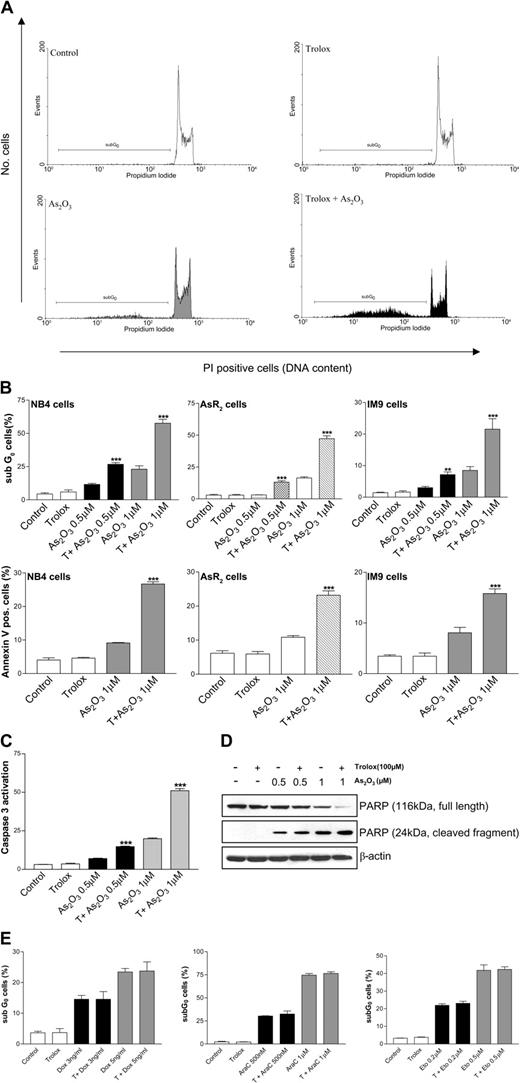
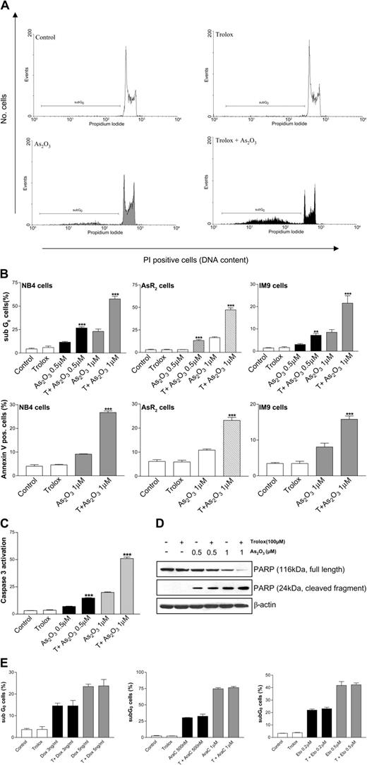
To evaluate whether the growth inhibitory effect observed upon combined treatment of As2O3 and trolox in NB4, AsR2, and IM9 cells was due to the induction of apoptosis, cells were treated for 48 hours, subsequently stained with PI, and analyzed by flow cytometry. As shown in Figure 2A-B, trolox enhanced As2O3-mediated apoptosis in the cell lines studied, at all concentrations of arsenic tested, while trolox alone had no effect on the apoptotic rate. To confirm an enhanced induction of apoptotic death, FITC-labeled annexin V, which detects phosphatidylserine residues appearing on the external surface of early apoptotic cells, was used. Consistent with the increase in the sub-G0 subpopulation after PI staining, trolox augmented the percentage of cells positive for annexin V (Figure 2B, lower panels). To further confirm the induction of apoptosis by the combination of As2O3 and trolox, we evaluated caspase-3 activation and PARP cleavage. Trolox significantly enhanced the percentage of cells with activated caspase-3 (Figure 2C) and cleaved PARP (Figure 2D). These results support the hypothesis that the combined treatment with As2O3 and trolox induced apoptosis in NB4 cells in a dose-dependent fashion. Similar results were obtained with AsR2 and IM9 cells (data not shown).
Trolox enhances arsenic-mediated apoptosis in NB4, AR2, and IM9 cells. (A-B) NB4, AsR2, and IM9 cells were treated with As2O3 and trolox (T) for 48 hours. Apoptosis was detected by PI staining. Flow cytometric histograms are shown in panel A. Quantitation of PI-positive cells in a hypotonic fluorochrome solution was performed. Apoptotic cells were also stained with annexin V–FITC and propidium iodide in binding buffer and quantified (B). Each bar represents an average of 3 independent samples, and standard deviation bars are shown. Asterisks indicate significant differences from As2O3-treated cells (**P < .01; ***P < .001). (C) Cells were treated as indicated for 48 hours. Caspase-3 activation was measured using Red-DEVD-FMK. Its binding to activated caspase-3 was analyzed by flow cytometry. Asterisks indicate significant differences (P < .001) from As2O3-treated cells. (D) Western blotting was performed to determine PARP protein levels after 48 hours of treatment. β-Actin was used to show equal loading of lanes. Results are representative of 3 independent experiments each performed in duplicate. (E) NB4 cells were treated with doxorubicin, AraC, or etoposide with or without trolox (T) for 48 hours. Apoptosis was detected by PI staining as described in “Materials and methods.” Each bar represents an average of 3 independent samples, and standard deviation bars are shown.
Trolox enhances arsenic-mediated apoptosis in NB4, AR2, and IM9 cells. (A-B) NB4, AsR2, and IM9 cells were treated with As2O3 and trolox (T) for 48 hours. Apoptosis was detected by PI staining. Flow cytometric histograms are shown in panel A. Quantitation of PI-positive cells in a hypotonic fluorochrome solution was performed. Apoptotic cells were also stained with annexin V–FITC and propidium iodide in binding buffer and quantified (B). Each bar represents an average of 3 independent samples, and standard deviation bars are shown. Asterisks indicate significant differences from As2O3-treated cells (**P < .01; ***P < .001). (C) Cells were treated as indicated for 48 hours. Caspase-3 activation was measured using Red-DEVD-FMK. Its binding to activated caspase-3 was analyzed by flow cytometry. Asterisks indicate significant differences (P < .001) from As2O3-treated cells. (D) Western blotting was performed to determine PARP protein levels after 48 hours of treatment. β-Actin was used to show equal loading of lanes. Results are representative of 3 independent experiments each performed in duplicate. (E) NB4 cells were treated with doxorubicin, AraC, or etoposide with or without trolox (T) for 48 hours. Apoptosis was detected by PI staining as described in “Materials and methods.” Each bar represents an average of 3 independent samples, and standard deviation bars are shown.
We then asked whether trolox could enhance the induction of apoptosis by other cytotoxic agents that induce ROSs. The anthracycline doxorubicin has been shown to generate genotoxic stress in a different number of cell types.35,36 1-β-d-Arabinofuranosylcytosine (AraC) is a nucleoside analog used in the treatment of acute myelogenous leukemia.37,38 Etoposide causes single- and double-strand DNA breaks when incubated with cells.39,40 We examined the possibility that the combination of trolox and doxorubicin, AraC, or etoposide might increase cell growth inhibition and apoptosis in NB4 cells. As shown in Figure 2E, these compounds induced apoptosis in a dose-dependent manner. However, no additional increase of cellular apoptosis was observed when the cells were cotreated with trolox. Trolox also had no effect on sensitivity to doxorubicin or AraC in AsR2 and IM9 cells (data not shown).
The combination of As2O3 and trolox results in increased cellular oxidative stress
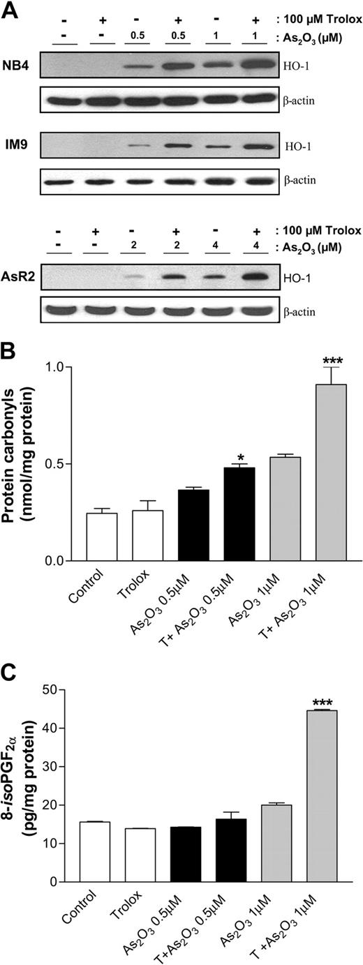
Oxidative damage has been postulated to be a key mechanism by which arsenic initiates the apoptotic process. Because trolox potentiates As2O3-induced apoptosis, it is possible that the combination treatment increases cellular oxidative stress. Therefore, we determined whether As2O3 affected various markers for oxidative stress and whether trolox could augment this effect. Heme oxygenase-1 (HO-1), which is the rate-limiting enzyme for heme degradation and has been widely described as a stress responsive protein,41 was not detected when trolox was used alone (Figure 3A). However, the combined treatment markedly enhanced As2O3-mediated HO-1 induction in all of the cell lines tested, suggesting that this combination increased the cellular oxidative stress. To document oxidative damage to cellular components, we analyzed lipids and proteins isolated from NB4 cells treated with As2O3 or As2O3 + trolox for 3 days. Protein carbonyls are generated by a variety of mechanisms and are sensitive indices of oxidative injury.42 Isoprostanes are chemically stable prostaglandin-like compounds that are produced independent of the cyclooxygenase (COX) enzyme by free radical–catalyzed peroxidation of arachidonic acid in situ in membrane phospholipids.43 F2-isoprostanes are a reliable marker of lipid peroxidation in vivo.44,45 Figure 3B-C shows that As2O3 alone induces protein oxidation and, to a lesser extent, lipid peroxidation. Oxidative damage to both proteins and lipids was found to be significantly higher when trolox and As2O3 were combined. Similar results were obtained in AR2 and IM9 cells (data not shown)
Trolox potentiates As2O3-mediated oxidative stress. (A) NB4, IM9, and AsR2 cells were treated with As2O3 and trolox for 24 hours. Western blot was used to determine HO-1 protein levels. β-Actin was used as a loading control. These data represent 3 independent experiments. (B) Protein carbonyl content was detected by ELISA in NB4 cells treated with As2O3 alone, trolox, or the combination for 3 days with the concentrations indicated. Data depicted are representative of 3 independent experiments each performed in duplicate. Asterisks indicate significant differences from As2O3-treated cells. (*P < .05; ***P < .001). (C) 8-iso PGF2α was detected in whole cells extracts from NB4 cells treated with the indicated compounds for 3 days. Standard deviation bars are shown. Asterisks indicate significant differences from As2O3-treated cells (P < .001).
Trolox potentiates As2O3-mediated oxidative stress. (A) NB4, IM9, and AsR2 cells were treated with As2O3 and trolox for 24 hours. Western blot was used to determine HO-1 protein levels. β-Actin was used as a loading control. These data represent 3 independent experiments. (B) Protein carbonyl content was detected by ELISA in NB4 cells treated with As2O3 alone, trolox, or the combination for 3 days with the concentrations indicated. Data depicted are representative of 3 independent experiments each performed in duplicate. Asterisks indicate significant differences from As2O3-treated cells. (*P < .05; ***P < .001). (C) 8-iso PGF2α was detected in whole cells extracts from NB4 cells treated with the indicated compounds for 3 days. Standard deviation bars are shown. Asterisks indicate significant differences from As2O3-treated cells (P < .001).
The cytotoxic effects observed when trolox and As2O3 are combined are not due to generation of extracellular H2O2
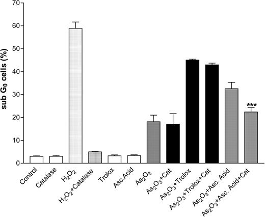
Several reports have demonstrated that ascorbic acid (AA), a known antioxidant compound, enhances As2O3-induced cytotoxicity in multiple myeloma cells. Clement et al24 reported that ascorbate-mediated killing in HL60 cells depends on the levels of H2O2 produced by the reaction of AA within the cell culture medium, and direct addition of H2O2 to the cells reproduced these results. Further, degradation of extracellular H2O2 by the addition of catalase, which catalyzes the decomposition of H2O2 to H2O and O2, blocked any additional toxicity from AA.24 They concluded that the extracellular H2O2 generated plays a major role in the synergy observed in vitro by As2O3 and AA. We asked whether the synergy observed between trolox and As2O3 was influenced by the generation of extracellular H2O2. If so, we would expect that the addition of catalase could block the generation of extracellular H2O2 and consequently decrease the apoptotic rate. Therefore, we treated NB4 cells for 3 days with As2O3, trolox, AA, and catalase as indicated in Figure 4. The addition of catalase (500 U/mL) prevented the induction of apoptosis by H2O2, suggesting that even a very large extracellular production of H2O2 by As2O3 and trolox could be blocked. Catalase significantly blunted the synergy of As2O3 with AA, confirming previous reports.22 In contrast, the addition of catalase did not protect cells treated with As2O3 + trolox.
The synergistic effects of trolox on arsenic-mediated apoptosis are not related to extracellular H2O2 production. Cells were treated with As2O3 (1 μM) and trolox or ascorbic acid (100 μM) for 48 hours. Catalase (500 U/mL, Cat) was added as indicated to degrade the extracellular H2O2 generated. Apoptosis was detected by PI staining and quantitated by flow cytometric measurement of PI-positive cells. Each bar represents an average of 3 independent samples, and standard deviation bars are shown. Asterisks indicate significant differences from As2O3 + AA–treated cells (P < .001).
The synergistic effects of trolox on arsenic-mediated apoptosis are not related to extracellular H2O2 production. Cells were treated with As2O3 (1 μM) and trolox or ascorbic acid (100 μM) for 48 hours. Catalase (500 U/mL, Cat) was added as indicated to degrade the extracellular H2O2 generated. Apoptosis was detected by PI staining and quantitated by flow cytometric measurement of PI-positive cells. Each bar represents an average of 3 independent samples, and standard deviation bars are shown. Asterisks indicate significant differences from As2O3 + AA–treated cells (P < .001).
Trolox enhances As2O3-mediated c-jun terminal kinase (JNK) activation
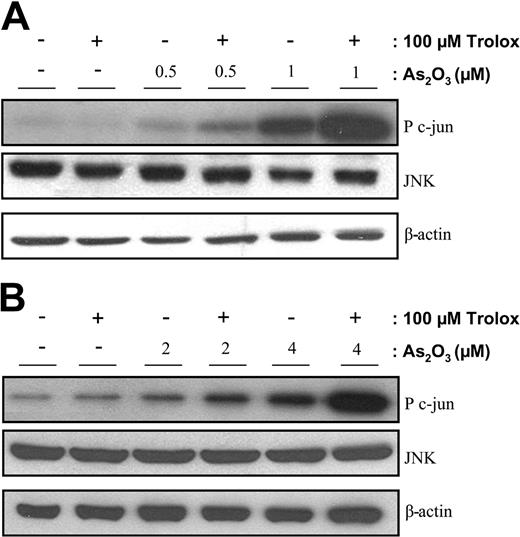
It has been demonstrated that JNK is activated in response to oxidative stress.46,47 We have reported that JNK activation is necessary for As2O3-induced apoptosis of NB4 cells.30 Therefore, we asked whether the activation of JNK in NB4 cells treated with As2O3 and trolox for 16 hours might play a role in the synergistic effect of these compounds. We used an immune complex assay with glutathione-S–transferase (GST)–c-jun as an exogenous substrate. Figure 5A shows that a 24-hour treatment of NB4 cells with as little as 0.5 μM As2O3 induces significant JNK activation leading to phosphorylation of c-jun. As expected, higher As2O3 concentrations increased JNK activation. Consistent with the idea that trolox enhances As2O3-mediated oxidative stress, we observed a further increase in JNK activity when cells are cotreated with As2O3 and trolox. As expected, the arsenic-resistant cell line AsR2 cells showed little activation of JNK following treatment with As2O3, even at doses sufficient to elicit robust activation of NB4 cells (Figure 5B). However, when trolox was added to the media, a considerable JNK activation was observed that correlated with apoptotic induction.
Trolox enhances As2O3-mediated JNK activation. Immune complex kinase assays were performed to measure JNK activity with extracts from NB4 (A) or AsR2 cells (B) treated with As2O3 and trolox for 16 hours as described in “Materials and methods.” Data depicted are representative of 3 independent experiments.
Trolox enhances As2O3-mediated JNK activation. Immune complex kinase assays were performed to measure JNK activity with extracts from NB4 (A) or AsR2 cells (B) treated with As2O3 and trolox for 16 hours as described in “Materials and methods.” Data depicted are representative of 3 independent experiments.
As2O3 induces the formation of trolox phenoxyl radicals
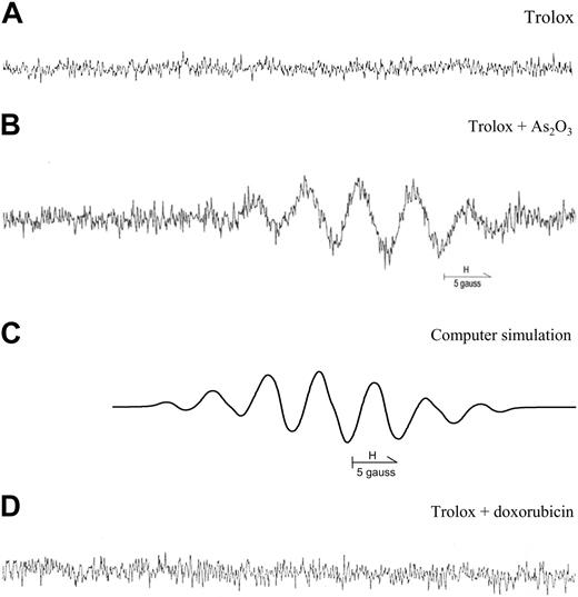
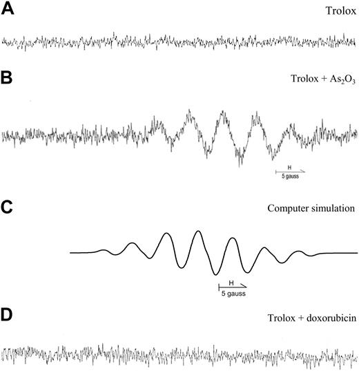
Electron paramagnetic resonance (EPR) is an important tool in experimental studies of systems containing unpaired electrons. We used EPR to directly assay the generation of trolox radicals. As shown in Figure 6B, addition of trolox to reaction mixtures containing As2O3 resulted in the observation of an intense 7-line EPR signal. The g-value (3477.530 G), the relative intensities, and the splittings all confirm the presence of the trolox phenoxyl radical. Its identity is further confirmed by the simulated spectrum (Figure 6C), which is based on the published coupling constants for this radical.48 This signal is not generated by trolox alone (Figure 6A) nor in the presence of doxorubicin (Figure 6D), suggesting the requirement of As2O3 and its hydration products for the formation of this radical.
Electronic paramagnetic resonance detection of the trolox phenoxyl radical. EPR spectra of trolox in the reaction system containing 1 mM Trolox, 5% (vol/vol) DMSO (A), and 0.02 mM As2O3 (B) or 0.2 μg/mL doxorubicin (C). (D) Computer simulation of spectrum in panel B obtained using the hyperfine splitting constants: aH (CH3) = 4.56 G; aH (CH3) = 4.86 G; aH (CH3) = 0.23 G; aH (CH2) = 0.37 G; aH′ (CH2) = 0.76 G.
Electronic paramagnetic resonance detection of the trolox phenoxyl radical. EPR spectra of trolox in the reaction system containing 1 mM Trolox, 5% (vol/vol) DMSO (A), and 0.02 mM As2O3 (B) or 0.2 μg/mL doxorubicin (C). (D) Computer simulation of spectrum in panel B obtained using the hyperfine splitting constants: aH (CH3) = 4.56 G; aH (CH3) = 4.86 G; aH (CH3) = 0.23 G; aH (CH2) = 0.37 G; aH′ (CH2) = 0.76 G.
Trolox does not potentiate As2O3 effects in nonmalignant cells
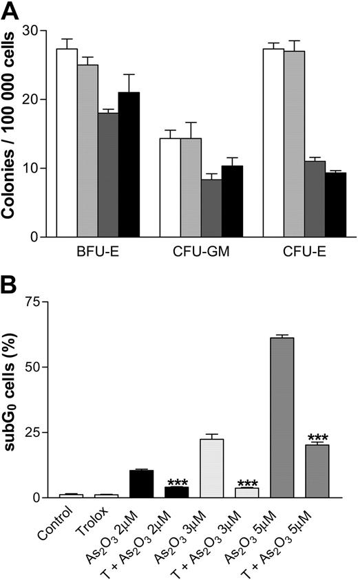
We sought to determine the effects of As2O3 combined with trolox in normal human hematopoietic colony-forming cells and mouse embryonic fibroblasts. Normal human PBMCs were isolated, grown in methylcellulose, and treated with As2O3, trolox, or the combination for 2 weeks. Figure 7A shows that 1 μM As2O3 inhibited CFU-Es by approximately 62%, but had minimal effect on CFU-GM or BFU-E colony formation. Treatment with trolox alone did not inhibit colony formation and trolox did not enhance As2O3 inhibition of CFU-GMs, BFU-Es, or CFU-Es. Mouse embryonic fibroblasts were treated with different concentrations of As2O3 for 3 days, stained with PI, and analyzed by flow cytometry. Interestingly, trolox significantly decreased As2O3-mediated apoptosis at all doses studied (Figure 7B).
The synergistic effects of trolox on arsenic-mediated apoptosis are unique to cancer cells. (A) Normal human PBMCs were isolated from 3 healthy donors using a Ficoll gradient. Colony-forming ability of PBMCs treated with As2O3 and trolox was assessed by counting CFU-Es, CFU-GMs, and BFU-Es after 15 days. Results are representative of 3 independent experiments each performed in triplicate. (B) Mouse embryonic fibroblasts were treated with As2O3 with or without trolox for 3 days. Apoptosis was detected by PI staining and quantitated by flow cytometry measurement of PI-positive cells. Each bar represents an average of 3 independent samples. Asterisks indicate significant differences from As2O3-treated cells (P < .001).
The synergistic effects of trolox on arsenic-mediated apoptosis are unique to cancer cells. (A) Normal human PBMCs were isolated from 3 healthy donors using a Ficoll gradient. Colony-forming ability of PBMCs treated with As2O3 and trolox was assessed by counting CFU-Es, CFU-GMs, and BFU-Es after 15 days. Results are representative of 3 independent experiments each performed in triplicate. (B) Mouse embryonic fibroblasts were treated with As2O3 with or without trolox for 3 days. Apoptosis was detected by PI staining and quantitated by flow cytometry measurement of PI-positive cells. Each bar represents an average of 3 independent samples. Asterisks indicate significant differences from As2O3-treated cells (P < .001).
Discussion
The induction of apoptosis by As2O3 has been linked to the accumulation of free radicals and subsequent induction of oxidative stress. Intracellular oxidative status has been shown to be important for As2O3 sensitivity, and strategies to alter the redox environment may allow normally As2O3-resistant cells to become susceptible to As2O3-mediated apoptosis.
In part because of differences in cellular redox environments, As2O3 is less active in most malignant cells than in APL,49 prompting a search for agents that enhance As2O3 efficacy. One such agent is the widely used antioxidant, ascorbic acid. AA has been shown to potentiate As2O3-mediated cytotoxicity in HL60 cells, as well as in su-DHL-4,22 8226/S, and U266 cells.23 There is some evidence that the time at which AA is administered in relation to exposure to oxidative stress has an impact on AA pro-oxidant capacity in vitro.50 Cotreatment of Chinese hamster ovary AS52 cells with AA and a radical generating system (RGS) resulted in a significant increase in cell death compared with treatment with RGS alone.50 However, when AS52 cells were pretreated for 24 hours with AA and then challenged with RGS, the cells were protected. Ascorbate-generated H2O2 may be responsible for the enhancement of As2O3 cytotoxicity in vitro, because the latter is attenuated by coadministration of catalase. Furthermore, the propensity for AA to generate H2O2 in vitro appears to be substantially influenced by the composition of the culture media.22,24 The effects of AA on arsenic-mediated cytotoxicity in vivo are conflicting. No additional benefits were observed by combining As2O3 with ascorbic acid in the treatment of murine T-cell leukemia.51 On the other hand, the combined effects of AA and As2O3 increased the survival time of BDF1 mice injected with P388D1 lymphoma cells.22
On the basis of the ascorbate experience, we evaluated various “antioxidant” compounds for their ability to enhance As2O3-mediated apoptosis. Our initial experiments performed with the human APL cell line, NB4, revealed that trolox had a strong potentiating effect on As2O3-mediated growth inhibition and apoptosis. In contrast, melatonin and resveratrol, compounds reported to have antioxidant or pro-oxidant capacities depending on the cellular redox status,52,53 were not able to substantially enhance As2O3-mediated cytotoxicity (data not shown). NB4-derived AsR2, IM9, myeloma cells, and a variety of breast cancer cells were all sensitized to As2O3 when trolox was added to the culture media, revealing a broad capacity of trolox to increase As2O3-mediated cytotoxicity in different tumor cells. We showed that the potentiation of growth inhibition by trolox involved induction of apoptotic cell death as monitored by caspase-3 activation and PARP cleavage. In contrast, trolox was not able to enhance apoptosis mediated by other common chemotherapeutic drugs (doxorubicin, AraC, or etoposide). These data are consistent with previous reports that trolox does not influence doxorubicin-induced oxidative damage.54,55 In addition, cisplatin, a major antineoplastic drug for the treatment of solid tumors,56 has been shown to induce apoptosis in LLC-PK1 cells that can be significantly inhibited by trolox cotreatment.26 Trolox also prevented cisplatin-induced ototoxicity when applied locally in guinea pigs' round windows.57
Although the mechanism by which trolox enhances As2O3-mediated apoptosis in malignant cells remains unknown, here we provide evidence that trolox potentiates As2O3-induced oxidative stress. The ho-1 gene is highly sensitive to induction by oxidative stress, and HO-1 activity degrades cellular heme to products (biliverdin, bilirubin) with proven antioxidant properties.58 Our findings that HO-1 protein is substantially up-regulated in tumor cell lines following treatment with As2O3 and trolox implicate oxidative stress as a killing mechanism in this system. Augmented levels of protein carbonyls and isoprostanes in these cells further support this conclusion. However, we could not find any differences in the intracellular GSH content upon As2O3 and trolox treatment in any of the cells studied (data not shown) suggesting that, unlike ascorbic acid, the synergy observed between trolox and As2O3 is not due to changes in GSH levels.
To our knowledge, there are only a few reports on the pro-oxidant capacity of trolox. Synergistic effects between selenite and trolox resulting in enhanced superoxide production and cytotoxicity have been reported. In a mechanism similar to that proposed for As2O3, cytotoxicity of selenium is believed to involve simultaneous thiol oxidation and superoxide production.59 This is supported by data showing that only selenium compounds that generate superoxide can synergize with trolox. In addition, the combination of Cu2+ and trolox resulted in increased ROS generation and cytotoxicity in astrocytes.60 Trolox exhibits prooxidant properties when Cu2+ is used to mediate low-density lipoprotein oxidation, but in the absence of Cu2+, trolox shows an antioxidant capacity.61 Thus, redox-cycling may be an important feature in the pro-oxidant mechanism of trolox.
Our EPR experiments suggest that As2O3 may bioactivate trolox to a potentially tumoricidal phenoxyl radical (trolox·). These observations suggest that As2O3 not only generates ROSs, but also induces the formation of trolox·, which contributes to the intracellular pool of reactive oxygen species and consequently enhances cellular oxidative stress. Consistent with the hypothesis that trolox· plays a role in the augmentation of As2O3-induced apoptosis, the combination of trolox and doxorubicin was not synergistic and did not generate the trolox radical. Therefore, trolox may be useful to increase As2O3-mediated cytotoxicity, but it may not be effective when combined with other chemotherapeutic drugs. These observations do not preclude the possibility that other mechanisms may also be involved. Plasma membrane CoQ reductase (PMQR), the enzyme responsible for reducing trolox· to trolox, may be affected by As2O3, thereby augmenting cellular oxidative stress.62 Experiments are in progress to elucidate the rates and mechanisms for the reactions between trolox and As2O3 under aerobic and anaerobic conditions and the potential role of PMQR in the generation of trolox·.
We have previously demonstrated that in stress-activated kinase (SEK)-/- murine embryonic fibroblast cells, the activation of c-jun N terminal kinase in response to As2O3 is dampened. The reduced JNK response in these cells or in NB4 cells treated with dicumarol (a known JNK inhibitor) is associated with resistance to As2O3-induced apoptosis.30 These results indicated that JNK activation is required for sensitivity to As2O3 in these cells. We observed that trolox enhances As2O3-mediated JNK activation, which is consistent with our previous conclusion that JNK is an important mediator in As2O3-induced oxidative damage and apoptosis. We treated SEK+/+ and SEK-/- cells with the combination of As2O3 and trolox and analyzed the induction of apoptosis. As expected, SEK-/- cells were more resistant to As2O3 than SEK+/+, and trolox alone had no effect on apoptosis in either the SEK+/+ or SEK-/- cells. Interestingly, trolox protected these nonmalignant cells from As2O3-mediated toxicity, although care should be taken when extrapolating these data from murine to human cells.
Consistent with this finding, we found that trolox does not enhance cytotoxicity of As2O3 in colony-forming assays using human hematopoietic peripheral blood mononuclear cells. We show here that the combination of As2O3 and trolox does not increase the As2O3-mediated reduction in CFU-Es, CFU-GMs, and BFU-Es. It has been also found that arsenic damage to supercoiled φX174 DNA and DNA in peripheral human lymphocytes in culture is inhibited by trolox.63 Therefore, cytotoxic enhancement accruing from trolox exposure may be specific to tumor cells.
In this study, we provide preclinical evidence for the potential efficacy of As2O3 and trolox combination therapy in APL and other malignancies intrinsically less sensitive to As2O3 monotherapy. It is important to note that standard treatment of APL patients with 0.15 mg/kg per day As2O3 yields a maximum concentration of 1 to 2 μM As2O3 in the plasma,64,65 which is similar to the in vitro doses reported here. Given the low toxicity of trolox and As2O3 and considering the synergy we observed between the 2 drugs, our results justify the study of this combination in in vivo models of human leukemias and other malignancies.
Prepublished online as Blood First Edition Paper, October 5, 2004; DOI 10.1182/blood-2004-05-1772.
Supported by a predoctoral fellowship award from MCET-FRSQ-CIHR Strategic Training Program and operating grants from the Canadian Institute for Health Research (CIHR) and the Natural Sciences and Engineering Research Council of Canada (NSERC). W.H.M. Jr is an investigator of the CIHR. D.S.B. is a Canadian Research Chair.
The publication costs of this article were defrayed in part by page charge payment. Therefore, and solely to indicate this fact, this article is hereby marked “advertisement” in accordance with 18 U.S.C. section 1734.
We thank David Hamilton for his helpful comments and Alessandra Padovani for critical reading of this manuscript.


