Abstract
The 4A11 antigen is a unique cytokine-inducible antigen up-regulated on rheumatoid arthritis (RA) synovial endothelial cells (ECs) compared with normal ECs. Previously, we showed that in soluble form, this antigen, Lewisy-6/H-5-2 (Ley/H) or its glucose analog, 2-fucosyl lactose (H-2g), induced the expression of EC intercellular adhesion molecule-1 (ICAM-1) and leukocyte-endothelial adhesion through the Janus kinase 2 (JAK2)–signal transducer and activator of transcription 3 (STAT3) pathway. Currently, we show that H-2g induces release of EC angiogenic basic fibroblast growth factor (bFGF) and vascular endothelial growth factor (VEGF), an effect inhibited by decoy nuclear factor κB (NFκB) oligodeoxynucleotide (ODN). JAK2 and phosphoinositide-3 kinase (PI3K) are 2 upstream kinases of NFκB activated by H-2g, as confirmed by an inhibitor of kappa B kinase (IKKβ) assay. In vitro, H-2g induces vascular sprouting in the rat aortic ring model, whereas blockade of JAK2, PI3K, or NFκB inhibits sprouting. Likewise, in the in vivo mouse Matrigel plug angiogenesis assay, chemical inhibitors and antisense or decoy ODNs of JAK2, PI3K, or NFκB decrease angiogenesis, confirming the importance of these pathways in H-2g–induced EC signaling. The critical role of Ley/H involvement in angiogenesis and its signaling pathways may provide new targets for therapy of diseases characterized by pathologic neovascularization.
Introduction
Angiogenesis is critical in vasculoproliferative processes, including tumor growth and inflammatory states such as psoriasis and rheumatoid arthritis (RA).1 Many bioactive molecules such as interleukin 1 (IL-1), IL-8, and tumor necrosis factor-α (TNF-α), as well as cell adhesion molecules such as soluble E-selectin and intercellular adhesion molecule-1 (ICAM-1) induce angiogenesis. Most of these mediators are glycoconjugates (glycoproteins or glycolipids). These chemoattractants or soluble adhesion molecules participate in cell-cell interactions with circulating leukocytes and endothelial cells (ECs),2 and play an essential role in the development of neovascularization.3-5
We have shown that the soluble form of E-selectin mediates angiogenesis via its endothelial ligand, sialyl Lewis (Le)x.6,7 To further examine EC function in inflammation, we raised a monoclonal antibody (mAb), 4A11, to adherent human RA synovial tissue (ST) cells.8 mAb 4A11 recognizes the glycoconjugate blood group antigen, Lewis (Le)y-6/H-5-2 (Ley/H), which is expressed on ECs predominantly in lymphoid organs, ST, and skin. This antigen is EC selective, being found mainly on endothelium and epithelium. It is also cytokine-inducible and up-regulated in soluble form in RA synovial fluid compared with noninflammatory osteoarthritis synovial fluid. Ley and H-5-2 are structurally related, in that Ley is the result of fucose addition by α-(1,3)-fucosyltansferase, to the nonterminal N-acetylglucosamine of H-5-2. In soluble form, the 4A11 antigen, or its glucose analog, 2-fucosyl lactose (H-2g), is angiogenic.9
Basic fibroblast growth factor (bFGF) and vascular endothelial growth factor (VEGF) are 2 potent angiogenic cytokines.10 VEGF is secreted by a number of cell types and acts in a paracrine fashion.7 VEGF plays a key role and has effects on all of the main steps in angiogenesis, including EC mitogenesis, chemotaxis, vascular permeability, and proteolysis. Both VEGF and its endothelial-specific receptors are up-regulated in the setting of hypoxia, potentially enabling localization of the effect to areas of hypoxia or ischemia.11 bFGF is also important in angiogenesis.12 In spite of the historic name, “fibroblast” growth factor, it is in fact a potent mitogen for many cell types including ECs, smooth muscle cells, and cells of mesenchymal, neural, and epithelial origin. FGFs regulate key migratory and proliferative processes involved in angiogenesis. As VEGF and bFGF are both chemotactic and mitogenic for ECs, they induce matrix-resorbing activity and proteolytic systems that are necessary for matrix degradation involved in neovascularization.
Nuclear factor κB (NFκB) plays a pivotal role in the coordinated transactivation of a series of genes regulating cytokines and adhesion molecules that are involved in the onset of inflammation and angiogenesis. NFκB is a heterodimeric protein and is constitutively expressed in cells. Once activated, NFκB translocates from the cytoplasm to the nucleus to activate gene transcription. Cytokines such as VEGF and bFGF have NFκB binding sites at or near their promoters. Thus, NFκB activation regulates VEGF and bFGF expression.13 Inactivated NFκB dimers exist in the cytoplasm through interactions with an inhibitor, IκB. The inactive trimer reacts to stimulatory signals by targeted phosphorylation, and subsequent ubiquination and degradation of IκB. IKKβ is one of the kinases in this pathway that induces IκB phosphorylation. Once IκB is released, the NFκB dimer is able to enter the nucleus and bind DNA.14,15
Here we report that H-2g induces angiogenesis through up-regulation of EC bFGF and VEGF expression. NFκB is activated by H-2g, and NFκB in turn induces EC bFGF and VEGF expression. Moreover, H-2g activates Janus kinase 2 (JAK2) and phosphoinositide-3 kinase (PI3K) upstream of NFκB, with resultant EC chemotaxis in vitro. Using the in vitro “aortic ring” assay and in vivo “Matrigel plug” assay for angiogenesis, we show that NFκB is a key factor in H-2g–induced angiogenesis. Taken together, soluble 4A11 antigen or its glucose analog, H-2g, effect angiogenesis by inducing bFGF and VEGF expression and signaling through JAK2, PI3K, and NFκB pathways. Potential treatment strategies through the inhibition of PI3K and JAK2-NFκB pathways to target H-2g signals may provide a useful approach for therapy of angiogenesis-driven diseases, such as tumor growth and RA.
Materials and methods
Reagents
H-2g was purchased from Sigma-Aldrich (St Louis, MO). The PI3K inhibitor, LY294002, and the Erk1/2 inhibitor, PD98059, were purchased from Calbiochem (San Diego, CA). The JAK2 inhibitor, AG-490, was purchased from LC Laboratories (Woburn, MA). VEGF and bFGF enzyme-linked immunosorbent assay (ELISA) kits were from R&D Systems (Minneapolis, MN). The sequences of the oligodeoxynucleotide (ODNs) employed in this study were as follows: JAK2 antisense: AAG GCA GGC CAT TCC CAT; JAK2 sense: ATG GGA ATG GCC TGC CTT; PI3K antisense: GTA CTG GTA CCC CTC AGC ACT CAT; PI3K sense: ATG AGT GCT GAG GGG TAC CAG TAC; Erk1/2 antisense: GCC GCC GCC GCC GCC AT; Erk1/2 sense: ATG GCG GCG GCG GCG GC.16 The corresponding sense ODN was used as a control for each antisense ODN. The ODNs were synthesized and purified by the Northwestern University Biotechnology Laboratory (Chicago, IL) and modified with phosphorothioate. Decoy NFκB ODN: 5′-CCT TGAAGG GAT TTC CCT CC-3′/5′-GGA GGG AAA TCC CTT CAA GG-3′, and scrambled NFκB ODN: 5′-TTG CCG TAC CTG ACT TAG CC-3′/5′-GGC TAA GTC AGG TAC GGC AA-3′ were synthesized by Integrated DNA Technologies (IDT; Coralville, IA). LipofectAMINE was from Invitrogen (Carlsbad, CA). The radioisotope γ-[32P] adenosine triphosphate (ATP; 3000 Ci/mmol) was obtained from PerkinElmer Life Sciences (Wellesley, MA). Rabbit anti-human PI3K polyclonal antibody (p85) was from Upstate Biotechnology (Lake Placid, NY). The in vitro IKKβ kinase assay kit was from Clontech Laboratories (Palo Alto, CA). Both recombinant human (rh) TNF-α (specific activity 1.3 × 10⁷ U/mg) and human recombinant IL-4 (huIL-4; specific activity 5 × 10⁶ U/mg) were from Peprotech (Rocky Hill, NJ). C57/BL6 mice were from the National Cancer Institute (NCI; Bethesda, MD). Growth factor–reduced (GFR) Matrigel was purchased from Becton Dickinson (Bedford, MA).
Cell culture
Human microvascular endothelial cells (HMVECs) were purchased from BioWhittaker (Walkersville, MD), and maintained in growth factor complete endothelial basal medium (EBM) supplemented with 10% fetal bovine serum (FBS). Cells were between passages 3 and 12, and did not display discernable phenotypic changes when observed before each assay. Cells were maintained at 37°C, 5% CO2.
HMVEC chemotaxis
HMVEC chemotaxis was performed using a 48-well Boyden chemotaxis chamber (Neuroprobe, Cabin John, MD) as described previously.6 HMVECs (4 × 10⁴ cells/25 μL EBM and 0.1% FBS) were plated in the bottom wells of the chambers with a polyvinypyrolidone-free polycarbonate filter (8-μm pore size; Nucleopore, Pleasant, CA). The chambers were inverted and incubated in a humidified incubator with 5% CO2/95% air at 37°C for 2 hours, allowing HMVECs to attach to the membrane. The chambers were inverted again and H-2g, phosphate-buffered saline (PBS), or positive control bFGF in PBS were added, and the chamber was further incubated for 2 hours at 37°C. For inhibition assay, HMVECs were pretreated with the inhibitors (50 μM JAK2 inhibitor, AG490; 20 μM PI3K inhibitor, LY294002; or 20 μM Erk1/2 inhibitor, PD98059, for 2 hours). Inhibitors were present in the lower chambers, along with HMVECs, during the chemotaxis assay. The membranes were removed, fixed in methanol for one minute, and stained with Diff-Quik (VWR International, West Chester, PA). The data are expressed as the number of cells migrating through the membrane per well (the sum of 3 high-power × 40 fields per well, averaged for each quadruplicate well). For antisense and sense ODN assays, HMVECs (5.5 × 10⁵/well) were seeded in 6-well plates in complete EBM with 10% FBS. Upon 70% confluence, cells were LipofectAMINE-transfected with antisense or sense ODNs to JAK2, PI3K, or Erk1/2 for 4 hours. Cells were washed with prewarmed PBS once, fed with fresh media, and cultured overnight. Chemotaxis assays were conducted as described in this paragraph.
VEGF and bFGF ELISA assays
HMVECs were plated at 15 000 cells per well in 24-well plates. After 12 hours of incubation, the medium was changed to RPMI 1640 with 2% FBS. Cells were then treated with H-2g and inhibitors. Medium was collected after 12 hours and cell debris spun down. Supernatant (200 μL) from each well was stored at -80°C for ELISA assay. To examine if NFκB was involved in VEGF and bFGF expression, HMVECs were transfected with either decoy or scrambled NFκB ODNs17 prior to H-2g exposure. Decoy NFκB ODN and scrambled NFκB ODN were incubated at 94°C for 5 minutes to be denatured, and then annealed at -1.0°C/min in GeneAmp PCR system 9700 (PerkinElmer, Wellesley, MA).
Western blotting
HMVECs were allowed to achieve quiescence by incubation in EBM containing 0.1% FBS for 2 hours, and were pretreated with 50 μM AG490, 20 μM PD098059, or 20 μM LY294002 for another 2 hours. Chemical signaling inhibitor concentrations were maintained through the culture period. After pretreatments, HMVECs were exposed to 100 nM H-2g in the presence and absence of 50 μM AG490, 20 μM LY294002, and 20 μM PD98059 for the specified times at 37°C. An IL-4 (10 ng/mL)/TNF-α (1.15 nM) combination was used as the positive control. The cells were then lysed in a buffer containing 20 nM HEPES (N-2-hydroxyethylpiperazine-N′-2-ethanesulfonic acid), pH 7.4, 2 nM EDTA (ethylenediaminetetraacetic acid), 1 nM β-dithiothreitol, 50 nM glycerophosphate, 1% Triton X-100, 20 μg/mL aprotinin, 1 μg/mL leupeptin, 1 nM sodium orthovanadate, and 400 μM phenylmethylsulfonyl fluoride (PMSF). The samples were resolved on 10% sodium dodecyl sulfate (SDS)–polyacrylamide gels and Western blotted.18
IKKβ kinase in vitro assays
HMVECs (1 × 10⁶ cells/well) were plated in 100-mm Petri dishes in complete EBM with 10% FBS. Once the cells were 80% to 90% confluent, they were further incubated with complete EBM containing 0.1% FBS for 2 hours and were pretreated with 50 μM AG490, 20 μM PD098059, or 20 μM LY294002 for another 2 hours. HMVECs were then stimulated with 100 nM H-2g for 15 minutes at 37°C. Cells were then washed with PBS. Cell lysate buffer (1.2 mL) was added. Cells were homogenized in an ice-cold dounce tissue grinder (30 seconds per time, twice). Cells were centrifuged at 12 000g at 4°C for 10 minutes. The protein content of each sample was quantitated using a bicinchoninic acid (BCA) protein assay kit and normalized to 1 μg/μL. From each sample, 500 μL lysate was incubated with 5 μg IKKβ antibody, and mixed for one hour at 4°C with gentle rocking. Protein G agarose conjugate (60 μL; 50% slurry in PBS) was added to each sample and further incubated overnight at 4°C. The beads were spun down at 12 000g for 5 minutes and washed twice with 1× IKKβ in vitro kinase buffer (supplied with the kit). The last wash was removed. The pellet was mixed by gently flicking the tube. Next, 2 μL γ-[32P] ATP and 4 μL substrate (IκB) were added, the reaction mixed well, and incubated at 30°C for 30 minutes. The sample (20 μL) was resolved on a 12% SDS–polyacrylamide gel electrophoresis, and the gel was dried and exposed to x-ray film.
Rat aortic ring sprouting growth assay
Aortic arches were removed from female Lewis rats weighing 140 g to 160 g, cleaned of surrounding connective tissue, and sliced.19 Aortic rings were placed on a Matrigel drop in 48-well plates and covered with an additional drop of Matrigel. Serum-free EGM-medium (Invitrogen; 400 μL) with various concentrations of H-2g was added to each well. bFGF (10 nM) was used as a positive control. Aortic rings were incubated 48 hours at 37°C in 5% CO2 to allow microvessel sprouting from the adventitial layer, which was fully apparent in positive controls after 24 hours. Rings were fixed and stained with Diff-Quik and observed under an inverted microscope. Sprouting was measured using the following scale: 0 indicated no sprouting; 0.25, isolated sprouting; 0.5, sprouting in 20% to 50% of the arterial circumference; 1, sprouting in 50% to 75% of the circumference; 1.5, sprouts in 100% of the circumference; and 2, 100% of the artery circumference occupied by sprouts longer than one-third of the length of the average radius of the rings. Each condition was tested in 6 wells. The experiment was repeated 4 times. Results were expressed as mean plus or minus standard error (SE).
Matrigel plug in vivo angiogenesis assay
To examine the effects of H-2g on angiogenesis in vivo, Matrigel plug assays were performed.20 C57/BL6 mice were anesthetized by Metofane inhalation. Each mouse was shaved on its ventral aspect and given a subcutaneous injection of sterile Matrigel (500 μL/injection). Matrigel containing PBS served as the negative control; Matrigel containing bFGF (1 ng/mL) served as the positive control; and Matrigel with H-2g (1 μM) was the test substance. In the inhibition study, 50 μM AG490, 20 μM LY294002, or 20 μM PD98059 or 20 μg/mL decoy NFκB ODN was added along with H-2g in the Matrigel before it was injected. After 7 to 10 days, the mice were killed by Metofane inhalation and cervical dislocation and the Matrigel plugs were dissected out and analyzed by hemoglobin measurement,21,22 since hemoglobin levels correlate with blood vessel growth.23 Hemoglobin levels were normalized to the weight of the plugs.
Results
Signaling profile of H-2g–induced HMVEC chemotaxis, and VEGF and bFGF expression
Our previous work showed that H-2g is a potent angiogenic mediator.8 To examine the signaling pathways by which H-2g mediates angiogenesis, we performed HMVEC chemotaxis assays. H-2g induced a dose-dependent increase in HMVEC chemotaxis, reaching significance at 10 nM to 1000 nM (P < .05, Figure 1A), compared with negative control PBS. To further study the role of different signaling pathways that may be involved in H-2g–mediated HMVEC chemotaxis, HMVECs were pretreated with the respective signaling inhibitors: a JAK2 inhibitor, AG490, 50 μM; a PI3K inhibitor, LY294002, 20 μM; and an Erk1/2 inhibitor, PD98059, 20 μM, for 2 hours prior to performing the chemotaxis assays. The JAK2 inhibitor, AG490, was most effective in inhibiting H-2g–mediated HMVEC chemotaxis by 98% (P < .05), whereas the PI3K inhibitor, LY294002, showed 65% inhibition (P < .05); the Erk1/2 inhibitor, PD98059; and the Src inhibitor, PP2, induced 15% and 13% inhibition, respectively, which were not statistically significant (Figure 1B). To further confirm the results obtained in the HMVEC chemotaxis studies using chemical inhibitors, we performed assays using HMVECs transiently LipofectAMINE-transfected with sense and antisense ODNs to JAK2, PI3K, and Erk1/2. JAK2 antisense ODN reduced H-2g–mediated HMVEC chemotaxis (93%, P < .05) significantly. PI3K antisense ODN also showed a significant inhibition (59%, P < .05). However, Erk1/2 and Src antisense ODNs showed weaker inhibition (19%, not significant) or no inhibition, which is consistent with results obtained with the chemical inhibitors (Figure 1C). These studies suggested that H-2g may be mediating HMVEC chemotaxis predominantly through the JAK2 pathway and partially through the PI3K pathway.
H-2g induces HMVEC chemotaxis through JAK2 and PI3K pathways but not through Src and MAPK. (A) H-2g induced a dose-dependent increase in HMVEC chemotaxis compared with negative control PBS. (B) For inhibition studies, HMVECs were pretreated with the chemical inhibitors (50 μM AG490, 20 μM LY294002, 20 μM PD98059, or 20 μM PP2) for 2 hours at 37°C and then assayed in 48-well chemotaxis chambers in response to 100 nM of H-2g. H-2g induced HMVEC chemotaxis through the JAK2 and PI3K pathways. (C) HMVECs were transfected with JAK2, Src, PI3K, and Erk1/2 sense (▪) or antisense (□) ODNs. The data confirmed that JAK2 and PI3K are involved in H-2g–induced HMVEC chemotaxis, but Src and MAPK are not. Results are expressed as the number of cells migrating through the membrane per well plus or minus the standard error of the mean (SEM) from 6 independent experiments. *Represents a significant difference (*P < .05) between the respective groups.
H-2g induces HMVEC chemotaxis through JAK2 and PI3K pathways but not through Src and MAPK. (A) H-2g induced a dose-dependent increase in HMVEC chemotaxis compared with negative control PBS. (B) For inhibition studies, HMVECs were pretreated with the chemical inhibitors (50 μM AG490, 20 μM LY294002, 20 μM PD98059, or 20 μM PP2) for 2 hours at 37°C and then assayed in 48-well chemotaxis chambers in response to 100 nM of H-2g. H-2g induced HMVEC chemotaxis through the JAK2 and PI3K pathways. (C) HMVECs were transfected with JAK2, Src, PI3K, and Erk1/2 sense (▪) or antisense (□) ODNs. The data confirmed that JAK2 and PI3K are involved in H-2g–induced HMVEC chemotaxis, but Src and MAPK are not. Results are expressed as the number of cells migrating through the membrane per well plus or minus the standard error of the mean (SEM) from 6 independent experiments. *Represents a significant difference (*P < .05) between the respective groups.
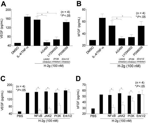
We hypothesized that H-2g may mediate angiogenesis by inducing HMVEC VEGF and bFGF release. We conducted ELISAs to examine if H-2g up-regulates VEGF and bFGF in HMVEC-conditioned media. After 12 hours of culturing HMVECs with H-2g, both VEGF and bFGF were significantly increased (2- to 3-fold) in the conditioned media. When cocultured with chemical inhibitors, the JAK2 inhibitor, AG490, inhibited both VEGF and bFGF expression induced by H-2g by 96% and 90%, respectively (P < .05; Figure 2A). The PI3K inhibitor LY294002 inhibited both VEGF and bFGF expression induced by H-2g by 85% and 87%, respectively (P < .05). However, the Erk1/2 inhibitor, PD98059, did not have a significant inhibitory effect on H-2g–induced bFGF or VEGF expression (Figure 2B). This suggests that HMVEC VEGF and bFGF expression induced by H-2g is mainly through the JAK2 and PI3K pathways. Furthermore, we transfected HMVECs with decoy and scrambled NFκB ODNs, and the decoy NFκB decreased both VEGF and bFGF expression induced by H-2g by 98% and 91%, respectively, compared with the effect of scrambled ODN transfection (P < .05). JAK2 antisense ODN transfection decreased HMVEC VEGF and bFGF expression induced by H-2g by 88% and 79%, respectively (P < .05). For PI3K antisense ODN transfection, VEGF and bFGF expression induced by H-2g was down-regulated by 56% and 34%, respectively, compared with PI3K sense ODN transfection, P < .05 (Figure 2C-D). These results demonstrated that JAK2 and PI3K are activated by H-2g, which in turn up-regulates bFGF and VEGF expression. In addition, NFκB is activated in the nucleus and is involved in EC VEGF and bFGF expression induced by H-2g.
H-2g induces HMVEC VEGF, and bFGF through JAK2, PI3K, and NFκB. HMVECs were pretreated with chemical inhibitors: AG490, LY294002, and PD98059. HMVECs were then exposed to H-2g (100 nM) for 12 hours. ELISAs of the supernatants showed that H-2g induced both VEGF (A) and bFGF (B) expression and that the JAK2 inhibitor, AG490, or the PI3K inhibitor, LY294002, inhibited H-2g–induced EC VEGF and bFGF. Next, HMVECs were transiently transfected with JAK2, PI3K, Erk1/2 antisense ODN, or control sense ODN, or with decoy NFκB or scrambled NFκB. At 16 hours after transfection, HMVEC VEGF and bFGF ELISAs were performed. Decoy NFκB and antisense JAK2 and PI3K ODNs (□) inhibited HMVEC VEGF and bFGF expression compared with their control ODNs (▪) (C-D). Results are expressed as the mean number of cells migrating through the membrane per well plus or minus SEM from 4 independent experiments. *Represents a significant difference (P < .05) between the respective groups.
H-2g induces HMVEC VEGF, and bFGF through JAK2, PI3K, and NFκB. HMVECs were pretreated with chemical inhibitors: AG490, LY294002, and PD98059. HMVECs were then exposed to H-2g (100 nM) for 12 hours. ELISAs of the supernatants showed that H-2g induced both VEGF (A) and bFGF (B) expression and that the JAK2 inhibitor, AG490, or the PI3K inhibitor, LY294002, inhibited H-2g–induced EC VEGF and bFGF. Next, HMVECs were transiently transfected with JAK2, PI3K, Erk1/2 antisense ODN, or control sense ODN, or with decoy NFκB or scrambled NFκB. At 16 hours after transfection, HMVEC VEGF and bFGF ELISAs were performed. Decoy NFκB and antisense JAK2 and PI3K ODNs (□) inhibited HMVEC VEGF and bFGF expression compared with their control ODNs (▪) (C-D). Results are expressed as the mean number of cells migrating through the membrane per well plus or minus SEM from 4 independent experiments. *Represents a significant difference (P < .05) between the respective groups.
H-2g activates EC NFκB via JAK2 and PI3K
We hypothesized that H-2g activated EC JAK2, PI3K, and Erk1/2 upstream of NFκB. To test this, we conducted Western blots to examine the signaling mechanism leading to NFκB activation. HMVECs were allowed to achieve quiescence by incubation in EBM containing 0.1% FBS for 2 hours and were pretreated with AG490 and LY249002, and then treated with H-2g for another 20 minutes. By this method, we showed substantial inhibition of NFκB (p65) phosphorylation, whereas PD98059, a MAP kinase inhibitor, did not inhibit NFκB activation. We confirmed that equal amounts of protein samples were loaded in each lane by stripping the blots and reprobing with rabbit anti-human total β-actin antibody (Figure 3A-B). We next verified the signaling pathways by transfecting HMVECs with antisense JAK2, PI3K, and Erk 1/2 ODNs to confirm that antisense ODNs blocked NFκB expression compared with transfection using sense ODNs (Figure 3C-D).
H-2g–induced NFκB activation in HMVECs is inhibited by JAK2 and PI3 kinase inhibitors, and antisense ODNs directed against JAK2 and PI3K. HMVECs were stimulated with H-2g (100 nM) for 15 minutes. (A) In the inhibition study, HMVECs were pretreated with AG490 (50 μM), LY294002 (20 μM), or PD98059 (20 μM) for 2 hours before stimulation with H-2g, and inhibitors were also present during the stimulation with H-2g. AG490 and LY294002 inhibited H-2g–induced EC NFκB while PD98059 had no effect. (B) Transient transfection of HMVECs with sense and antisense ODNs to JAK2, PI3K, and Erk1/2 confirmed the results obtained using chemical inhibitors: JAK2 and PI3K antisense ODNs inhibited H-2g–induced EC NFκB. Results are representative of 4 independent experiments.
H-2g–induced NFκB activation in HMVECs is inhibited by JAK2 and PI3 kinase inhibitors, and antisense ODNs directed against JAK2 and PI3K. HMVECs were stimulated with H-2g (100 nM) for 15 minutes. (A) In the inhibition study, HMVECs were pretreated with AG490 (50 μM), LY294002 (20 μM), or PD98059 (20 μM) for 2 hours before stimulation with H-2g, and inhibitors were also present during the stimulation with H-2g. AG490 and LY294002 inhibited H-2g–induced EC NFκB while PD98059 had no effect. (B) Transient transfection of HMVECs with sense and antisense ODNs to JAK2, PI3K, and Erk1/2 confirmed the results obtained using chemical inhibitors: JAK2 and PI3K antisense ODNs inhibited H-2g–induced EC NFκB. Results are representative of 4 independent experiments.
We have shown that H-2g induced stimulation of up-regulation of p65 NFκB. An essential step in the stimulus-induced activation of the canonical NFκB pathway is the phosphorylaton of IκB proteins by the IKKs, release of active IκB from NFκB, and resultant NFκB activation.24 We next used an in vivo kinase assay based on IKK activation to determine if NFκB activation was also induced coincidently. Our results showed that IKK activity was induced by H-2g. Inhibitors of JAK2 and PI3K, but not of Erk1/2, inhibited the H-2g–induced EC IKK activity (Figure 4A). Similarly, antisense ODNs directed to JAK2 or PI3K blocked IKK activity, whereas Erk1/2 antisense ODN did not (Figure 4B).
H-2g induces IκB phosphorylation by IKKβ labeled with γ–[32P] ATP, which in turn causes NFκB activation in HMVECs. The effect is inhibited by JAK2 and PI3 kinase inhibitors, and antisense ODNs directed against JAK2 and PI3K. HMVECs were stimulated with H-2g (100 nM) for 15 minutes. (A) In the inhibition study, HMVECs were pretreated with AG490 (50 μM), LY294002 (20 μM), or PD98059 (20 μM) for 2 hours before stimulation with H-2g, and inhibitors were also present during the stimulation with H-2g. AG490 and LY294002 inhibited H-2g–induced EC NFκB while PD98059 had no effect. (B) Transient transfection of HMVECs with sense (▪) and antisense (□) ODNs to JAK2, PI3K, and Erk1/2 confirmed the results obtained using chemical inhibitors: JAK2 and PI3K antisense ODNs inhibited H-2g–induced EC IKKβ activation. Nontransfected cells were used as a control. Results are representative of 4 independent experiments. * indicates a significant difference (P < .05) between the respective groups.
H-2g induces IκB phosphorylation by IKKβ labeled with γ–[32P] ATP, which in turn causes NFκB activation in HMVECs. The effect is inhibited by JAK2 and PI3 kinase inhibitors, and antisense ODNs directed against JAK2 and PI3K. HMVECs were stimulated with H-2g (100 nM) for 15 minutes. (A) In the inhibition study, HMVECs were pretreated with AG490 (50 μM), LY294002 (20 μM), or PD98059 (20 μM) for 2 hours before stimulation with H-2g, and inhibitors were also present during the stimulation with H-2g. AG490 and LY294002 inhibited H-2g–induced EC NFκB while PD98059 had no effect. (B) Transient transfection of HMVECs with sense (▪) and antisense (□) ODNs to JAK2, PI3K, and Erk1/2 confirmed the results obtained using chemical inhibitors: JAK2 and PI3K antisense ODNs inhibited H-2g–induced EC IKKβ activation. Nontransfected cells were used as a control. Results are representative of 4 independent experiments. * indicates a significant difference (P < .05) between the respective groups.
Rat aortic ring and Matrigel plug assays show H-2g induced angiogenesis in vitro and in vivo
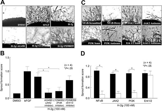
No single assay represents angiogenesis fully. For instance, EC chemotaxis represents one aspect of the process of newly forming blood vessels. Here we used a rat aortic ring in vitro culture assay25 to examine H-2g–induced angiogenesis in vitro. Using the criteria described in “Materials and methods,” we show that H-2g stimulated microvessel sprouting from the adventitia of cultured rat aortic rings in a dose-dependent manner. Consistently, when we incorporated the inhibitors (50 μM AG490, 20 μM LY294002, and the antisense ODNs directed against JAK2 and PI3K [10 μg/mL]), microvessel sprouting was also inhibited. Incorporated decoy NFκB (10 μg/mL) ODN also inhibited the sprouting, whereas control scrambled NFκB ODN had no effect. Figure 5A depicts photographs of the “microvessel sprouting” and the effect of chemical inhibitors, Figure 5B shows semiquantitative data collected by using the criteria described in “Materials and methods.” Figure 5C shows the microvessel sprouting, and the effect of antisense ODNs directed against JAK2, PI3K, and Erk1/2. Figure 5D shows semiquantitative vessel formation data. Thus, using this model, H-2g induces angiogenesis through JAK2, PI3K, and NFκB pathways, but not through the MAPK pathway.
H-2g–induced rat aortic ring angiogenesis in vitro is inhibited by inhibitors of JAK2 and PI3 kinase. Rat aortic rings were placed on a Matrigel drop in 48-well plates and covered with an additional drop of Matrigel. Serum-free EGM medium (400 μL) with various concentrations of H-2g was added to each well. bFGF (10 nM) was used as a positive control. (A) H-2g (100 nM) induced rat aortic ring sprouting.AG490, a JAK2 inhibitor, and LY294002, a PI3K inhibitor, inhibited this sprouting. (B) Quantitatively, JAK2 blocking resulted in 94% inhibition of EC sprouting, whereas PI3K inhibition resulted in 82% inhibition. (C) We incorporated antisense and sense ODNs (10 μg/mL) in the Matrigel, and in the media throughout the experiments. Consistent with the chemical inhibitor effect, JAK2 and PI3K antisense ODNs inhibited microvessel sprouting, as did NFκB decoy ODNs. (D) Quantitatively, NFκB decoy ODN transfection resulted in 87% inhibition, compared with scrambled NFκB ODN; JAK2 antisense ODN transfection resulted in 78% inhibition compared with JAK2 sense ODN; and PI3K antisense ODN transfection resulted in 76% inhibition compared with PI3K sense ODN (decoy or antisense ODN transfection, □; scrambled or sense ODN transfection, ▪). Results represent 4 independent experiments. Results are expressed as the mean number of sprouting plus or minus SEM from 4 independent experiments. *Represents a significant difference (P < .05) between the respective groups. The pictures were taken with a Nikon Coolpix 4500 camera (Nikon, Tokyo, Japan) and an Olympus CK40 microscope (Olympus, Melville, NY), objective × 40/0.55 numerical aperture. Photoshop 5.5 (Adobe, San Jose, CA) was then used to process the images.
H-2g–induced rat aortic ring angiogenesis in vitro is inhibited by inhibitors of JAK2 and PI3 kinase. Rat aortic rings were placed on a Matrigel drop in 48-well plates and covered with an additional drop of Matrigel. Serum-free EGM medium (400 μL) with various concentrations of H-2g was added to each well. bFGF (10 nM) was used as a positive control. (A) H-2g (100 nM) induced rat aortic ring sprouting.AG490, a JAK2 inhibitor, and LY294002, a PI3K inhibitor, inhibited this sprouting. (B) Quantitatively, JAK2 blocking resulted in 94% inhibition of EC sprouting, whereas PI3K inhibition resulted in 82% inhibition. (C) We incorporated antisense and sense ODNs (10 μg/mL) in the Matrigel, and in the media throughout the experiments. Consistent with the chemical inhibitor effect, JAK2 and PI3K antisense ODNs inhibited microvessel sprouting, as did NFκB decoy ODNs. (D) Quantitatively, NFκB decoy ODN transfection resulted in 87% inhibition, compared with scrambled NFκB ODN; JAK2 antisense ODN transfection resulted in 78% inhibition compared with JAK2 sense ODN; and PI3K antisense ODN transfection resulted in 76% inhibition compared with PI3K sense ODN (decoy or antisense ODN transfection, □; scrambled or sense ODN transfection, ▪). Results represent 4 independent experiments. Results are expressed as the mean number of sprouting plus or minus SEM from 4 independent experiments. *Represents a significant difference (P < .05) between the respective groups. The pictures were taken with a Nikon Coolpix 4500 camera (Nikon, Tokyo, Japan) and an Olympus CK40 microscope (Olympus, Melville, NY), objective × 40/0.55 numerical aperture. Photoshop 5.5 (Adobe, San Jose, CA) was then used to process the images.
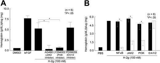
To confirm that H-2g induces angiogenesis in vivo via the JAK2, PI3K, and NFκB pathways, we used a Matrigel plug angiogenesis assay. H-2g plus AG490, LY294002, or PD98059 was incorporated in the Matrigel and the mixture injected into mice. Similar to the in vitro aortic ring angiogenesis assay, AG490 and LY294002 significantly inhibited H-2g–mediated blood vessel formation (95% and 91%, respectively, P < .05) in the Matrigel in vivo assay (Figure 6A). However, the MAPK inhibitor PD98059 did not show significant inhibition of H-2g–mediated blood vessel formation. We next incorporated 10 μg/mL NFκB decoy ODN and JAK2, PI3K, and Erk1/2 antisense ODNs in the Matrigel along with H-2g prior to injection into mice. NFκB decoy ODN, as well as JAK2 and PI3K antisense ODNs, inhibited H-2g–mediated blood vessel formation by 100%, 85%, and 86%, respectively (P < .05), confirming the results of the rat aortic ring studies. The Matrigel in vivo assay consistently showed that H-2g induces angiogenesis in vivo through JAK2, PI3K, and NFκB activation (Figure 6B).
H-2g–induced angiogenesis in vivo occurs mainly through NFκB and also through JAK2 and PI3K. Matrigel with H-2g and test substance was injected into C57/BL6 mice, and 7 to 10 days later the plugs were removed, homogenized, and hemoglobin was measured. (A) AG490 (50 μM) resulted in 100% inhibition of H-2g–induced hemoglobin production and LY294002 (20 μM) inhibited 92% of H-2g–induced hemoglobin production. (B) We incorporated decoy or antisense (□) and scrambled or sense (▪) ODNs (10 μg/mL) in the Matrigel together with 1 μM H-2g, and results were consistent with the chemical inhibitor effect. JAK2 and PI3K antisense ODNs inhibited blood vessel growth by 70% and 63%, respectively. Incorporating NFκB decoy ODN resulted in 90% inhibition of blood vessel growth compared with incorporation of NFκB scrambled ODN. Results are expressed as the mean of hemoglobin (g/dL) per plug (mg) plus or minus SEM from 6 mice. *Represents a significant difference (P < .05) between the respective groups.
H-2g–induced angiogenesis in vivo occurs mainly through NFκB and also through JAK2 and PI3K. Matrigel with H-2g and test substance was injected into C57/BL6 mice, and 7 to 10 days later the plugs were removed, homogenized, and hemoglobin was measured. (A) AG490 (50 μM) resulted in 100% inhibition of H-2g–induced hemoglobin production and LY294002 (20 μM) inhibited 92% of H-2g–induced hemoglobin production. (B) We incorporated decoy or antisense (□) and scrambled or sense (▪) ODNs (10 μg/mL) in the Matrigel together with 1 μM H-2g, and results were consistent with the chemical inhibitor effect. JAK2 and PI3K antisense ODNs inhibited blood vessel growth by 70% and 63%, respectively. Incorporating NFκB decoy ODN resulted in 90% inhibition of blood vessel growth compared with incorporation of NFκB scrambled ODN. Results are expressed as the mean of hemoglobin (g/dL) per plug (mg) plus or minus SEM from 6 mice. *Represents a significant difference (P < .05) between the respective groups.
Discussion
Ley/H as an antigen was originally found to be highly expressed in precursor blood cells and tumor cells. “Biologic missiles” for cancers have been proposed using this antigen as a target.26 However, the function of the antigen is poorly understood. We have shown that this antigen might not just act as a marker, but also function as an angiogenic factor.
Angiogenesis is critical in the development of tumors27 and inflammatory disorders such as RA.28,29 Angiogenesis involvement in RA progression and development is thought to be driven in part by cytokines.30,31 Glycoconjugates are involved in many aspects of cell biology, such as immunologic recognition, ligand-receptor binding, cell trafficking, and cell-cell interactions. Nguyen and coworkers32 identified a role for sialyl Lex in capillary morphogenesis. We have shown that soluble E-selectin can act as an angiogenic mediator via interacting with its EC ligand sialyl Lex.6 Due to the angiogenic properties of soluble E-selectin and also based on our previous work, we hypothesized that the 4A11 antigen, which contains Ley/H released by activated ECs, induces angiogenesis. Here, we show that H-2g, a glucose analog of soluble Ley/H antigen, elicits an angiogenic response through inducing potent angiogenic factors such as VEGF and bFGF. Furthermore, Ley has been found to be highly expressed on many types of cancer cells, such as breast, colon, ovarian, and lung cancer.33-36 As we have shown here, H-2g is involved in angiogenesis, perhaps suggesting this mechanism as a method augmenting cancer growth and metastasis. We have shown the 4A11 antigen to be up-regulated in RA ST compared with normal ST.8 Both RA and tumor growth require new blood vessel formation in their pathogenesis. In this study, our in vitro and in vivo data elucidate the signaling mechanisms of H-2g in angiogenesis.
We have examined the roles of different signaling pathways by which H-2g mediates angiogenesis, and our results show that H-2g mediates HMVEC chemotaxis and angiogenesis predominantly through the JAK2 and PI3K pathways. NFκB also plays a key role in the H-2g–induced signaling pathway. It is well established that the transcription factor NFκB is essential for VEGF and other angiogenic gene expression. For example, VEGF promoter activity is significantly decreased in cancer cells transfected with mutated IκB, which blocks NFκB activation.37,38 Based on results obtained from the in vitro IKK activity assay, IκB phosphorylation was inhibited by the JAK2 inhibitor AG490, and the PI3K inhibitor LY294002. Antisense ODNs of JAK2 and the PI3K were also able to inhibit IκB phosphorylated by IKK.
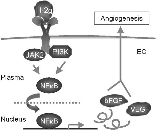
Signaling studies showed that H-2g, through JAK2, PI3K, and NFκB, induced angiogenic VEGF and bFGF expression. Next, we determined if angiogenesis was induced through these pathways. We employed both in vitro and in vivo assays to study angiogenesis. H-2g induced an 8.2-fold increase in EC tube formation compared with the vehicle control. AG490 and LY294002 significantly inhibited H-2g–mediated EC tube formation by 94% and 82%, respectively. Similarly, HMVECs transfected with JAK2, PI3K antisense ODNs, or decoy NFκB ODN showed significant inhibition of H-2g–mediated tube formation compared with HMVECs transfected with the JAK2 or PI3K sense ODNs or scrambled NFκB ODN. However, HMVECs transfected with an ERK1/2 antisense ODN or treated with an ERK1/2 inhibitor (PD98059), failed to show significant inhibition of tube formation. A model for signaling cascades of H-2g–induced endothelial angiogenesis is shown in Figure 7.
Proposed mechanism of action of H-2g signaling in endothelial cells (ECs).
Proposed mechanism of action of H-2g signaling in endothelial cells (ECs).
The penultimate step in the biosynthesis of human ABO blood group oligosaccharide antigens is catalyzed by α-(1,2)-fucosyltransferase(s), whose expression is determined by the H and secretor (SE) blood group loci (known as FUT1 and FUT2, respectively). These enzymes construct Fucα1 → 2Galβ-linkages (H determinants), which are essential precursors to the A and B antigens. In 1995, John Lowe's group39 detected that erythrocytes from rare Bombay blood group phenotype individuals are deficient in H determinants, and consequently A and B, the likely result of homozygosity for null alleles at the H locus. Since then, the Ley/H antigen has been proposed to play significant roles in many pathologic functions. To begin to investigate the regulation of Ley/H antigen in animal models, in 1997, the same group cloned FUT1 and FUT2 from rodent genomic DNA.40 Based on their fundamental work, we then are able to use rat aortic vessels and mice as our study models.
In the Matrigel in vivo assay, H-2g also induced a significant (9.5-fold) increase in blood vessel formation, which was inhibited by treatment with AG490 (95%, P < .05) or LY294002 (91%, P < .05). Similar results were obtained with the Matrigel in vitro tube formation assay, in which treatment with PD98059 (MAPK inhibitor) failed to significantly inhibit H-2g–mediated blood vessel formation in Matrigel plugs in vivo. These results suggest that H-2g–mediated blood vessel formation both in vitro and in vivo is predominantly mediated through the JAK2 and PI3K pathways, and that NFκB is also an important component of these pathways. Our study also suggests a potentially novel method to inhibit H-2g–mediated angiogenesis via JAK2 and PI3K pathways.
Previously, we have shown that soluble Ley/H or its analog, H-2g, induced ICAM-1 expression and myeloid-EC adhesion through JAK2-Stat3, Erk1/2, and PI3K.41 Our current data show that H-2g induces EC chemotaxis through specific signaling pathways, leading to EC chemotaxis and also angiogenesis. In contrast to the signaling pathways used by H-2g to induce ICAM-1 expression, H-2g induces angiogenesis via JAK2 and PI3K, but not MAPK pathways, indicating that the same molecule may employ different pathways to mediate endothelial-leukocyte adhesion versus angiogenesis. Through in vitro and in vivo assays, we found that JAK2 and PI3K are predominantly involved in angiogenesis. Both JAK2 and PI3K activated NFκB. NFκB is a key transcription factor involved in the control of inflammation through regulating the expression of several cytokines and adhesion molecules. For example, IL-1α, IL-1β, IL-6, IL-8, ICAM-1, VCAM-1, VEGF, and bFGF all contain an NFκB-responsive element in their promoter region.42,43 Tomita and coworkers17 reported a novel therapeutic strategy to suppress the inflammatory process in synovial cells by transfecting decoy ODN to block the binding of NFκB to its promoter sequence of target genes, thereby inhibiting the coordinated transactivation of genes of key cytokines and adhesion molecules involved in RA-associated angiogenesis.44 Their results suggest that decoy NFκB ODNs significantly inhibit synovial cell proliferation compared with that of scrambled decoy ODNs. Using decoy NFκB as a gene therapy reagent may be a very promising approach for treating angiogenesis-related diseases.17
On the other hand, angiogenesis is becoming an attractive therapeutic approach for cardiovascular diseases because the recently discovered families of regulators, both stimulators and inhibitors, have facilitated its practical application.45 It is now thought that a shift in the balance between stimulators and inhibitors in favor of the former triggers the angiogenic switch in endothelial cells, producing a distinct survival advantage.46 Here we report that H-2g activates EC NFκB mainly through JAK2 and PI3K. These signaling pathways, when triggered by H-2g, cause EC chemotaxis and angiogenesis in vitro and in vivo. These findings may suggest potentially new therapies via inhibiting signaling intermediates in angiogenesis-dependent diseases.
Prepublished online as Blood First Edition Paper, October 21, 2004; DOI 10.1182/blood-2004-08-3140.
Supported by the Gallagher Professorship for Arthritis Research, the William D. Robinson and Frederick G. L. Huetwell Endowed Professorship, and funds from the Veterans Affairs Research Service, National Institutes of Health, grants AI-40987, HL-58695, and AR-048267.
The publication costs of this article were defrayed in part by page charge payment. Therefore, and solely to indicate this fact, this article is hereby marked “advertisement” in accordance with 18 U.S.C. section 1734.


![Figure 4. H-2g induces IκB phosphorylation by IKKβ labeled with γ–[32P] ATP, which in turn causes NFκB activation in HMVECs. The effect is inhibited by JAK2 and PI3 kinase inhibitors, and antisense ODNs directed against JAK2 and PI3K. HMVECs were stimulated with H-2g (100 nM) for 15 minutes. (A) In the inhibition study, HMVECs were pretreated with AG490 (50 μM), LY294002 (20 μM), or PD98059 (20 μM) for 2 hours before stimulation with H-2g, and inhibitors were also present during the stimulation with H-2g. AG490 and LY294002 inhibited H-2g–induced EC NFκB while PD98059 had no effect. (B) Transient transfection of HMVECs with sense (▪) and antisense (□) ODNs to JAK2, PI3K, and Erk1/2 confirmed the results obtained using chemical inhibitors: JAK2 and PI3K antisense ODNs inhibited H-2g–induced EC IKKβ activation. Nontransfected cells were used as a control. Results are representative of 4 independent experiments. * indicates a significant difference (P < .05) between the respective groups.](https://ash.silverchair-cdn.com/ash/content_public/journal/blood/105/6/10.1182_blood-2004-08-3140/6/m_zh80060575840004.jpeg?Expires=1763600661&Signature=SqKL5V3bzV8AY9gplZkpvX8fNBpKknoGpekHHEaBriqlYJ63y84sDKBj~HseFofzx9EZ0ljv4FA4dU5I-Orba~eD8m1Antt6bjGYzo0CU46LEB~h93mcz-P5W1nZJiwwNl8k6YXynkfreB7KRaBcFQ6xnop5LCsTjhbVThsKdkFEDgjXkLqhgi7kZhiSljo8BkPdI053BGeaTrTieQwDurK56oHoOcDHpNkweLU0zQ0ncrPAst1LCEKRKLXme75PN1uubZnBbky30EL53pfMvRV7i2Ea59qX60UEdy5fjXCDE~Da2QMXtcOUV8sZugcuQn4IjnAC3JzHNF9JO~uc0Q__&Key-Pair-Id=APKAIE5G5CRDK6RD3PGA)


