Abstract
Background
The myocardial T2* technique has been validated as a reproducible non-invasive measurement of myocardial iron load and is now widely used for measurement of myocardial iron in iron overload diseases such as thalassaemia. The reduction in myocardial T2* seen in iron overload conditions is substantially greater than is seen in any other clinical circumstance, but there has been no direct comparison of myocardial T2* in normals and other conditions such as increasing age, myocardial infarction or impairment in left ventricular function. We aimed therefore to compare the findings in patients affected by these conditions with normals.
Method
A total of 38 patients in total were scanned using the myocardial T2* technique. Fifteen patients had normal hearts, 18 had impaired LV function and 6 had chronic myocardial infarction affecting the anteroseptal wall, where myocardial T2* measurements are normally made.
Results
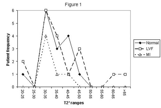
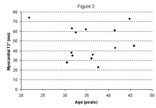
The mean myocardial T2* in normals was 36.0 +/− 6.4 ms, yielding a lower limit of normal of 23ms. In patients with impaired LV function, the mean myocardial T2* was 39.0 +/− 11.7ms (p= 0.37 vs normals). In patients with anteroseptal myocardial infarction, the mean myocardial T2* was 34.7ms +/− 3.9ms (p= 0.64 vs normals). The frequency distribution of the myocardial T2* values are shown in figure 1. These approximate to normal, and are very similar in distribution. In addition, the age distribution of myocardial T2* in the 15 normals is shown in figure 2. There was no significant relation between myocardial T2* and age (r2 = 0.066, p=0.82).
Conclusion
There is no significant reduction in myocardial T2* associated with fibrosis from chronic myocardial infarction, impairment of left ventricular function, or increasing age. This suggests that structural changes associated with remodelling, infarction and fibrosis, and ageing do not have significant effects on the absolute measure of myocardial T2*, and in particular do not cause a reduction below 20ms as is seen in myocardial overload conditions. Thus these date suggest that myocardial T2* is robust to these structural alterations, and that myocardial iron overload can be ascertained from reduced myocardial T2* values, in a similar manner to that which can be achieved in normals.
Author notes
Corresponding author


