Abstract
Background
The T2* technique has been validated as a non-invasive measurement of myocardial iron load. However, calibration of the technique against myocardial tissue remains to be determined. Since such calibration is easiest to perform using post-mortem tissue, it is useful to know the effects of storage of the tissue in formalin, and in particular whether iron leaks from the heart during storage, and whether fixation alters the myocardial T2* significantly.
Method
A heart was donated after death of a patient with sideroblastic anemia who had been poorly compliant with deferoxamine treatment and died in heart failure. The pre-mortem myocardial T2* was 5.2 ms indicating severe myocardial iron loading. The majority of the heart was assigned to a calibration study of myocardial iron, but 2 samples from the heart were available for this study.
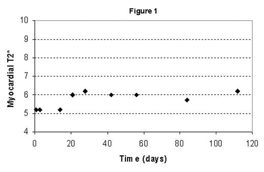
Sample 1 was an apical short-axis slice of the left ventricle. This was preserved in formalin for 3 days prior to measurement of the initial post-mortem myocardial T2* which was 5.2 ms (Siemens Sonata 1.5T MR scanner). The T2* of the slice was then measured weekly for 4 weeks; then 2 weekly for 8 weeks; and finally 4 weekly.
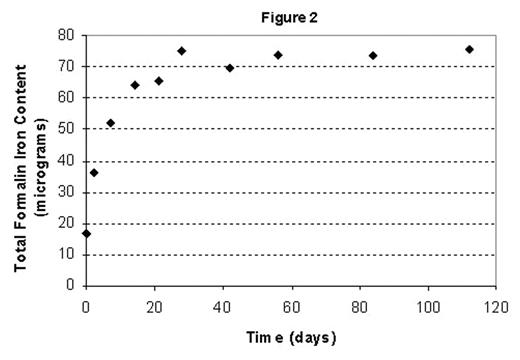
Sample 2 was a 7.35 g piece of apical myocardium which was stored in 280ml formaldehyde. A 1ml sample of formaldehyde solution was taken for iron analysis from sample 2 initially and at 2 days, and then weekly for 4 weeks; two-weekly for 6 weeks; and then 4-weekly for 12 weeks. The iron concentration in the formalin was measured using Inductive Coupled Plasma with Mass Spectometry (ICPMS).
Results
The results from sample 1 are shown as myocardial T2* against days elapsed. The T2* rose from a pre-mortem and post-mortem (equivalent values) of 5.2ms to a mean of 6ms over the first 20 days, and after this was stable (figure 1).
The results from sample 2, show an increase from 16.6 microgram to 74.9 microgram over the initial 3–4 weeks after which little significant additional accumulation occurred (figure 2). Estimation of the total iron content of the myocardial sample (prior to destructive iron measurement at the end of the study) suggested that the total iron leaked into the formalin was 0.44% of the iron in the myocardial sample.
Conclusion
There was a small increase in myocardial T2* over the first 20 days of formalin fixation, but little significant change after that. This was associated with a loss of approximately 75 micrograms of iron into the formalin fixation solution, which was estimated as only 0.44% of the iron in the myocardial sample. This suggests that formalin fixation has a small but measurable effect on myocardial T2* over time, and this is not explained by loss of iron into the fixation fluid.
Author notes
Corresponding author


