Kaposi sarcoma–associated herpesvirus (KSHV) latency-associated nuclear antigen-1 (LANA1) is essential for the maintenance and segregation of viral episomes in KSHV latently infected B cells. We report development of intracellular, rabbit-derived antibodies generated by phage display technology, which bind to N-terminal LANA1 epitopes and neutralize the chromosome-binding activity of LANA1. Although these cloned single-chain variable fragments (scFvs) show relatively low binding affinities for the LANA1 viral antigen in in vitro assays, they nonetheless outcompete KSHV-seropositive human sera for LANA1 epitope binding. In heterologous cells, intracellular intrabody expression inhibits LANA1-dependent plasmid maintenance of both an artificial plasmid containing KSHV LANA1 binding sequences and a bacterial artificial chromosome containing the entire KSHV genome. In KSHV naturally infected primary effusion lymphoma cells, intracellular intrabody expression causes a reduction or loss of the typical LANA1 punctate, nuclear pattern. This morphologically apparent LANA1 dispersion correlates to loss of viral episome by molecular analysis. These data suggest a novel approach to antiherpes viral therapy and confirm LANA1 is critical target for neutralization of KSHV viral latency.
Introduction
Kaposi sarcoma–associated herpesvirus (KSHV or human herpesvirus 8 [HHV-8]) is a double-stranded DNA gammaherpesvirus associated with KS, primary effusion lymphoma (PEL), and subsets of multicentric Castleman disease (MCD), an aggressive lymphoproliferative disorder.1-3 KSHV predominantly infects B lymphocytes and endothelial cells and persists as a closed, circular episome expressing few viral genes during latent replication.4 While KS rates among AIDS patients have dramatically decreased with the use of highly effective antiretroviral therapy,5 therapeutic options for MCD or PEL are limited, and these neoplastic diseases have high mortality rates.1,6,7 Persistent, latent KSHV infection is not amenable to common antiviral drugs targeting the lytic-phase herpesvirus-encoded DNA polymerase.8
One latent protein, the latency-associated nuclear antigen-1 (LANA1) encoded by open reading frame 73 (ORF73),9,10 is consistently expressed in all forms of KS-associated malignancies.11-14 LANA1 tethers the viral episome to cellular chromosomes during mitosis, allowing equal segregation of virus genome to daughter cells.12,14-17 LANA1 is a large nuclear protein with an N-terminal proline-rich domain, an internal glutamine-rich domain, and an acidic repeat domain followed by a leucine zipper motif.18,19 LANA1 binds through a C-terminal region to 2 adjacent sites within each KSHV terminal repeat (TR), which acts as the latent origin of replication for the viral episome.20,21 LANA1 N-terminal amino acids 5 to 22 form a chromosome binding sequence (CBS) that interacts with chromatin proteins and is sufficient to mediate a specific interaction of LANA1 with chromosomes during mitosis.16,22,23 A second region in the C-terminus (from amino acids 1129 to 1143) has been described as essential for heterochromatin association but is not sufficient alone to mediate this interaction.24,25 Thus, by binding the episome through its C-terminal domain and chromosome-associated proteins through its N-terminal domain, LANA1 acts as a physical bridge between the episome and cellular chromosomes during mitosis. In vitro assays for KSHV plasmid maintenance in heterologous cell types have been developed in which LANA1 expression is used to maintain an artificial plasmid with antibiotic selection markers containing the TR replication origins.12 In addition to its episome maintenance functions, LANA1 inhibits retinoblastoma protein (pRB1),26 p53,27 and glycogen synthase kinase-3β (GSK-3β)28 and is likely to contribute to KSHV-related tumorigenesis. It is, in many respects, functionally analogous to the well-studied large T antigen of simian virus 40 (SV40).
Because of LANA1's central role in maintaining KSHV, it is an attractive target for therapeutics designed to eliminate latent KSHV infection. Unfortunately, siRNA against LANA1 mRNA fails to inhibit KSHV replication,29 presumably due to the stability of LANA1 protein (J. Wiezorek, R. Sarid, Y. Chang, unpublished observation), which ensures that sufficient protein is available for viral replication under these conditions.
We have taken an alternative approach to neutralize LANA1 function through intrabody expression. Intrabodies (antibodies for intracellular applications) are single-chain variable region antibody fragments that can be expressed through transfection or by using a viral vector.30,31 The potential of intrabodies to neutralize the function of intracellular and extracellular proteins has been demonstrated in several applications.32-34 In this study, we generated 2 anti-LANA1 intrabodies by phage display technology specific for the N-terminal CBS domain, which efficiently inhibits KSHV maintenance in PEL cells. We show that loss of KSHV episome in these cells results in cell death without the induction of viral lytic reactivation. These findings suggest a novel therapeutic approach to inhibit KSHV latent viral replication.
Materials and methods
Expression and purification of LANA1 proteins
LANA1 was amplified by polymerase chain reaction (PCR) from pSG5 LANA1-FLAG 12 (a kind gift from Dr Kenneth Kaye, Harvard University, Cambridge, MA) into 2 separate fragments, LANA1000 (amino acids 1 to 337) and LANA2000 (amino acids 337 to 1162), due to the existence of a repeat region (region rich in proline and glycine) between 1000 bp and 2000 bp that complicated both amplification and purification of such a large protein. A polyhistidine tag (His6) was added at the N-terminal end of each fragment. Primers used were as follows: LANA11000: (sense [S]) 5′-ATGGCGCCCCCGGGAATGC-3′ and (antisense [AS]) 5′-CTAATGGTGATGGTGATGCTGCTCCTCATCTGTCTCCTGC-3′; LANA12000: (S) 5′-ATGCAGGAGACAGATGAGGAGGACGAGGAGG-3′ and (AS) 5′-CTAATGGTGATGGTGTGTCATTTCCTGTGGAGAGTCCCCAGGTGG-3′. These products were cloned into pBad-TOPO (Invitrogen, Carlsbad, CA) and expressed in bacteria by induction with arabinose. LANA1000 was purified by fast protein liquid chromatography (FPLC)–affinity chromatography using nickel–nitrilotriacetic acid (Ni-NTA) resin; and LANA2000 was purified by ionic exchange chromatography followed by molecular exclusion chromatography.
Phage display library
Two New Zealand white rabbits were immunized with purified LANA11000 and LANA12000 proteins. Spleens and bone marrows were harvested separately35 for total RNA using TRI Reagent (Molecular Research Center, Cincinnati, OH). First-strand cDNA was synthesized using Superscript (Invitrogen). Rabbit VL and VH sequences were combined with human CL and CH1 sequences by overlap PCR, as previously described.34 PCR fragments encoding a library of antibody fragments (Fabs) were digested with SfiI, purified, and cloned into pComb3X, 35 which contains an amber stop codon, His6 tag for purification, and hemagglutinin (HA) for detection.35 Fab fragments were expressed in fusion with protein III of M13 phage and screened against LANA1 proteins. A total of 88 anti-LANA1 Fabs were expressed, purified, and screened by enzyme-linked immunosorbent assay (ELISA) against LANA11000 and LANA12000 proteins. For the conversion of a LANA1-specific Fab into a single-chain variable domain fragment (scFv), specific oligonucleotide primers covering all known rabbit antibody family sequences were used to amplify VH and VL gene segments.35 The purified PCR products were assembled by overlapping PCR, and the resulting PCR products encoded different scFvs in which the VL region was linked with the VH region through an 18–amino acid peptide linker (SSGGGGSGGGGGGSSRSS). DNA fragments were gel purified, digested with SfiI, and cloned into pComb3X. LANA1 binding activity of the corresponding scFv was confirmed, and the genes encoding the scFv were cloned into pCDNA3.1/Zeo (+) (Invitrogen).35 ScFv proteins were purified using BD TALON Metal Affinity columns (Clontech, Palo Alto, CA) as previously described.33 Purified scFv proteins were analyzed by immunoblotting using standard methods. 4BL is an irrelevant scFv used as control.33
Mapping LANA1 epitopes
LANA1 epitopes were mapped using 171 cleavable, biotinylated (N-terminus SGSK) 17-mer peptides offset by 5 amino acids as previously described.36 Briefly, streptavidin ELISA plates (Roche Applied Science, Indianapolis, IN) were coated with 1.5 μg of each peptide. After blocking with 3% bovine serum albumin/phosphate-buffered saline (BSA/PBS), peptides 5 to 73 (amino acids 1 to 357) were incubated with purified scFv BM9 and BM10. Detection was performed by horseradish peroxidase (HRP)–conjugated anti-HA monoclonal antibody (high affinity 3F10; Roche Applied Science) at a dilution of 1:3000. Absorbance was measured on a spectrophotometer (Biophotometer; Eppendorf, Hamburg, Germany) at 405 nm.
Competition assay
Streptavidin ELISA plates (Roche Applied Science) coated overnight at 4°C with 1.8 μg of each peptide were blocked, washed, and then incubated for 2 hours at 37°C with sera that had been preincubated for 30 minutes with the different scFvs (0.1 μg). After blocking, unbound proteins were removed with wash buffer containing Tween 20, and an HRP-conjugated anti-HA monoclonal antibody (high affinity 3F10; RocheApplied Science) was added at a dilution of 1:3000 for 1 hour at 37°C. HRPactivity was detected by 2,2′-azino-bis(3-ethylbenzthiazdine-6)-sulforic acid (ABTS), and absorbance was measured on a spectrophotometer (Biophotometer; Eppendorf) at 405 nm.
Cell lines and transfection procedures
CM12 cells from a poorly defined but highly transfectable human epithelial cell line were cultured in Dulbecco modified Eagle medium (DMEM) (GibcoBRL, Carlsbad, CA) with 10% fetal calf serum (FCS) (GibcoBRL), 0.05 μg/mL penicillin/streptomycin, and 2 mM l-glutamine. BJAB cell lines stably transfected with KSHV Z6 and containing 8 copies of TR (FL-9/8TR), flag-tagged LANA1 (FL-9), and empty vector (pSG5) were kindly provided by Dr Kenneth Kaye.12 BC-1 (American Type Culture Collection [ATCC], Manassas, VA), BCBL1 (AIDS Research and Reference Reagent Program, Bethesda, MD), and BJAB were cultured in RPMI 1640 supplemented with 10% FCS, 1 mM sodium pyruvate, and 2 mM l-glutamine. All cell cultures were maintained at 37°C in 5% CO2. CM12 cells were transfected with KSHV–bacterial artificial chromosome-36 (KSHV-BAC36) plasmid (kind gift from S. J. Gao, San Antonio Cancer Institute, TX) either transiently or stably using Fugene 6 (Roche Applied Science) according to the manufacturer's instructions. Selection for stable KSHV-BAC36 was performed in 150 μg/mL hygromycin. BJAB (solution R, program T-01) and PEL cells (solution V, program T-01) were transfected (5 μg DNA and 1 × 10⁶ cells per transfection) using Amaxa nucleofection (Amaxa, Cologne, Germany). Transfection efficiency measured by fluorescence-activated cell sorter (FACS) analysis was 47% for BC-1 cells and 57.1% for BCBL-1 cells.
Immunofluorescence staining and antibodies
BJAB and KSHV-infected cells were placed on glass slides using a cytospin (Shandon, Pittsburgh, PA). Cells were fixed in 4% paraformaldehyde and permeabilized in 1% Triton X-100/PBS. Cells were incubated with the primary antibodies for LANA1 (CMA-810; 1:500 dilution) or His6 (Roche Applied Science; 1:1000 dilution) for 1 hour at room temperature in a humidified chamber, washed with 0.1% Tween 20/PBS, and incubated with species-appropriate fluorescein isothiocyanate (FITC)–(1:500 dilution) or rhodamine isothiocyanate (RITC)–(1:1000 dilution) conjugated secondary antibodies for 1 hour at room temperature. Slides were mounted with DAPI (4,6 diamidino-2-phenylindole) Vectashield (Vector Labs, Burlingame, CA) to stain nuclei, and cells were visualized using an Olympus AX70 epifluorescence microscope (Olympus, Melville, NY) with triple color filters, and a 20 × air objective. Numeric aperture varied automatically according to the light emitted. Images were captured with a Zeiss Axiocam camera (Zeiss, Göttingen, Germany). Cells were arrested in metaphase with 0.1 μg/mL colcemid (Irvine Scientific, Santa Ana, CA) for 24 hours and resuspended in 0.075 M KCl for 12 minutes to swell nuclei. Immunofluorescence assay (IFA) was performed as described.
Outgrowth assays
For selection of pSG5-LANA1 48 hours after transfection FL-9/8TR and FL-9 cell cultures were grown in medium containing 200 U/mL hygromycin (Invitrogen) to select for LANA1. For selection of Z6 cosmid, cell cultures were grown in medium containing 400 μg/mL neomycin (G418) (Invitrogen), and for selection of pcDNA3.1 Zeo (+)–derived constructs, cell cultures were grown in medium containing 200 μg/mL zeocin (Invitrogen) to select for scFv. Cell viability was determined using the cell proliferation reagent WST-1 (Roche Applied Science) according to the manufacturer's instructions. The activity of mitochondrial dehydrogenases, an indicator of cell viability, was measured in 10 000 cells by the conversion of the tetrazolium salt WST-1 to the red dye formazan. Formazan accumulation is detected by increased absorbance at 440 nm.
Gardella gel analysis
Gardella experiments were performed as described elsewhere.37 Briefly, cells were separated by Ficoll and ressuspended in 15% Ficoll/0.01% bromophenol blue/TBE (Tris–borate–ethylenediaminetetraacetic acid) buffer, loaded onto a vertical 0.8% agarose gel, and overlaid with lysis buffer (5% Ficoll, 1% sodium dodecyl sulfate [SDS], 1 mg/mL pronase, and 0.05% xylene cyanol green) After electrophoresis, DNA was transferred to nitrocellulose membranes by Southern blotting and detected with KSHV-TR 32P-labeled probes (Redi-Prime; Amersham Biosciences, Piscataway, NJ).
Transcription/translation in vitro and GST pulldown
In vitro transcription and translation (TNT System; Promega, San Luis Obispo, CA) of pCDNA3.1/scFv was performed according to the manufacturer's instructions. Radiolabeled scFv proteins were incubated overnight at 4°C with 3μg purified glutathione S-transferase (GST)–LANA1. Glutathione Sepharose beads (Amersham Biosciences) were washed with PBS/0.1% Nonidet P-40 (NP-40)/60 mM EDTA (ethylenediaminetetraacetic acid) together with antiproteases and added to labeled scFv/LANA1-GST mix, incubated at 4°C, and washed with PBS. Samples were analyzed by 12% SDS–polyacrylamide gel electrophoresis (PAGE) and subjected to autoradiography.
Results
Selection of LANA1-specific antibody fragments
A chimeric rabbit/human Fab library was generated from cDNA from bone marrow and spleen RNA from 2 immunized rabbits having a strong antibody response to a mixture of LANA1 protein fragments (amino acids 1 to 337 and 337 to 1162). An antibody library of 1 × 10⁸ independent clones was constructed and selected. Several Fab clones binding LANA1 protein fragments were then isolated and converted to single-chain variable fragments (scFvs) by PCR. An 18–amino acid linker fragment (SSGGGGSGGGGGGSSRSS) was cloned between the VL and VH fragments, connecting the C-terminal end of the VL fragment to the N-terminal end of the VH fragment. This linker allows the intrabody to be expressed from a single cloned cDNA and provides enhanced intracellular stability of the expressed antibody.33 Figure 1A summarizes the relative binding for the Fab clones prior to scFv construction. From the selected clones, we used 2 Fabs, BM9 and BM10, with highest relative binding to convert into scFvs, for further characterization and expression.
scFv specificity for LANA1
To test specificity of constructed scFvs, cell extracts from BJAB cell lines stably transfected with LANA1 and Z6 cosmid (containing 8 copies of the KSHV-TR)12 were separated by 6% SDS-PAGE and immunoblotted with purified anti-LANA1 scFvs (Figure 1B). BM9, but not BM10, recognized the major LANA1 doublet band of approximately 220 kDa in cell extracts from FL-9 and FL-9/8TR, BJAB cell lines constitutively expressing LANA1 protein. No bands were detected using a transfected cell line with empty control pSG5 vector. Similarly, immunoprecipitation of [35S]methionine-labeled BM9 and BM10 with GST-LANA1 fusion protein demonstrated interaction between LANA1 and BM9 but not BM10 (Figure 1C). Despite this evidence for in vitro BM9 scFv binding to LANA1, direct immunoprecipitation of LANA1 using the specific monoclonal antibody CMA-810, followed by immunoblot detection with either BM9 or BM10, failed to detect direct interaction, suggesting a weak recognition of the epitope by the scFv. To map anti-LANA1 scFv epitopes, LANA1 peptide screening using 171 biotinylated peptides was performed by ELISA using the purified BM9 and BM10 scFvs, as previously described, to determine dominant epitopes recognized by sera from KSHV-infected patients.36 BM9 and BM10 (Table 1; Figure 1E) bound to peptides encompassing the N-terminal sequence motif (amino acids 5 to 22) containing the chromosome binding site involved in tethering LANA1 to cellular chromosomes during mitosis22 as well as to a putative nuclear localization signal (NLS) of LANA1 (amino acids 24 to 30).16 Both scFvs also bound to peptide 43, which has not been described as relevant to LANA1 CBS or NLS functions. To determine if BM9 reacts to epitopes also recognized during natural KSHV infection, we competed the scFv with KS-positive human serum for binding to specific LANA1 peptides. In this assay, biotinylated peptides bound to streptavidin plates were incubated with BM9 with or without KSHV-positive serum overnight. As seen in Figure 1D, increasing optical absorbance values at higher dilutions of human serum indicate that native anti-LANA1 human antibodies compete with LANA1-specific BM9 for peptides encompassing N-terminal protein motif. These results show that single-chain antibodies that can bind LANA1 protein at its N-terminal end are capable of competing with anti-LANA1 sera for the antigen. Thus, LANA1 protein is specifically recognized in vitro by BM9. Taken together, these data show weak but specific LANA1 interaction with BM9. Despite this apparent low binding, the effectiveness of intrabodies expressed in vivo is highly dependent on antibody stability, which allows relatively low-binding antibodies to successfully neutralize intracellular functions.38 For this reason, neutralization of LANA1 function may be a more sensitive indicator of anti-LANA1 scFv specificity, and thus both BM9 and BM10 antibodies were examined in functional studies.
BM9 and BM10 scFvs bind to the N-terminal CBS domain of LANA1 with low but specific binding. (A) Representative enzyme-linked immunoassay activities for 10 Fab clones isolated by phage display from LANA1-immunized rabbits. Clones BM9 and BM10 having highest immunoreactivity were subcloned into single-chain Fv (scFv) and examined for LANA1 binding. (B) HA epitope-tagged BM9, but not BM10, scFv binds LANA1 on direct immunoblotting. FL-9/8TR and FL-9 are BJAB cell lines constitutively expressing LANA-1 protein, and pSG5 is a BJAB cell line transfected with empty vector alone. (C) In vitro binding of BM9 scFv to a GST-LANA1 fusion protein. In this assay, [35S]methionine-labeled BM9 scFv but not BM10 scFv directly interacted with GST-LANA1 fusion protein isolated with glutathione-linked beads. (D) Human serum competition assay: BM9 was mixed with the indicated dilutions of KSHV LANA1-seropositive human serum, and ELISAs were performed using plates coated with peptides P7, P8, P9, P19, and P43. BM9 competes with hyperimmune sera for peptide binding with P7, P8, and P43 but not with P19. Each point represents the value of binding minus nonspecific binding of scFv or human serum to uncoated wells blocked with 3% BSA/PBS. (E) Schematic representation of LANA1 and binding epitopes of BM9, BM10, and other cellular proteins that bind to its N-terminal domain.
BM9 and BM10 scFvs bind to the N-terminal CBS domain of LANA1 with low but specific binding. (A) Representative enzyme-linked immunoassay activities for 10 Fab clones isolated by phage display from LANA1-immunized rabbits. Clones BM9 and BM10 having highest immunoreactivity were subcloned into single-chain Fv (scFv) and examined for LANA1 binding. (B) HA epitope-tagged BM9, but not BM10, scFv binds LANA1 on direct immunoblotting. FL-9/8TR and FL-9 are BJAB cell lines constitutively expressing LANA-1 protein, and pSG5 is a BJAB cell line transfected with empty vector alone. (C) In vitro binding of BM9 scFv to a GST-LANA1 fusion protein. In this assay, [35S]methionine-labeled BM9 scFv but not BM10 scFv directly interacted with GST-LANA1 fusion protein isolated with glutathione-linked beads. (D) Human serum competition assay: BM9 was mixed with the indicated dilutions of KSHV LANA1-seropositive human serum, and ELISAs were performed using plates coated with peptides P7, P8, P9, P19, and P43. BM9 competes with hyperimmune sera for peptide binding with P7, P8, and P43 but not with P19. Each point represents the value of binding minus nonspecific binding of scFv or human serum to uncoated wells blocked with 3% BSA/PBS. (E) Schematic representation of LANA1 and binding epitopes of BM9, BM10, and other cellular proteins that bind to its N-terminal domain.
Functional analysis of intrabodies against LANA1
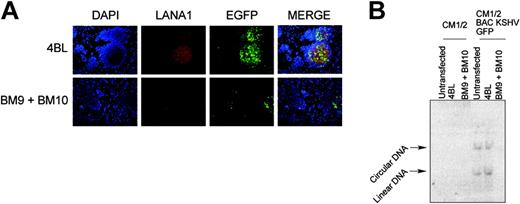
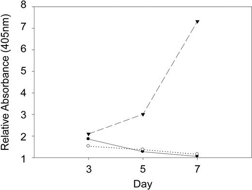
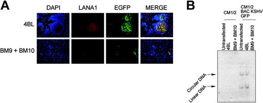
Functional studies were performed in cell lines stably transfected with LANA1 and Z6 (FL9/8TR) and in CM12 cells stably transfected with the KSHV-BAC36 clone.39 In FL-9/8TR cells, a hygromycin resistance marker is encoded on the pSG5 LANA1 plasmid and a neomycin resistance marker on the Z6 cosmid. The TR-multimer plasmid (KSHV Z6 cosmid) is maintained through transexpression of LANA1 protein. Expression of BM9 and BM10 results in inhibition of LANA1 function and consequent loss of the TR plasmid. When either BM9 and/or BM10 are transfected into FL-9/8TR, susceptibility to neomycin cell killing occurs within 5 days of scFv expression, whereas cells transfected with empty vector (pcDNA 3.1) continue to replicate, reflecting the loss and maintenance of the TR plasmid, respectively (Figure 2A). Cells expressing either BM9 and/or BM10 have shown loss of TR plasmid by day 14 of culture compared with cells expressing an irrelevant anti-HIV scFv, 4BL (data not shown). These results were confirmed by cotransfection of the anti-LANA1 intrabodies with a cloned KSHV-BAC36 expressing enhanced green fluorescent protein (EGFP). When BM9 or BM10 are transfected together with KSHV-BAC36 DNA into CM12 cells, rapid loss of EGFP fluorescence occurs within 48 hours (Figure 3A) with corresponding loss of the episomal KSHV-BAC36 as shown by Gardella gel electrophoresis (Figure 3B).
Expression of BM9 or BM10 inhibits KSHV plasmid maintenance in a FL-9 heterologous cell assay. Outgrowth assays of FL-9/8TR BJAB cells stably expressing LANA1 under a hygromycin resistance marker and together with a KSHV-TR plasmid under a neomycin resistance marker show that BM9 (•) and BM10 (zeocyn selection) (○), but not pcDNA3.1 vector (▴), induce inhibition of LANA1 and consequent loss of TR plasmid. Cells were transfected with the intrabodies or empty vector and placed under hygromycin, neomycin (LANA1-TR plasmid), and zeocyn (intrabody/vector) selection on day 0 (48 hours after transfection). Cell counts were performed in 24 replicas using the cell proliferation reagent WST-1 and measured by spectrophotometer at 405 nm.
Expression of BM9 or BM10 inhibits KSHV plasmid maintenance in a FL-9 heterologous cell assay. Outgrowth assays of FL-9/8TR BJAB cells stably expressing LANA1 under a hygromycin resistance marker and together with a KSHV-TR plasmid under a neomycin resistance marker show that BM9 (•) and BM10 (zeocyn selection) (○), but not pcDNA3.1 vector (▴), induce inhibition of LANA1 and consequent loss of TR plasmid. Cells were transfected with the intrabodies or empty vector and placed under hygromycin, neomycin (LANA1-TR plasmid), and zeocyn (intrabody/vector) selection on day 0 (48 hours after transfection). Cell counts were performed in 24 replicas using the cell proliferation reagent WST-1 and measured by spectrophotometer at 405 nm.
Intrabody neutralization of LANA1 in PEL cells
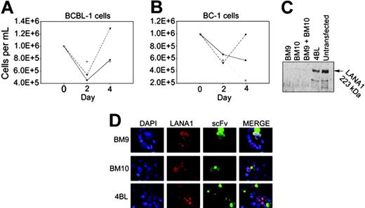
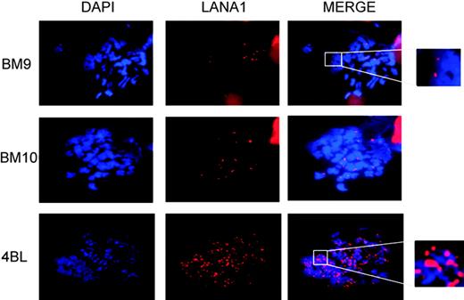
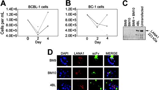
To determine if anti-LANA1 intrabody expression in naturally infected PEL cells inhibits KSHV episome maintenance, BM9 and BM10 were transfected into BC-1 (infected with Epstein-Barr virus [EBV] and KSHV) and BCBL-1 cells (infected with KSHV alone). LANA1 knockdown by expression of anti-LANA1 scFvs BM9 and BM10 in both PEL cell lines show marked short-term growth inhibition when anti-LANA1 scFvs are expressed compared with the irrelevant control intrabody, 4BL, cloned in the same plasmid backbone (Figure 4A-B). This corresponds to dispersion of LANA1 protein expression as depicted by IFA (in red) when anti-LANA1–specific scFvs are expressed (in green) (Figure 4D), and confirmed by Western blot (Figure 4C). ScFv expression in BC-1 cells has a greater effect in inhibiting cell growth than in BCBL-1. This may indicate that KSHV is the essential oncogenic virus driving these tumors. As previously described, LANA1 has a punctate nuclear pattern in PEL cells due to the colocalization of LANA1 with KSHV episomes when attached to cellular chromosomes. Using cytogenetically prepared mitotic spreads of PEL cells arrested in metaphase with colchicine treatment, the number of LANA1-positive punctate signals averages 40 per cell. Expression of anti-LANA1 scFv disperses LANA1 and significantly reduces the number of LANA1 nuclear dots per mitotically arrested cell (Figure 5; Table 2). When an irrelevant antibody is expressed, no apparent effect is observed in the number of LANA1 chromosomally associated dots. These results strongly correlate with the loss of the TR plasmid when coexpressed with BM9 and BM10, as described.
BM9 or BM10 expression promotes loss of KSHV plasmid in CM12 during transient transfection. Gardella gel electrophoresis followed by Southern blot hybridization with a TR probe shows loss of viral genome from CM12 cells stably transfected with KSHV-BAC36 GFP and transiently transfected with BM9 and BM10. Transfection with the negative control 4BL intrabody has no effect on viral persistence.
BM9 or BM10 expression promotes loss of KSHV plasmid in CM12 during transient transfection. Gardella gel electrophoresis followed by Southern blot hybridization with a TR probe shows loss of viral genome from CM12 cells stably transfected with KSHV-BAC36 GFP and transiently transfected with BM9 and BM10. Transfection with the negative control 4BL intrabody has no effect on viral persistence.
Expression of BM9 and BM10 inhibits PEL cell growth and reduces endogenous expression of LANA1 protein. Short-term growth curves for BCBL-1 (A) and BC-1 (B) cells transfected with BM9 (•), BM10 (○), and 4BL (♦) scFvs and selected with zeocyn show growth inhibition with anti-LANA1 scFv expression contrary to 4BL, an irrelevant HIV-specific intrabody. (C) Immunoblotting for LANA1 in BCBL-1 cells transfected with BM9, BM10, and 4BL shows a diminished protein level when BM9 and BM10 are expressed compared with 4BL and untransfected control cells. (D) Distribution of LANA1 and anti-LANA1 scFvs in BC-1 cells with immunofluorescence microscopy after simultaneous detection with mLANA1 and monoclonal HA. LANA1's speckled pattern becomes dispersed when BM9 and BM10 are expressed. scFv is shown in green (FITC) and LANA1 in red (rhodamine). Magnification × 1000.
Expression of BM9 and BM10 inhibits PEL cell growth and reduces endogenous expression of LANA1 protein. Short-term growth curves for BCBL-1 (A) and BC-1 (B) cells transfected with BM9 (•), BM10 (○), and 4BL (♦) scFvs and selected with zeocyn show growth inhibition with anti-LANA1 scFv expression contrary to 4BL, an irrelevant HIV-specific intrabody. (C) Immunoblotting for LANA1 in BCBL-1 cells transfected with BM9, BM10, and 4BL shows a diminished protein level when BM9 and BM10 are expressed compared with 4BL and untransfected control cells. (D) Distribution of LANA1 and anti-LANA1 scFvs in BC-1 cells with immunofluorescence microscopy after simultaneous detection with mLANA1 and monoclonal HA. LANA1's speckled pattern becomes dispersed when BM9 and BM10 are expressed. scFv is shown in green (FITC) and LANA1 in red (rhodamine). Magnification × 1000.
Discussion
Intracellular expression and antigen targeting using cloned scFvs represents a novel tool for dissecting biologic processes. Unlike technologies that target nucleic acids, intrabodies operate at the posttranslational level and can be directed to relevant subcellular compartments and precise epitopes on target proteins, potentially blocking only one of several possible functions of an expressed protein. Moreover, this technique holds advantages over other knockdown technologies, such as RNAi or ribozyme cleavage, for long-lived cellular proteins. In this study, we demonstrate that intrabodies neutralize the function of LANA1, which reduces viral persistence in infected cells.
Although antiviral drugs targeting the KSHV lytic replication can inhibit viremia, these agents do not eliminate persistent latent KSHV. During latency, most viral genes are silent, and thus there are few viral proteins that can be targeted to eliminate latent viral infection. Unlike most other KSHV proteins, LANA1 expression appears to be required for virus-infected cells to maintain the viral episome and to silence cellular antiviral responses mediated through regulatory proteins such as p53. RNAi has been described as a means to inhibit KSHV latency and shows therapeutic promise.29 RNAi against LANA1, however, is ineffective possibly due to the long half-life of this protein, allowing even small amounts of LANA1 message to code for sufficient protein to allow maintenance of the viral episome. Intrabody technology might therefore hold advantages in eliminating persistent KSHV infection by directly targeting the active viral protein.
Nuclear dispersion of LANA1 by BM9 and BM10 expression. Quantitation of LANA1 speckles in metaphase spreads demonstrates diminished chromatin-associated LANA1 in cells expressing BM9 or BM10 compared with 4BL. Magnification × 1000.
Nuclear dispersion of LANA1 by BM9 and BM10 expression. Quantitation of LANA1 speckles in metaphase spreads demonstrates diminished chromatin-associated LANA1 in cells expressing BM9 or BM10 compared with 4BL. Magnification × 1000.
In this study, we generated anti-LANA1 single-chain antibodies from immunized rabbits having weak binding to LANA1. Despite this, both BM9 and BM10 cause episome loss when expressed in KSHV-infected cells. This LANA1 neutralization is most likely to be related to N-terminal specificity for the intrabodies because this region is required for LANA1 interaction with cellular chromatin-associated proteins. LANA1 is a multifunctional viral protein that inhibits pRB1, p53, and GSK-3β signaling.26-28,40-42 Although we have not formally examined whether BM9 and BM10 neutralization may also inhibit these or other cellular signaling pathways, our data show that at least a disruption of the mitotic bridge between cellular chromatin and the viral episome occurs. In either case, cellular growth of PEL cells expressing active antiLANA1 intrabodies is markedly reduced. Ongoing studies are being conducted to determine precisely how neutralization causes this effect.
The effectiveness of these relatively low-affinity scFvs in inhibiting latent KSHV infection provides a proof of principle for the feasibility of using engineered intracellular antibodies as a novel therapy against latent viral infections. Intrabody fragments could be delivered through gene therapy allowing direct treatment of latently infected cells. In situ lysis of latently infected cells may provoke a protective immune response against viral antigens that could extend the efficacy of this form of treatment. The development of high-affinity anti-LANA1 scFv antibodies might result in even more efficacious anti-KSHV agents.
Prepublished online as Blood First Edition Paper, August 9, 2005; DOI 10.1182/blood-2005-04-1627.
Supported by grants from the Fundação para a Ciência e Tecnologia, Portugal (POCTI/33096/MGI/2000 and PSIDA/MGI/49729/2003) and the National Institutes of Health/National Cancer Institute. S.C.-R and F.A.S. are recipients of a doctoral fellowship from Fundação para a Ciência e Tecnologia, Portugal.
The publication costs of this article were defrayed in part by page charge payment. Therefore, and solely to indicate this fact, this article is hereby marked “advertisement” in accordance with 18 U.S.C. section 1734.
We thank Sofia Coelho for technical assistance and Dr Kenneth Kaye (Harvard University) for the kind gift of reagents.

![Figure 1. BM9 and BM10 scFvs bind to the N-terminal CBS domain of LANA1 with low but specific binding. (A) Representative enzyme-linked immunoassay activities for 10 Fab clones isolated by phage display from LANA1-immunized rabbits. Clones BM9 and BM10 having highest immunoreactivity were subcloned into single-chain Fv (scFv) and examined for LANA1 binding. (B) HA epitope-tagged BM9, but not BM10, scFv binds LANA1 on direct immunoblotting. FL-9/8TR and FL-9 are BJAB cell lines constitutively expressing LANA-1 protein, and pSG5 is a BJAB cell line transfected with empty vector alone. (C) In vitro binding of BM9 scFv to a GST-LANA1 fusion protein. In this assay, [35S]methionine-labeled BM9 scFv but not BM10 scFv directly interacted with GST-LANA1 fusion protein isolated with glutathione-linked beads. (D) Human serum competition assay: BM9 was mixed with the indicated dilutions of KSHV LANA1-seropositive human serum, and ELISAs were performed using plates coated with peptides P7, P8, P9, P19, and P43. BM9 competes with hyperimmune sera for peptide binding with P7, P8, and P43 but not with P19. Each point represents the value of binding minus nonspecific binding of scFv or human serum to uncoated wells blocked with 3% BSA/PBS. (E) Schematic representation of LANA1 and binding epitopes of BM9, BM10, and other cellular proteins that bind to its N-terminal domain.](https://ash.silverchair-cdn.com/ash/content_public/journal/blood/106/12/10.1182_blood-2005-04-1627/2/m_zh80240587460001.jpeg?Expires=1767864194&Signature=Inh1jHjUpAOwWWcWkLD2tKe-9uHGxneizpHIKHt8z0LwSMji9RMhgW0SNinU0Bi1YPQ9fw7dozsrzCn9yrZemhuNRWmkJHILX3E24PE20U2apzX9NiRKMwJCzl82RZjSguVpOIJF1KyZap~OFVf5Tfn8IG4FfG9p-azC~AqSpztG3uBz3mzep8yyYEY4RcTinRgfJJ2gW18WoOrc8gW9ygB9RqaY8rGxh3pHxDMGvJ89~4eMVWfWAyR9pnZoM6ic5~KQajdTFb9aLCglkJDFYWt8wJN7YLjUPcby~i2ypp9Hw33XYVgjJXn9YLa9KN2gAgO0O65cK4G9SDpz46e3Dw__&Key-Pair-Id=APKAIE5G5CRDK6RD3PGA)


![Figure 1. BM9 and BM10 scFvs bind to the N-terminal CBS domain of LANA1 with low but specific binding. (A) Representative enzyme-linked immunoassay activities for 10 Fab clones isolated by phage display from LANA1-immunized rabbits. Clones BM9 and BM10 having highest immunoreactivity were subcloned into single-chain Fv (scFv) and examined for LANA1 binding. (B) HA epitope-tagged BM9, but not BM10, scFv binds LANA1 on direct immunoblotting. FL-9/8TR and FL-9 are BJAB cell lines constitutively expressing LANA-1 protein, and pSG5 is a BJAB cell line transfected with empty vector alone. (C) In vitro binding of BM9 scFv to a GST-LANA1 fusion protein. In this assay, [35S]methionine-labeled BM9 scFv but not BM10 scFv directly interacted with GST-LANA1 fusion protein isolated with glutathione-linked beads. (D) Human serum competition assay: BM9 was mixed with the indicated dilutions of KSHV LANA1-seropositive human serum, and ELISAs were performed using plates coated with peptides P7, P8, P9, P19, and P43. BM9 competes with hyperimmune sera for peptide binding with P7, P8, and P43 but not with P19. Each point represents the value of binding minus nonspecific binding of scFv or human serum to uncoated wells blocked with 3% BSA/PBS. (E) Schematic representation of LANA1 and binding epitopes of BM9, BM10, and other cellular proteins that bind to its N-terminal domain.](https://ash.silverchair-cdn.com/ash/content_public/journal/blood/106/12/10.1182_blood-2005-04-1627/2/m_zh80240587460001.jpeg?Expires=1767864195&Signature=HF6EC5JN7DC5HBmxvw6MYZXNjCFHrbvBvbuCzOywluxWOHottILThs2Mn3865fOlT-zhZzKhQ1E9Bu3xv37cTsg4V2HwWApYvfM5wi84uhfUCVhgXyFXyC1rZTsCSEMTLiYtLXeCef1ySiTwjX1RMGKMAorDXkzLBxwOtw6eApNDVpWjb3Lg~fWivQI7LU2g6sLyDBoTypd2M2EXJ1VkLnCW1eWHamU1lp9BAqqa~~75CXg41Fblt45i7CvkZQXJQuSfkan~wVI5ghLIadhXMjN3UO0Ivmd5oxZ9CKK8w6JddZ~tnJZvOt-hD~pLHk6Mi8H3HxTsOTNK25cBuCn2yQ__&Key-Pair-Id=APKAIE5G5CRDK6RD3PGA)

