Anaplastic large-cell lymphoma (ALCL) is frequently associated with the 2;5 translocation and expresses the NPM-ALK fusion protein, which possesses a constitutive tyrosine kinase activity. We analyzed SHP1 tyrosine phosphatase expression and activity in 3 ALK-positive ALCL cell lines (Karpas 299, Cost, and SU-DHL1) and in lymph node biopsies (n = 40). We found an inverse correlation between the level of NPM-ALK phosphorylation and SHP1 phosphatase activity. Pull-down and coimmunoprecipitation experiments demonstrated a SHP1/NPM-ALK association. Furthermore, confocal microscopy performed on ALCL cell lines and biopsy specimens showed the colocalization of the 2 proteins in cytoplasmic bodies containing Y664-phosphorylated NPM-ALK. Dephosphorylation of NPM-ALK by SHP1 demonstrated that NPM-ALK was a SHP1 substrate. Downregulation of SHP1 expression by RNAi in Karpas cells led to hyperphosphorylation of NPM-ALK, STAT3 activation, and increase in cell proliferation. Furthermore, SHP1 overexpression in 3T3 fibroblasts stably expressing NPM-ALK led to the decrease of NPM-ALK phosphorylation, lower cell proliferation, and tumor progression in nude mice. These findings show that SHP1 is a negative regulator of NPM-ALK signaling. The use of tissue microarrays revealed that 50% of ALK-positive ALCLs were positive for SHP1. Our results suggest that SHP1 could be a critical enzyme in ALCL biology and a potential therapeutic target.
Introduction
ALK-positive anaplastic large-cell lymphomas (ALCLs) are characterized by the expression of a hybrid protein, associating the cytoplasmic portion of the ALK tyrosine kinase (anaplastic lymphoma kinase) with a partner protein. This hybrid kinase is often encoded by the NPM-ALK fusion gene resulting from the (2;5)(p23; q35) chromosomal translocation. However, several reports have shown that the ALK gene at 2p23 may also be involved in a number of variant translocations.1,2 The dimerization of NPM-ALK through NPM mimics ligand binding and leads to the constitutive activation of the ALK tyrosine kinase through tyrosine transphosphorylation and autophosphorylation.3,4 Phosphorylated NPM-ALK promotes cell survival and tumorigenesis through the activation of multiple SH2-domain–containing proteins, including the phospholipase C γ1 (PLCγ1), PI-3Kinase/AKT, and the JAK/STAT pathways, that control cell-cycle progression.5-11 It is likely that the oncogenic potential of NPM-ALK may be related with its kinase capability and consequently its tyrosine phosphorylation level.1,12 As previously reported, pp60c-src of the Src-family kinases interacts with NPM-ALK, participates in its phosphorylation, and could be an important actor in NPM-ALK–mediated mitogenicity.13 Although the mechanism of NPM-ALK activation is well documented, the downregulation process is totally unknown.
Phosphorylation of proteins on tyrosine residues is regulated by the activities of protein tyrosine kinases (PTKs) and protein tyrosine phosphatases (PTPs). Deregulation of the balance that exists between these 2 groups of enzymes can trigger the accumulation of tyrosine-phosphorylated proteins, which, in turn, can lead to abnormal cell proliferation and tumorigenesis. Two SH2-domain–containing phosphotyrosine phosphatases, SHP1 and SHP2, are widely implicated in the regulation of signaling pathways involved in cell proliferation, differentiation, and survival.14 It is well established that these 2 PTPs are involved in the pathogenesis of lymphoma, leukemia, and other types of cancers.15,16 Despite their high degree of homology, SHP1 and SHP2 have completely distinct expression profiles and functions. In contrast to SHP2, which is ubiquitously expressed, SHP1 is mainly found in hematopoietic cells.17 Multiple binding partners and substrates of SHP1 have been identified. For example, SHP1 acts as a negative regulator of several signal transduction proteins, including cytokine receptors, by dephosphorylating the receptor itself and/or receptor-associated kinases such as Src and JAK family kinases.18,19 This PTP can also downregulate the activation of STATs.20 The association of the murine motheaten phenotype with severe hematopoietic dysregulation and loss of SHP1 tyrosine phosphatase activity stresses a critical role for SHP1 in the regulation of hematopoietic-cell growth and differentiation.21 The dysfunctional regulation of SHP1 causes abnormal T-lymphocyte proliferation and induces various types of leukemias and lymphomas.22-25 Furthermore, recent reports have clearly shown that the absence of SHP1 or a drop in the level of SHP1 protein expression is frequent in malignant T cells, including ALCL, and results from the methylation of the SHP1 gene promoter.26-28 Because the expression of SHP1 reverses the malignant phenotype, this phosphatase has been proposed as a tumor suppressor gene candidate in various hematopoietic cancers.29
The purpose of this study was to investigate the role of SHP1 in NPM-ALK–mediated cell proliferation in ALCL. We report, for the first time, an association between SHP1 and NPM-ALK in 2 ALK-positive ALCL cell lines, Karpas 299 and Cost, and in lymph node biopsies of ALCL. We have shown that the NPM-ALK oncoprotein is a substrate of SHP1 and that SHP1 is involved in the negative regulation of ALK-positive cell proliferation. In addition, immunohistochemical analysis of lymph node biopsies of ALCL using tissue microarrays has shown that SHP1 is expressed in half of these tumors. Our data suggest that SHP1 could be a critical negative regulator of the oncogenic potential of NPM-ALK in ALCL.
Materials and methods
Reagents, antibodies, and cell culture
ALK-positive Karpas 29930 and SU-DHL1 and ALK-negative FE-PD cell lines31 were a gift from Dr K. Pulford (Oxford, United Kingdom). The ALK-positive Cost cell line was generated in our laboratory from a patient who developed a small-cell variant of ALK-positive ALCL.32 These cell lines were maintained in culture as previously described.13 Stable NPM-ALK–transfected NIH3T3 fibroblast clones were obtained and cultivated in DMEM (Gibco BRL, Carlsbad, CA) as described.33
Passive lysis buffer and the MTS proliferation assay kit were purchased from Promega (Lyon, France). Protein A and G sepharose beads were obtained from Sigma (Steinheim, Germany). Paranitrophenyl-phosphate (P-NPP) was purchased from Calbiochem (La Jolla, CA). Glutathione sepharose beads were from Amersham Biosciences (Uppsala, Sweden). pGEX-KG prokaryote expression vectors containing full-length SHP1 wild-type (WT) or SHP1 C455S (C/S) cDNA and pcDNA3 eukaryote expression vector containing full-length SHP1 WT were kindly provided by Dr C. Susini (INSERM U531, Toulouse, France).
Polyclonal anti-SHP1 and monoclonal anti-GST were purchased from Santa Cruz Biotechnology (Santa Cruz, CA). Anti–phosphotyrosine 4G10 monoclonal antibody was obtained from Upstate Biotechnology (Lake Placid, NY). The monoclonal antibody against the cytoplasmic part of ALK (ALKc) was kindly provided by Dr B. Falini (Perugia, Italy). The ALK1 monoclonal antibody and horseradish peroxidase–conjugated antimouse and antirabbit secondary antibodies were purchased from Dako (Glostrup, Denmark). Anti-STAT3, phospho-STAT3 (Y705), and phospho-ALK (Y1604) polyclonal antibodies were obtained from Cell Signaling Technology (Ozyme, France). Alexa Fluor 594 goat anti–mouse IgG and Alexa Fluor 488 goat anti–rabbit IgG were obtained from Molecular Probes (Eugene, OR). CyTM3-conjugated goat anti–rabbit IgG was obtained from Jackson ImmunoResearch Laboratories (Bar Harbor, ME). SHP1 siRNA and control siRNA were designed and purchased from Eurogentec (Angers, France). Cell line nucleofector kit T and R were from Amaxa Biosystem (Koeln, Germany).
Cell lysate preparation
Cells (107) were lysed with 750 μL of passive lysis buffer containing 1 mM sodium orthovanadate, 10 μg/mL leupeptin, and 10 μg/mL aprotinin. After 30 minutes on ice, cells lysates were sonicated 3 times for 10 seconds and centrifuged at 18 000g for 3 minutes. Orthovanadate was omitted for lysates used for phosphatase activity analysis. In some experiments, cells were pretreated with 500 μM of sodium orthovanadate for 1 hour at 37°C before lysis.
Immunoprecipitation and immunoblotting
Cell lysates (700 μL) were precleared at 4°C for 30 minutes using 50 μL protein G or A sepharose beads (50% vol/vol). Supernatants were then incubated with 5 μg of the appropriate antibodies for 1 hour at 4°C on a rocking platform and incubated with 50 μL protein G or A sepharose beads (50% vol/vol) for one additional hour. Immune complexes were washed 3 times with RIPA buffer.34 Immunoprecipitates or cell lysates were resolved by 9% sodium dodecyl sulfate–polyacrylamide gel electrophoresis (SDS-PAGE), electrotransferred to nitrocellulose membranes, and detected by immunoblotting with the appropriate antibodies using the enhanced chemiluminescence (ECL) lighting system (Perkin Elmer, Shelton, CT).
Binding assays using GST fusion proteins
Glutathione S-transferase (GST), GST-SHP1 WT, or GST-SHP1 C/S was purified by affinity chromatography using glutathione sepharose beads according to the manufacturer's recommendations. Lysates from 107 cells were precleared for 30 minutes with 50 μL glutathione sepharose beads (50% vol/vol) and then incubated with 5 μg of immobilized GST fusion proteins for 3 hours at 4°C under gentle shaking. After 4 washings at 4°C with RIPA buffer, the complexes were submitted to SDS-PAGE and analyzed by immunoblotting using ALKc antibody.
In vitro phosphatase assay and NPM-ALK dephosphorylation
Tyrosine phosphatase assays were performed using P-NPP as substrate. Cells were lysed with passive lysis buffer without sodium orthovanadate. SHP1 immunoprecipitations were performed as described under “Immunoprecipitation and immunoblotting.” The immune complexes were washed twice with RIPA buffer without phosphatase inhibitors and twice with phosphatase assay buffer (62 mM HEPES, pH 5; 6.25 mM EDTA; 12.5 mM dithiothreitol). Immune complexes were incubated in 200 μL phosphatase assay buffer with a final concentration of 25 mM of P-NPP for 30 minutes at 30°C under shaking. After centrifugation (4500g, 4°C) for 3 minutes, 800 μL of 1N NaOH was added to the supernatants and the optical density (OD) was measured at 410 nm. The corresponding immune complexes, bound to protein A–sepharose, were submitted to SDS-PAGE and immunodetection of SHP1 was performed by Western blotting.
In-gel phosphatase assays were performed according to the procedure of Burridge and Nelson with some modifications as previously described.34
For in vitro NPM-ALK dephosphorylation, NPM-ALK immunoprecipitates were obtained from Karpas 299 cells pretreated with sodium orthovanadate (500 μM for 1 hour). NPM-ALK immunoprecipitates were incubated in 50 μL phosphatase assay buffer for 1 hour at 30°C under shaking with or without SHP1 immunoprecipitates from Karpas cells (without sodium orthovanadate). In another experiment, antiphosphotyrosine immunoprecipitates obtained from Karpas or FE-PD cells pretreated with sodium orthovanadate were incubated in the same conditions with 5 μg GST-SHP1 WT or GST-SHP1 C/S fusion proteins. The reactions were stopped by the addition of 10 μL 5 × SDS sample buffer and the phosphoproteins were detected by immunoblotting with the antiphosphotyrosine antibody.
SHP1 RNA interference and cell transfection
Karpas 299 cells (2 × 10⁶) were resuspended in 100 μL of T solution (Cell line nucleofector kit T; Amaxa) and 200 nM of SHP1 siRNA duplex (5′ GCAGGAGGUGAAGAACUUG 3′) or control siRNA duplex. Cell suspensions were then submitted to the nucleofection program T16 according to the Amaxa protocol for cell suspension using the Amaxa Nucleofector apparatus. Cells were then transferred into 12-well plates containing 2 mL of IMDM + 10% FCS.
NIH3T3 fibroblasts stably expressing NPM-ALK33 and NPM-ALK–negative NIH3T3 fibroblasts were transfected with 5 μg of pcDNA3 or pcDNA3-SHP1 by nucleofection program U30 (R solution) according to the Amaxa protocol for adherent cells.
Cell proliferation assay
Transfected Karpas 299 cells with SHP1 siRNA or control siRNA and NIH3T3 fibroblasts expressing or not expressing NPM-ALK transfected with pcDNA3 or pcDNA3-SHP1 were cultivated overnight at 37°C in IMDM or DMEM, respectively, containing 10% FCS. Karpas cells were diluted at a final concentration of 2 × 10⁴ cells/mL in IMDM supplemented with 0.5% or 5% FCS and NIH3T3 at a final concentration of 4 × 10⁴ cells/mL in DMEM + 10% FCS. Cells (100 μL) were seeded in triplicate in 96-well plates. Twenty microliters MTS (3-(4,5-dimethylthiazol-2-yl)-5-(3(carboxymethoxyphenyl)-2-(4-sulfophenyl)-2H-tetrazolium) was then added to each well and incubated at 37°C, 4 hours for Karpas cells or 2 hours for NIH3T3. The OD (490 nm) was measured at 0, 24, 48, 72, and 96 hours after seeding.
Tumorigenicity of NIH3T3 cells transfected with pcDNA3 or pcDNA3-SHP1
A total of 2 × 10⁶ NPM-ALK–positive NIH3T3 fibroblasts transfected with pcDNA3 or pcDNA3-SHP1 were injected subcutaneously into 10 nude mice. Three days after the inoculation, tumors were observed. Mice were killed 7 days after the graft and the weight of the tumors was assessed.
Confocal immunofluorescence microscopy
The ALCL cells were left to adhere on poly-l-lysine–coated slides for 5 minutes. Adherent cells were fixed with 3% paraformaldehyde for 10 minutes at room temperature. Cells were permeabilized for 10 minutes with 0.2% Triton X-100 in phosphate-buffer saline (PBS) containing 10 mM HEPES, 3% bovine serum albumin (BSA). Nonspecific sites were saturated with 3% BSA and 3% FCS in the same buffer for 1 hour. The Karpas 299 cells were incubated for 1 hour at room temperature with 5 μg/mL ALK1 monoclonal antibody or polyclonal anti–phospho-ALK or polyclonal anti-SHP1 antibody alone or in combination. In some experiments, Karpas 299 and SU-DHL1 cells were incubated with the polyclonal phospho-STAT3 (Y705) antibody. After 3 washings with PBS containing 10 mM HEPES and 3% BSA, cells were incubated with the appropriate secondary antibodies (Alexa Fluor 488–conjugated goat antirabbit antibody or CyTM3-congugated goat antirabbit antibody or Alexa Fluor 594 goat antimouse antibody) for 45 minutes at a dilution of 1:300 in the same buffer. Samples were mounted in fluorescence mounting medium (DakoCytomation, Trappes, France). Slides were examined under a Zeiss confocal microscope (LSM 510; Axiovert 100; Oberkochen, Germany) equipped with a 63 × 1.4 oil-immersion Plan-Apochromat objective lens. For tissue biopsy analysis, sections of 5 μm were deparaffinized and subjected to heat antigen retrieval before immunostaining as described above. Unprocessed data have been analyzed with Zeiss LSM-Image software.
NPM-ALK tyrosine phosphorylation and SHP1 phosphatase activity in Cost and Karpas 299 ALK-positive ALCL-derived cell lines. (A) Western blotting analysis of tyrosine phosphorylation status with the 4G10 antiphosphotyrosine antibody in total cell extracts from Cost and Karpas 299 cell lines. (B) NPM-ALK immunoprecipitation (IP) from 107 cells with the ALK1 antibody followed by immunoblotting (IB) with the 4G10 antiphosphotyrosine antibody (i). Nitrocellulose membrane was stripped and reprobed with the ALKc antibody to assess NPM-ALK loading (ii). (C) Detection of SHP1 by Western blotting performed on Cost and Karpas 299 cell lysates showing a stronger expression of SHP1 in Karpas cells compared with Cost cells. (D) For the SHP1 phosphatase activity assay, anti-SHP1 immunoprecipitates from Cost and Karpas cell lines were incubated for 30 minutes at 30°C with P-NPP as a substrate. The OD of supernatants was measured at 410 nm and immune complexes were submitted to immunoblotting with an anti-SHP1 antibody. SHP1 phosphatase activities, expressed in OD measurements, were related to the same quantity of SHP1 protein (mean ± SD of 3 independent experiments; statistically significant difference [Student t test] was observed, ** P < .01). (E) SHP1 immunoprecipitates from Cost and Karpas cell lines were submitted to an in-gel phosphatase assay as described in “Materials and methods.” The higher level of SHP1 phosphatase activity in Karpas compared with Cost cells is in agreement with the results shown in panels C and D. Data shown in this figure are representative of 3 independent experiments.
NPM-ALK tyrosine phosphorylation and SHP1 phosphatase activity in Cost and Karpas 299 ALK-positive ALCL-derived cell lines. (A) Western blotting analysis of tyrosine phosphorylation status with the 4G10 antiphosphotyrosine antibody in total cell extracts from Cost and Karpas 299 cell lines. (B) NPM-ALK immunoprecipitation (IP) from 107 cells with the ALK1 antibody followed by immunoblotting (IB) with the 4G10 antiphosphotyrosine antibody (i). Nitrocellulose membrane was stripped and reprobed with the ALKc antibody to assess NPM-ALK loading (ii). (C) Detection of SHP1 by Western blotting performed on Cost and Karpas 299 cell lysates showing a stronger expression of SHP1 in Karpas cells compared with Cost cells. (D) For the SHP1 phosphatase activity assay, anti-SHP1 immunoprecipitates from Cost and Karpas cell lines were incubated for 30 minutes at 30°C with P-NPP as a substrate. The OD of supernatants was measured at 410 nm and immune complexes were submitted to immunoblotting with an anti-SHP1 antibody. SHP1 phosphatase activities, expressed in OD measurements, were related to the same quantity of SHP1 protein (mean ± SD of 3 independent experiments; statistically significant difference [Student t test] was observed, ** P < .01). (E) SHP1 immunoprecipitates from Cost and Karpas cell lines were submitted to an in-gel phosphatase assay as described in “Materials and methods.” The higher level of SHP1 phosphatase activity in Karpas compared with Cost cells is in agreement with the results shown in panels C and D. Data shown in this figure are representative of 3 independent experiments.
Detection of SHP1 phosphatase in ALCL lymph node biopsies by immunohistochemistry using TMA
A large number of ALCL cases have been collected with available paraffin blocks. Forty-eight cases, of which 40 cases were positive for the ALK protein, were used to prepare tissue microarray (TMA). These tumors were fixed in formalin (most cases) or in alcohol-based Bouin fixative. Random representative 1-mm cores were obtained using the Manual Tissue Arrayer (Alphelys, Plaisir, France). Sections (5 μm) were cut from each TMA, deparaffinized, and subjected to heat antigen retrieval. Sections were then stained with rabbit polyclonal anti-SHP1 antibody. Antibody binding was detected as previously described, using a Nikon Eclipse 80i microscope equipped with either a Plan UW 1 ×/0.04 or a PlanFluor 4 ×/0.13 objective lens (Nikon, Tokyo, Japan).32 The percentage of positive cells was evaluated and cases were considered positive when more than 10% of neoplastic cells were stained for SHP1.
Results
High SHP1 phosphatase activity is correlated with a low level of NPM-ALK tyrosine phosphorylation
Since the level of NPM-ALK tyrosine phosphorylation could be important for its oncogenic potential, we analyzed this level in 2 different ALK-positive ALCL cell lines (Cost and Karpas 299 cell lines) by anti-pY Western blotting. The tyrosine phosphorylation profile of total cell lysates, as shown in Figure 1A, revealed a major 80-kDa phosphoprotein corresponding to NPM-ALK with a signal that was stronger in Cost cells than in Karpas cells. In addition, a 70-kDa phosphoprotein was only detected in Karpas cells. This protein was not ZAP-70 as suggested by the negative result observed in the Western blot using anti–ZAP-70 antibody (data not shown). The identity of this protein remains to be determined. Two 55- to 60-kDa phosphoproteins were also present in the two cell lines. These proteins were identified as Src family kinases, as demonstrated through membrane reprobing with an anti–Src kinase antibody (data not shown). To confirm the identity of the 80-kDa protein, we performed NPM-ALK immunoprecipitations from the two cell lines with the ALK1 antibody. We analyzed the level of tyrosine phosphorylation by anti-pY Western blotting. As expected, the 80-kDa phosphoprotein detected in the cell lysates was NPM-ALK. Figure 1B clearly shows that the level of NPM-ALK tyrosine phosphorylation was higher in Cost than in Karpas cells, suggesting a difference in the kinase/phosphatase balance between these 2 ALK-positive ALCL cell lines.
In parallel, we evaluated SHP1 protein activity in the two cell lines. The result of the Western blot using an anti-SHP1 antibody, performed on total cell lysates, showed that the level of SHP1 protein expression was higher in Karpas than in Cost cells (Figure 1C). In addition, results from in vitro phosphatase assays using P-NPP as an exogenous substrate indicated a significant difference between SHP1 phosphatase activities in the two cell lines. Indeed, specific SHP1 activity (for an equal quantity of immunoprecipitated tyrosine phosphatase) was approximately 1.8-fold higher in Karpas than in Cost cells (Figure 1D). These data were confirmed by an in-gel phosphatase assay showing approximately a 2.5-fold increase in phosphatase activity for SHP1 immunoprecipitated from Karpas cells (Figure 1E). Overall, these data suggest an inverse correlation between SHP1 activity and NPM-ALK tyrosine phosphorylation.
SHP1 is associated with NPM-ALK
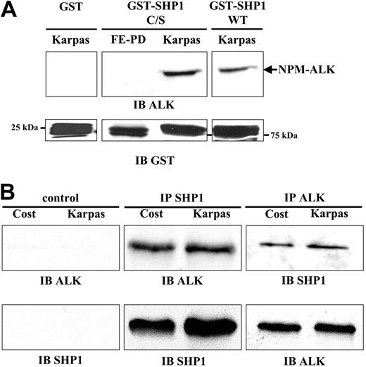
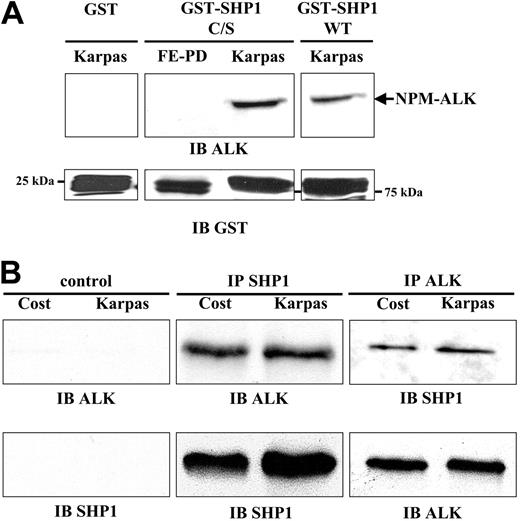
In vitro pull-down experiments using the recombinant wild-type SHP1 (GST-SHP1 WT) or the catalytically inactive C455S SHP1 (GST-SHP1 C/S) showed that NPM-ALK interacted with the 2 recombinant proteins (Figure 2A top panel lanes 3-4). These results were obtained from both Karpas 299 and Cost (not shown) cell lysates. The specificity of this interaction was demonstrated by the absence of NPM-ALK in the pull-down experiment using GST alone (Figure 2A top panel lane 1) and in the pull-down performed from an ALK-negative ALCL cell line (FE-PD) lysate incubated with the GST-SHP1 C/S (Figure 2A top panel lane 2). A Western blot anti-GST showed an equal input of recombinant proteins (Figure 2A bottom panel).
Association between SHP1 and NPM-ALK. (A) Pull-down experiments were performed with 5 μg of GST, GST-SHP1 C/S, or GST-SHP1 WT fusion proteins incubated with total cell extracts from 107 Karpas 299 cells (lanes 1, 3, and 4) or from 107 FE-PD cells (lane 2) pretreated with orthovanadate (500 μM for 1 h). Association of NPM-ALK with these fusion proteins was analyzed by immunoblotting with the ALKc antibody (top panel). The input recombinant proteins were detected with anti-GST antibody (bottom panel). Results are representative of 3 independent experiments. (B) Whole-cell extracts from 107 Cost or Karpas 299 cells were immunoprecipitated with anti-SHP1 antibody and immunoblotted with the ALKc antibody (top middle panel). To assess the quantity of immunoprecipitated SHP1, membranes were reprobed with anti-SHP1 antibody (bottom middle panel). NPM-ALK immunoprecipitates (using the ALK1 antibody) were subjected to immunoblotting with anti-SHP1 antibody (top right panel). Membranes were then reprobed with the ALKc antibody (bottom right panel). Immunoprecipitations were performed without addition of antibody as negative control (left panels). Data are representative of 5 independent experiments.
Association between SHP1 and NPM-ALK. (A) Pull-down experiments were performed with 5 μg of GST, GST-SHP1 C/S, or GST-SHP1 WT fusion proteins incubated with total cell extracts from 107 Karpas 299 cells (lanes 1, 3, and 4) or from 107 FE-PD cells (lane 2) pretreated with orthovanadate (500 μM for 1 h). Association of NPM-ALK with these fusion proteins was analyzed by immunoblotting with the ALKc antibody (top panel). The input recombinant proteins were detected with anti-GST antibody (bottom panel). Results are representative of 3 independent experiments. (B) Whole-cell extracts from 107 Cost or Karpas 299 cells were immunoprecipitated with anti-SHP1 antibody and immunoblotted with the ALKc antibody (top middle panel). To assess the quantity of immunoprecipitated SHP1, membranes were reprobed with anti-SHP1 antibody (bottom middle panel). NPM-ALK immunoprecipitates (using the ALK1 antibody) were subjected to immunoblotting with anti-SHP1 antibody (top right panel). Membranes were then reprobed with the ALKc antibody (bottom right panel). Immunoprecipitations were performed without addition of antibody as negative control (left panels). Data are representative of 5 independent experiments.
To demonstrate the association between NPM-ALK and SHP1, coimmunoprecipitation experiments were performed from Cost and Karpas 299 cell lysates. The detection of NPM-ALK in SHP1 immunoprecipitates demonstrated an association between the 2 proteins (Figure 2B top middle panel). The same membrane reprobed with an anti-SHP1 antibody showed a larger amount of SHP1 immunoprecipitated from Karpas cells compared with Cost cells (Figure 2B bottom middle panel), in agreement with the higher expression of the phosphatase in these cells (Figure 1C). In a reciprocal experiment, immunoprecipitation with ALK1 antibody followed by Western blotting using an anti-SHP1 antibody confirmed a physical association between the 2 proteins (Figure 2B top right panel). However, coimmunoprecipitation of NPM-ALK and SHP1 did not occur if antibodies were omitted (Figure 2B left panels) and when orthovanadate was not added to the buffer solution used during the preparation of cell lysates (data not shown).
NPM-ALK and SHP1 colocalize in cytoplasmic bodies of ALCL cells
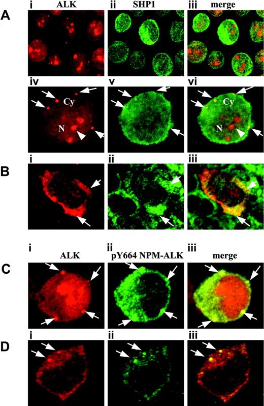
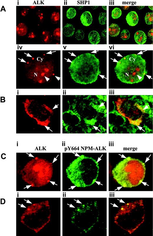
To visualize the interaction between SHP1 and NPM-ALK in Karpas 299 and Cost cells, the 2 proteins were stained with their respective antibodies. As previously described, confocal microscopy revealed a cytoplasmic, nuclear, and nucleolar NPM-ALK staining in Karpas cells (Figure 3Ai,Aiv,Ci). Interestingly, we also found a concentration of NPM-ALK in small cytoplasmic granules (arrows) associated with a diffuse cytoplasmic staining. Of note, these granules had a tendency to be close to the cell membrane. By collecting serial optical sections, we found approximately 10 granules per cell in Karpas 299 cell line and we did not observe a significant cell-to-cell variation in this cell line. In Cost cells, we found a larger number of the same granules (up to 15 per cell) that were of smaller size than in Karpas cells (data not shown). SHP1 staining revealed fluorescent spots that were essentially located within the cytoplasm of cells with a weak diffuse nuclear staining (Figure 3Aii,v). SHP1 staining overlapped with the same NPM-ALK cytoplasmic bodies, indicating a specific colocalization of the SHP1 tyrosine phosphatase in these structures (Figure 3Aiii,vi). Furthermore, cell labeling with the antibody directed against NPM-ALK phosphorylated on tyrosine 664 (PLCγ1 binding site) showed a strong concentration of phospho-Y664 NPM-ALK in the same cytoplasmic granules (Figure 3Cii-iii). Interestingly, these findings (ie, colocalization of SHP1, NPM-ALK, and phospho-Y664 NPM-ALK) were also observed in tissue microarrays from ALCL lymph node biopsy samples (Figure 3Bii-iii,3Dii-iii). Taken together, these data stress that SHP1 is involved in the tyrosine dephosphorylation of NPM-ALK, which might take place in distinct cytoplasmic compartments.
NPM-ALK is a SHP1 substrate
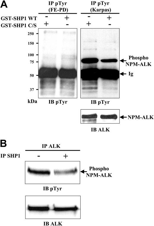
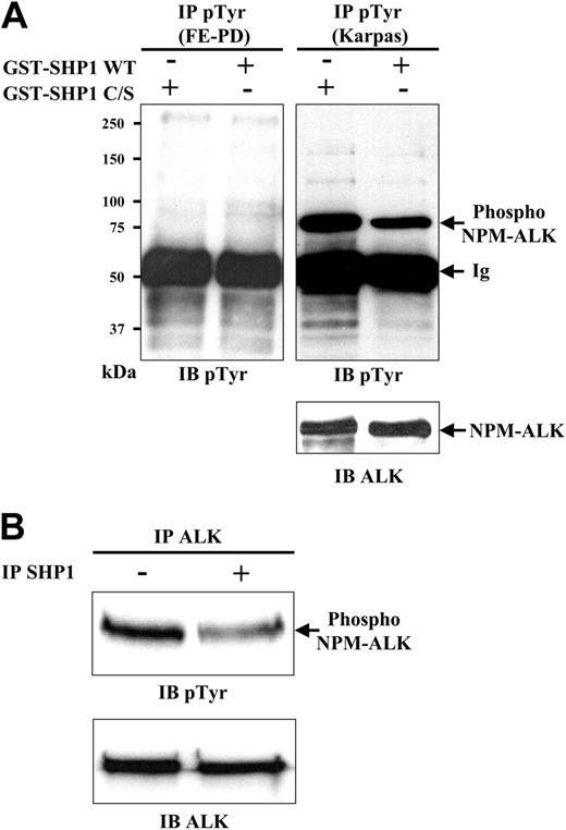
To demonstrate that NPM-ALK is a potential SHP1 substrate, we examined the capacity of SHP1 to dephosphorylate the NPM-ALK protein in vitro. We isolated phosphoproteins from orthovanadate-treated Karpas 299 cells by immunoprecipitation using the anti-pY antibody. Proteins were then incubated with GST-SHP1 WT or C/S recombinant proteins under in vitro PTPase assay conditions (see “Materials and methods”) and immunoblotting was performed using an anti-pY antibody. As illustrated in Figure 4A, a marked reduction in NPM-ALK tyrosine phosphorylation was observed with the GST-SHP1 WT in comparison with the mutated inactive GST-SHP1 C/S (Figure 4A top right panel). Other minor phosphoproteins could be detected, indicating that some of them were also dephosphorylated by active SHP1. A control has been made with ALK-negative cell line (FE-PD) showing no signal at 80 kDa. These results indicate that NPM-ALK is the main substrate of SHP1 in vitro. To confirm these data, NPM-ALK immunoprecipitates from Karpas cells were incubated with SHP1 immunoprecipitates. As shown in Figure 4B, the level of NPM-ALK tyrosine phosphorylation dramatically decreased after SHP1 incubation. Altogether, these data demonstrate that NPM-ALK is a SHP1 substrate in vitro.
Confocal microscopy showing colocalization of SHP1 and phosphorylated NPM-ALK into cytoplasmic granules. (A) Karpas 299 cells (A, low-power magnification; C, high-power magnification) and tissue sections (B,D) from the ALCL tissue microarrays were stained with ALK1 and anti-SHP1 (A-B) or with ALK1 and anti-pY664 NPM-ALK (C-D) antibodies. Antibody binding was visualized with conjugated goat antimouse or goat antirabbit, respectively, as described in “Materials and methods.” ALK1 antibody (red) shows diffuse and granular (arrows) cytoplasmic (Cy) staining associated with a strong nuclear (N) and nucleolar (arrowhead) staining (Ai,Aiv,Bi,Ci,Di). Comparable cytoplasmic staining pattern is observed after SHP1 staining (Aii,Av,Bii,Cii,Dii) and the 2 signals are colocalized (merge; Aiii,Avi,Biii, Ciii,Diii). Note that SHP1 (green) is essentially detected in the cytoplasm with a weak associated nuclear staining. Anti–NPM-ALK phosphorylated on Y664 staining (green) is restricted to the cytoplasm and clearly concentrated into cytoplasmic granules (arrows; C-D). The colocalization of the 2 signals is shown (Ciii,Diii). Note that a comparable cytoplasmic staining is seen on Karpas cells (C) and on tissue sections (D) after staining with ALK1 and anti-pY664 NPM-ALK antibody. Results shown are representative of 3 independent experiments. Total magnification is 63 × for Ai-iii; 170 × for Aiv-vi and all subpanels of B, C, and D.
Confocal microscopy showing colocalization of SHP1 and phosphorylated NPM-ALK into cytoplasmic granules. (A) Karpas 299 cells (A, low-power magnification; C, high-power magnification) and tissue sections (B,D) from the ALCL tissue microarrays were stained with ALK1 and anti-SHP1 (A-B) or with ALK1 and anti-pY664 NPM-ALK (C-D) antibodies. Antibody binding was visualized with conjugated goat antimouse or goat antirabbit, respectively, as described in “Materials and methods.” ALK1 antibody (red) shows diffuse and granular (arrows) cytoplasmic (Cy) staining associated with a strong nuclear (N) and nucleolar (arrowhead) staining (Ai,Aiv,Bi,Ci,Di). Comparable cytoplasmic staining pattern is observed after SHP1 staining (Aii,Av,Bii,Cii,Dii) and the 2 signals are colocalized (merge; Aiii,Avi,Biii, Ciii,Diii). Note that SHP1 (green) is essentially detected in the cytoplasm with a weak associated nuclear staining. Anti–NPM-ALK phosphorylated on Y664 staining (green) is restricted to the cytoplasm and clearly concentrated into cytoplasmic granules (arrows; C-D). The colocalization of the 2 signals is shown (Ciii,Diii). Note that a comparable cytoplasmic staining is seen on Karpas cells (C) and on tissue sections (D) after staining with ALK1 and anti-pY664 NPM-ALK antibody. Results shown are representative of 3 independent experiments. Total magnification is 63 × for Ai-iii; 170 × for Aiv-vi and all subpanels of B, C, and D.
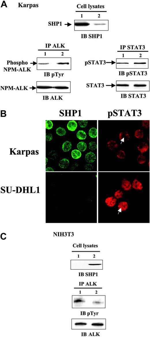
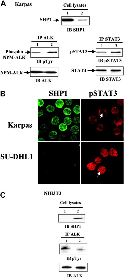
To confirm that NPM-ALK is a SHP1 substrate in ALCL cells, we performed experiments to downregulate SHP1 expression in Karpas 299 cells using the RNA interference (RNAi) technique as described in “Materials and methods.” SHP1 protein expression started to decrease by 24 hours and reached minimal levels (20% of control) at 48 hours after transfection (data not shown). We controlled the specificity of the SHP1 siRNA duplex by analyzing the expression of other widely expressed cytoplasmic tyrosine phosphatases such as SHP2 and PTP1B by Western blotting. We observed no changes in the expression of these phosphatases after SHP1 siRNA treatment (data not shown). Western blotting using anti-SHP1 antibody performed from Karpas 299 cell lysates showed a striking decrease in the level of SHP1 expression in cells transfected with the SHP1 siRNA (Figure 5A top panel; cell lysates) associated with a strong decrease (80%) in phosphatase activity (data not shown). To measure the impact of SHP1 on the negative regulation of NPM-ALK tyrosine phosphorylation, immunoprecipitations of NPM-ALK were performed from control siRNA–transfected cells and SHP1 siRNA–modified cells. As reported in Figure 5A (left panel; IP ALK, IB pTyr), NPM-ALK tyrosine phosphorylation was higher in cells transfected with SHP1 siRNA than in control cells. Furthermore, the downregulation of SHP1 by siRNA treatment led to the increase in STAT3 phosphorylation (Figure 5A right panel; IP STAT3, IB pSTAT3). As activation of STAT3 requires tyrosine phosphorylation followed by its translocation into the nucleus, we studied the localization of this transcription factor in 2 ALCL cell lines expressing or not expressing SHP1 (ie, Karpas 299 and SU-DHL1, respectively). While pSTAT3 was essentially located in the cytoplasm of the SHP1-positive cells (Karpas 299), we observed a strong signal in the nucleus of SU-DHL1 that did not express SHP1 (Figure 5B). These results indicate that SHP1 tyrosine phosphatase modulates the level of NPM-ALK tyrosine phosphorylation and the activation of its main target, namely STAT3, in NPM-ALK–positive ALCL cell lines. To confirm these data, we overexpressed SHP1 in NIH3T3 fibroblasts stably expressing NPM-ALK. The Western blotting anti-SHP1 performed from cell lysates demonstrated the overexpression of the phosphatase in cells transfected with the pcDNA3-SHP1 (Figure 5C left panel; cell lysates). An in vitro phosphatase assay performed on immunoprecipitated SHP1 from transfected NIH3T3 fibroblasts showed that the overexpressed SHP1 was active (data not shown). As expected, we observed a significant decrease of the NPM-ALK phosphorylation (Figure 5C right panel; IP ALK, IB pTyr) when SHP1 was overexpressed. Altogether, these results clearly indicate the implication of SHP1 in the dephosphorylation of NPM-ALK followed by the downregulation of STAT3 activation, suggesting the SHP1 role as a negative regulator of the oncogenic potential of NPM-ALK.
NPM-ALK dephosphorylation by SHP1 in vitro. (A) Antiphosphotyrosine immunoprecipitates were performed from 107 Karpas 299 cells (right panel) and FE-PD cells (left panel) pretreated with orthovanadate (500 μM for 1 hour). Immunoprecipitated phosphoproteins were incubated in phosphatase buffer with GST-SHP1 WT or GST-SHP1 C/S fusion proteins for 1 hour at 30°C and submitted to antiphosphotyrosine immunoblotting (top panels). The membrane was reprobed with the ALKc antibody to assess NPM-ALK loading (bottom panel). Data are representative of 3 independent experiments. (B) NPM-ALK immunoprecipitates from 107 Karpas cells, pretreated with orthovanadate, were incubated in the same PTP assay conditions (see above), with or without SHP1 immunoprecipitates performed on untreated Karpas 299 cells. Proteins in immune complexes were separated by SDS-PAGE and immunoblotted with antiphosphotyrosine antibody (top panel). Note that an equal amount of NPM-ALK was immunoprecipitated in each experiment (bottom panel). The data are representative of 2 independent experiments.
NPM-ALK dephosphorylation by SHP1 in vitro. (A) Antiphosphotyrosine immunoprecipitates were performed from 107 Karpas 299 cells (right panel) and FE-PD cells (left panel) pretreated with orthovanadate (500 μM for 1 hour). Immunoprecipitated phosphoproteins were incubated in phosphatase buffer with GST-SHP1 WT or GST-SHP1 C/S fusion proteins for 1 hour at 30°C and submitted to antiphosphotyrosine immunoblotting (top panels). The membrane was reprobed with the ALKc antibody to assess NPM-ALK loading (bottom panel). Data are representative of 3 independent experiments. (B) NPM-ALK immunoprecipitates from 107 Karpas cells, pretreated with orthovanadate, were incubated in the same PTP assay conditions (see above), with or without SHP1 immunoprecipitates performed on untreated Karpas 299 cells. Proteins in immune complexes were separated by SDS-PAGE and immunoblotted with antiphosphotyrosine antibody (top panel). Note that an equal amount of NPM-ALK was immunoprecipitated in each experiment (bottom panel). The data are representative of 2 independent experiments.
NPM-ALK dephosphorylation by SHP1 in ALCL and NIH3T3 cells. (A) Karpas 299 cells (6 × 10⁶) were transfected with 200 nM control siRNA (lane 1) or 200 nM SHP1 siRNA (lane 2). After 48 hours of cell culture, 107 cells were lysed and immunoblotting with anti-SHP1 antibody was performed from 25 μL of each lysate (top panel). NPM-ALK was immunoprecipitated with the ALK1 antibody and immune complexes were analyzed by immunoblotting with 4G10 antiphosphotyrosine antibody (top left panel). To assess the quantity of immunoprecipitated NPM-ALK, membrane was reprobed with the ALKc antibody (bottom left panel). Forty-eight hours after transfection with control siRNA (lane 1) or SHP1 siRNA (lane 2), STAT3 immunoprecipitations were performed and the immune complexes were submitted to Western blot using phospho-STAT3 (Tyr705) antibody (top right panel). The membrane was reprobed with anti-STAT3 antibody to assess STAT3 loading (bottom right panel). (B) Karpas 299 and SU-DHL1 cells were stained with anti-SHP1 or phospho-STAT3 antibodies and were observed by confocal microscope as described in “Materials and methods.” A strong nuclear staining for STAT3 is seen in SHP1-negative SU-DHL1 cells, whereas SHP1-positive Karpas cells show a weak doubtful nuclear staining. Total magnification is 63 ×. (C) NIH3T3 fibroblasts stably expressing NPM-ALK were transfected with 5 μg of pcDNA3 (lane 1) or pcDNA3-SHP1 (lane 2). After 15 hours of culture, SHP1 overexpression was detected from lysates of transfected NIH3T3 fibroblasts by anti-SHP1 Western blot (left panel). Transfected cells (5 × 10⁶) were subjected to NPM-ALK immunoprecipitation and immune complexes were analyzed as in panel A. Results reported in this figure are representative of 3 independent experiments.
NPM-ALK dephosphorylation by SHP1 in ALCL and NIH3T3 cells. (A) Karpas 299 cells (6 × 10⁶) were transfected with 200 nM control siRNA (lane 1) or 200 nM SHP1 siRNA (lane 2). After 48 hours of cell culture, 107 cells were lysed and immunoblotting with anti-SHP1 antibody was performed from 25 μL of each lysate (top panel). NPM-ALK was immunoprecipitated with the ALK1 antibody and immune complexes were analyzed by immunoblotting with 4G10 antiphosphotyrosine antibody (top left panel). To assess the quantity of immunoprecipitated NPM-ALK, membrane was reprobed with the ALKc antibody (bottom left panel). Forty-eight hours after transfection with control siRNA (lane 1) or SHP1 siRNA (lane 2), STAT3 immunoprecipitations were performed and the immune complexes were submitted to Western blot using phospho-STAT3 (Tyr705) antibody (top right panel). The membrane was reprobed with anti-STAT3 antibody to assess STAT3 loading (bottom right panel). (B) Karpas 299 and SU-DHL1 cells were stained with anti-SHP1 or phospho-STAT3 antibodies and were observed by confocal microscope as described in “Materials and methods.” A strong nuclear staining for STAT3 is seen in SHP1-negative SU-DHL1 cells, whereas SHP1-positive Karpas cells show a weak doubtful nuclear staining. Total magnification is 63 ×. (C) NIH3T3 fibroblasts stably expressing NPM-ALK were transfected with 5 μg of pcDNA3 (lane 1) or pcDNA3-SHP1 (lane 2). After 15 hours of culture, SHP1 overexpression was detected from lysates of transfected NIH3T3 fibroblasts by anti-SHP1 Western blot (left panel). Transfected cells (5 × 10⁶) were subjected to NPM-ALK immunoprecipitation and immune complexes were analyzed as in panel A. Results reported in this figure are representative of 3 independent experiments.
Modulation of SHP1 expression modifies the proliferation of NPM-ALK–positive cells
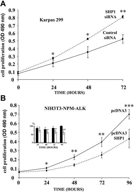
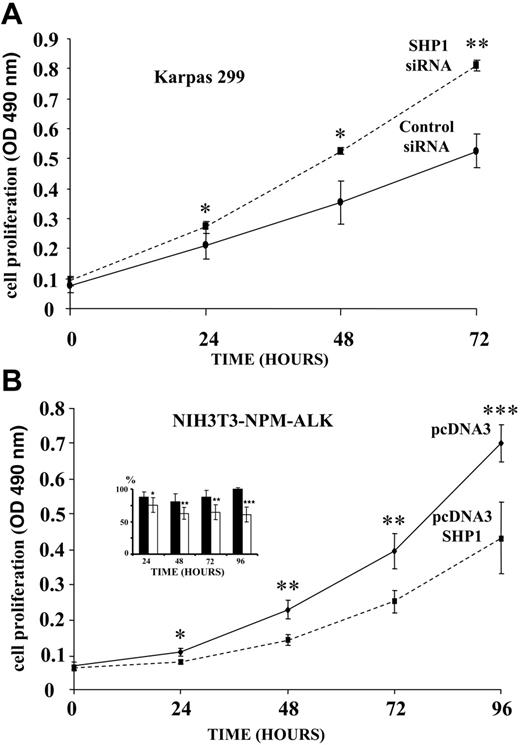
To assess the biologic role of SHP1 in cell proliferation, SHP1 expression was downregulated in Karpas 299 cells using the RNAi technique and cells were subjected to MTS proliferation assays in culture medium supplemented with 5% or 0.5% FCS (see “Materials and methods”). Figure 6A shows that SHP1 siRNA–treated lymphoma cells cultured in IMDM + 5% FCS grow faster than control siRNA cells. Experiments performed with Karpas cells cultured with 0.5% FCS showed similar results (data not shown). These findings demonstrate that the decrease in SHP1 is associated with an enhancement of cell growth in ALK-positive lymphoma cells.
Effect of SHP1 activity on the proliferation of NPM-ALK–positive cells. (A) Karpas 299 cells were transfected with SHP1 siRNA or control siRNA and cultivated overnight in IMDM containing 10% FCS. Cells were then diluted in IMDM containing 5% FCS and seeded in triplicate at 2 × 10³ cells per well in 96-well plates. Cell proliferation was determined each day for 3 days using an MTS proliferation assay and the OD (490 nm) was measured. (B) NPM-ALK–positive NIH3T3 fibroblasts were transfected with pcDNA3 or pcDNA3-SHP1 plasmids. After overnight culture in DMEM containing 10% FCS, fibroblasts (4 × 10³ cells/well) were seeded in triplicate in 96-well plates. Cell proliferation was measured each day for 4 days as described in panel A. The histogram (inset) represents the effect of SHP1 expression on the proliferation of NIH3T3 stably transfected with NPM-ALK (□) or NIH3T3 transfected with empty vector (▪). Each value is expressed as percentage of control cells that did not express SHP1. Results are mean ± SD; Student t test, *P < .05, **P < .01, ***P < .005 of 3 (Karpas) or 4 (NIH3T3) independent experiments performed in triplicate.
Effect of SHP1 activity on the proliferation of NPM-ALK–positive cells. (A) Karpas 299 cells were transfected with SHP1 siRNA or control siRNA and cultivated overnight in IMDM containing 10% FCS. Cells were then diluted in IMDM containing 5% FCS and seeded in triplicate at 2 × 10³ cells per well in 96-well plates. Cell proliferation was determined each day for 3 days using an MTS proliferation assay and the OD (490 nm) was measured. (B) NPM-ALK–positive NIH3T3 fibroblasts were transfected with pcDNA3 or pcDNA3-SHP1 plasmids. After overnight culture in DMEM containing 10% FCS, fibroblasts (4 × 10³ cells/well) were seeded in triplicate in 96-well plates. Cell proliferation was measured each day for 4 days as described in panel A. The histogram (inset) represents the effect of SHP1 expression on the proliferation of NIH3T3 stably transfected with NPM-ALK (□) or NIH3T3 transfected with empty vector (▪). Each value is expressed as percentage of control cells that did not express SHP1. Results are mean ± SD; Student t test, *P < .05, **P < .01, ***P < .005 of 3 (Karpas) or 4 (NIH3T3) independent experiments performed in triplicate.
To confirm the role of SHP1 in the downregulation of cell proliferation, we compared the proliferation of NPM-ALK–positive NIH3T3 fibroblasts expressing or not expressing SHP1. As shown in Figure 6B, NIH3T3 fibroblasts overexpressing SHP1 showed a significant decrease in their proliferation rate. Interestingly, the proliferation of NIH3T3 transfected with empty vector that did not express NPM-ALK was not significantly modified by the overexpression of SHP1 (Figure 6B histogram). This result indicates that the dysregulation of SHP1 expression might affect 3T3 fibroblast growth mainly through the dephosphorylation of NPM-ALK.
SHP1 expression decreases the tumorigenicity of NPM-ALK–positive NIH3T3 fibroblasts grafted in nude mice
To evaluate the role of SHP1 in the in vivo–transforming properties of NPM-ALK, NIH3T3 fibroblasts, expressing or not expressing SHP1, were grafted subcutaneously in athymic nude mice. NPM-ALK–positive NIH3T3 controls (negative for SHP1) induced rapidly growing subcutaneous tumors within 3 days, whereas the occurrence of tumors originating from cells expressing SHP1 was delayed and they were of smaller size (0.67 ± 0.38 g vs 0.36 ± 0.28 g; mean ± SD; Student t test, P = .03). These results indicate that SHP1 downregulates the oncogenic potential of NPM-ALK in vivo.
Detection of SHP1 phosphatase in lymph node biopsies of ALCL by immunohistochemistry using tissue microarrays (TMAs)
Of the 40 cases expressing the NPM-ALK protein, 21 cases (52.5%) were found to be positive for SHP1 (Figure 7). According to the World Health Organization (WHO) classification,35 19 cases (7 positive for SHP1) were of the common type and 9 cases (5 positive for SHP1) of the small-cell variant. Twelve cases (9 positive for SHP1) were difficult to classify, as several morphologic features were combined in the same lymph node biopsy. SHP1 staining was restricted to the cytoplasm and was diffuse, although some granules, similar to those observed with the confocal microscope, could be seen. In approximately 40% of the positive tumors, SHP1 staining was heterogeneous, with some strongly positive cells, whereas others showed a moderate to weak staining (Figure 7 middle panel). Only 2 of 8 cases of ALK-negative ALCL were positive for SHP1.
Immunohistochemical detection of SHP1 phosphatase using tissue microarrays (TMAs). (Left) ALCL positive for SHP1 protein showing a strong diffuse cytoplasmic staining. (Middle) Case showing heterogeneous SHP1 expression, some strongly positive cells among negative cells (arrows). (Right) ALCL negative for SHP1; only reactive lymphocytes admixed with lymphoma cells are positive for SHP1 and used as an internal control. All these cases are ALK-positive ALCLs (original magnifications ×8 [top] and ×50 [bottom]).
Immunohistochemical detection of SHP1 phosphatase using tissue microarrays (TMAs). (Left) ALCL positive for SHP1 protein showing a strong diffuse cytoplasmic staining. (Middle) Case showing heterogeneous SHP1 expression, some strongly positive cells among negative cells (arrows). (Right) ALCL negative for SHP1; only reactive lymphocytes admixed with lymphoma cells are positive for SHP1 and used as an internal control. All these cases are ALK-positive ALCLs (original magnifications ×8 [top] and ×50 [bottom]).
Discussion
The SHP1 tyrosine phosphatase has already been described as a negative regulator of hematopoietic-cell proliferation and proposed as a tumor suppressor gene candidate in lymphoma, leukemia, and other types of cancers.15,22-24,26,27,36 In the present study, we evaluated the involvement of SHP1 in the pathogenesis of ALK-positive ALCL. To our knowledge, only one other study mentions SHP1 expression and its loss in several ALK-positive ALCL cell lines and tumors.28 In contrast to Khoury et al,28 we found that the ALK-positive Karpas 299 cell line strongly expressed SHP1, as detected by Western blotting and immunofluorescence using a specific anti-SHP1 antibody. There is no clear explanation for this discrepancy although it could be due to different cell-culture conditions. Using tissue microarrays from ALK-positive ALCL lymph nodes, we found that 50% of these tumors were positive for SHP1. These results differ from those reported by Khoury et al28 who found only 18% of SHP1-positive cases. As shown by this group, the loss of SHP1 expression was not related to NPM-ALK status, as ALCLs are positive or negative for SHP1 regardless of NPM-ALK expression. Indeed, the variation of SHP1 expression, in particular, the absence of expression, seems to be related to the methylation of its promoter as demonstrated using the 5-aza-2′-deoxycytidine DNA methyltransferase inhibitor leading SHP1 re-expression 24 hours after treatment (unpublished personal data).
With the exception of the study of Khoury et al,28 little is known about the implication of the SHP1 tyrosine phosphatase in the signaling pathways activated by NPM-ALK. In the present study, several lines of evidence demonstrate that SHP1 associates with NPM-ALK. This association was demonstrated in Cost and Karpas 299 cell lines by pull-down and coimmunoprecipitation experiments. In addition, we observed that the SHP1/NPM-ALK association was dependent on the tyrosine phosphorylation status of NPM-ALK. This suggests that the SH2 domains of SHP1 could be involved in the interaction of SHP1 with one or several phosphorylated tyrosines on NPM-ALK. It is interesting to note that the C-terminal part of NPM-ALK contains a consensus sequence of immunoreceptor tyrosine-based inhibitory motifs (ITIMs) known to mediate SHP1 association with various signaling proteins.37 We therefore suggest that the potentially phosphorylated IEYGPL motif, which corresponds exactly to the ITIM consensus sequence ((V/I/L)XYXX(L/V)), could be the binding site for SHP1. However, we cannot exclude the possibility that the SHP1/NPM-ALK interaction is mediated by one or several proteins that form a complex containing SHP1 and NPM-ALK. Our findings, using confocal microscopy, provide additional evidence of the association of SHP1 with NPM-ALK, either directly or through a complex involving other proteins. Interestingly, we detected granular round-shaped cytoplasmic structures containing both SHP1 and phosphorylated Y664 NPM-ALK proteins in cultured ALK-positive ALCL cell lines and also in human tumoral tissues. To our knowledge, these granules, which might be dynamic sites of NPM-ALK dephosphorylation, have never been reported in cell lines or in tissue sections from patients with ALCL. These cytoplasmic structures could potentially represent major signaling sites that could play a crucial role in oncogenic events occurring in ALK-positive lymphomas. Furthermore, phosphorylated Y664 NPM-ALK was found to be restricted to the cytoplasm, without any associated nuclear localization. This finding is in agreement with previous studies performed with the 4G10 pan-antiphosphotyrosine antibody, indicating a pool of nonphosphorylated NPM-ALK localized in the nucleus of ALCL cells.38 Further experiments using confocal microscopy and biochemical approaches must be performed to characterize these cytoplasmic structures.
The interaction between SHP1 and NPM-ALK results in the dephosphorylation of NPM-ALK. To measure the impact of SHP1 expression and activity on the regulation of NPM-ALK tyrosine phosphorylation, we used 2 complementary approaches: the down-regulation of SHP1 in Karpas cells by RNAi technique and the overexpression of SHP1 by transfection in NIH3T3 fibroblasts stably expressing NPM-ALK. Our results demonstrate an inverse correlation between SHP1 activity and the level of NPM-ALK tyrosine phosphorylation. Furthermore, our results indicate the implication of SHP1 in the regulation of neoplastic-cell proliferation and in tumor progression in mice. Most importantly, we demonstrate that the modulation of SHP1 expression and activity are correlated by a significant modification in the activation state of STAT3 known to be the main target of the oncogenic protein NPM-ALK.7,11 This is the first demonstration of the implication of SHP1 in the negative regulation of the NPM-ALK oncogenic potential in vivo that could be mediated by its downstream key effector STAT3.
Several SHP1 substrates have already been described in hematopoietic cells. In particular, SHP1 is associated with the BCR-ABL oncogenic tyrosine kinase.39-41 SHP1 has also been shown to downregulate Src-family kinases such as Lck, Fyn,42 and ZAP-70.43,44 SHP1 also plays an important role in the negative regulation of the signaling pathway involving the JAK family kinases19,45 and STATs.46 As these pathways are activated by NPM-ALK, it is possible that SHP1 also acts on several other signaling molecules downstream from the NPM-ALK oncogenic protein. Furthermore, other tyrosine phosphatase(s) might be involved in NPM-ALK dephosphorylation. Interestingly, the catalytic domain of ALK shows a high homology with the insulin receptor kinase domain, including a conserved YY motif. In its phosphorylated form, this sequence has been described as an efficient PTP1B substrate and we cannot exclude that NPM-ALK could also be a target for this ubiquitously expressed protein tyrosine phosphatase.47
Finally, we must ask the question as to the clinical relevance of these results. Karpas 299 and Cost cell lines are representative of 2 different morphologic variants of ALK-positive ALCL.48 Indeed, Karpas 299 cells were isolated from a common type ALCL30 whereas Cost cells were generated from a small-cell variant ALCL.32 Even though Karpas and Cost cell lines express the SHP1 phosphatase, there are clear differences between the two cell lines regarding SHP1 catalytic activity. Indeed, it is important to note that SHP1 is less active in Cost cells derived from an ALK-positive small-cell variant ALCL. Interestingly, this low SHP1 activity is correlated with a high level of NPM-ALK tyrosine phosphorylation, suggesting a possible association between the aggressiveness of the ALCL and the degree of SHP1 phosphatase activation. Overall, our results suggest that SHP1 could be a critical enzyme in ALCL biology and a potential therapeutic target.
Supported by the Association pour la Recherche sur le Cancer, the Canceropôle Grand-Sud-Ouest, and the Conseil Régional de Midi-Pyrénées. J.-F.H. is supported by the Ligue Contre le Cancer comité du Gers, France.
J.-F.H. and A.R. performed all experimental procedures and, together with J.R.-T., controlled and analyzed the data; L.L. and G.D. reviewed the immunostaining and analyzed the results of TMA; J.R.-T. designed the research; and J.R.-T. and G.D. wrote the manuscript.
The publication costs of this article were defrayed in part by page charge payment. Therefore, and solely to indicate this fact, this article is hereby marked “advertisement” in accordance with 18 U.S.C. section 1734.
Prepublished online as Blood First Edition Paper, February 9, 2006; DOI 10.1182/blood-2005-06-2421.
We sincerely thank Dr S. Mueller-Valitutti for her advice and help with the confocal microscope. We thank Dr T. Al Saati and F. Capilla (Experimental Histopathology Platform of IFR30) for making TMA and J. Boyes for her help. We are also grateful to Dr C. Susini (INSERM U531) for her generous gift of SHP1 constructs and to Dr F. Amrstrong for making 3T3 fibroblasts stably expressing NPM-ALK. The authors wish to thank Drs M. Allouche and E. Espinos for fruitful discussions and Dr J. P. Jaffrezou for correcting the manuscript.

![Figure 1. NPM-ALK tyrosine phosphorylation and SHP1 phosphatase activity in Cost and Karpas 299 ALK-positive ALCL-derived cell lines. (A) Western blotting analysis of tyrosine phosphorylation status with the 4G10 antiphosphotyrosine antibody in total cell extracts from Cost and Karpas 299 cell lines. (B) NPM-ALK immunoprecipitation (IP) from 107 cells with the ALK1 antibody followed by immunoblotting (IB) with the 4G10 antiphosphotyrosine antibody (i). Nitrocellulose membrane was stripped and reprobed with the ALKc antibody to assess NPM-ALK loading (ii). (C) Detection of SHP1 by Western blotting performed on Cost and Karpas 299 cell lysates showing a stronger expression of SHP1 in Karpas cells compared with Cost cells. (D) For the SHP1 phosphatase activity assay, anti-SHP1 immunoprecipitates from Cost and Karpas cell lines were incubated for 30 minutes at 30°C with P-NPP as a substrate. The OD of supernatants was measured at 410 nm and immune complexes were submitted to immunoblotting with an anti-SHP1 antibody. SHP1 phosphatase activities, expressed in OD measurements, were related to the same quantity of SHP1 protein (mean ± SD of 3 independent experiments; statistically significant difference [Student t test] was observed, ** P < .01). (E) SHP1 immunoprecipitates from Cost and Karpas cell lines were submitted to an in-gel phosphatase assay as described in “Materials and methods.” The higher level of SHP1 phosphatase activity in Karpas compared with Cost cells is in agreement with the results shown in panels C and D. Data shown in this figure are representative of 3 independent experiments.](https://ash.silverchair-cdn.com/ash/content_public/journal/blood/107/10/10.1182_blood-2005-06-2421/2/m_zh80100695400001.jpeg?Expires=1768460059&Signature=HbT2IhtIsorZ8rT~9VUO7Wy53hPMFJx24LINnw5MiTemmuGvK6gG~kTbopyE3T7QzzokYagFaxSANeg2zbW1QD5Owl3ZYdPAiVNzeyxLMjMDvF-HYErzHu0xltP9kdWpfpKADUr541AbeAvRm23yGtFqqe7hd3WGqSVHxzuOAXis5OJvniBvk-putPxIhKYnehNXA8hIaz-oBFcTxAyqEVdsKPF94qv-spinS0n730C9R1j7CDza5b0t9lsSlK-PYAHdWKoIhL3whk7Kw5ROwtYjYH226UH7aYwgHYhUwfDjVwpHJwNrWIS1dOmYgvtrCSozVcvTd~CnjRo1vIQJcw__&Key-Pair-Id=APKAIE5G5CRDK6RD3PGA)
![Figure 7. Immunohistochemical detection of SHP1 phosphatase using tissue microarrays (TMAs). (Left) ALCL positive for SHP1 protein showing a strong diffuse cytoplasmic staining. (Middle) Case showing heterogeneous SHP1 expression, some strongly positive cells among negative cells (arrows). (Right) ALCL negative for SHP1; only reactive lymphocytes admixed with lymphoma cells are positive for SHP1 and used as an internal control. All these cases are ALK-positive ALCLs (original magnifications ×8 [top] and ×50 [bottom]).](https://ash.silverchair-cdn.com/ash/content_public/journal/blood/107/10/10.1182_blood-2005-06-2421/2/m_zh80100695400007.jpeg?Expires=1768460059&Signature=GEieCORl465YwIimBAtZFSKZcqzRNm-lKLEc1gUy-PBiHB-5khnso1VvIBIEOdL-utzuePj4kO9efSPWGgEMtF~A3ymKDjdtSbML-IrpUk~eLCH3HlAmMzoo1PBKzy8pbQRM37F7jcfCfNat3Ig87j4KYzjRmkdJCl~PtQqPm6GB1zMJ50cZO2KdyQc2AoGt1a3fD9-qYWLOsKiDIMvdCTYo3Wvg0s0Eo~m0KvOOVVjq4e1j4lJk9Z-F3~jhUGCDc0rmGSlrGj6DdBLkiDuv9OmsH7kOKo8jgOjE1LPt3JmuncRLL2sYlkEXdwlQFnqRgbIgMimcfK~BXyoPupURQg__&Key-Pair-Id=APKAIE5G5CRDK6RD3PGA)

![Figure 1. NPM-ALK tyrosine phosphorylation and SHP1 phosphatase activity in Cost and Karpas 299 ALK-positive ALCL-derived cell lines. (A) Western blotting analysis of tyrosine phosphorylation status with the 4G10 antiphosphotyrosine antibody in total cell extracts from Cost and Karpas 299 cell lines. (B) NPM-ALK immunoprecipitation (IP) from 107 cells with the ALK1 antibody followed by immunoblotting (IB) with the 4G10 antiphosphotyrosine antibody (i). Nitrocellulose membrane was stripped and reprobed with the ALKc antibody to assess NPM-ALK loading (ii). (C) Detection of SHP1 by Western blotting performed on Cost and Karpas 299 cell lysates showing a stronger expression of SHP1 in Karpas cells compared with Cost cells. (D) For the SHP1 phosphatase activity assay, anti-SHP1 immunoprecipitates from Cost and Karpas cell lines were incubated for 30 minutes at 30°C with P-NPP as a substrate. The OD of supernatants was measured at 410 nm and immune complexes were submitted to immunoblotting with an anti-SHP1 antibody. SHP1 phosphatase activities, expressed in OD measurements, were related to the same quantity of SHP1 protein (mean ± SD of 3 independent experiments; statistically significant difference [Student t test] was observed, ** P < .01). (E) SHP1 immunoprecipitates from Cost and Karpas cell lines were submitted to an in-gel phosphatase assay as described in “Materials and methods.” The higher level of SHP1 phosphatase activity in Karpas compared with Cost cells is in agreement with the results shown in panels C and D. Data shown in this figure are representative of 3 independent experiments.](https://ash.silverchair-cdn.com/ash/content_public/journal/blood/107/10/10.1182_blood-2005-06-2421/2/m_zh80100695400001.jpeg?Expires=1768548996&Signature=xG9qbaJVMsCjWNw8HQIWrd9jK-gtJznRAUZc8xkIa1RTfEg8r0vau3vG4o1tZvjg99Nj7E3yVC-atxwls9gEserol3n7U3hntjBjXMR0ps6tOHZh3fsEnOdqveS5JRDHsPhQfs9~9dX19GprAinsHqO-59BzpPeKwrlXPJCSXttIIpVWQYGCXihQLYFqt2i-9CyvFf7tjr6PVnJ0txMUTpSRr3nUCioRjFEh8y2T9VyUgysiJnCxLmbT0s9xjRJ3eGH7KQ-lTCn5NshMdDPwnIctL3V7y~C6Cnpo2xiysQZufdehOPDOqgEdlkbXUWE61cWAtmg33dlbKKtzm-RGOA__&Key-Pair-Id=APKAIE5G5CRDK6RD3PGA)
![Figure 7. Immunohistochemical detection of SHP1 phosphatase using tissue microarrays (TMAs). (Left) ALCL positive for SHP1 protein showing a strong diffuse cytoplasmic staining. (Middle) Case showing heterogeneous SHP1 expression, some strongly positive cells among negative cells (arrows). (Right) ALCL negative for SHP1; only reactive lymphocytes admixed with lymphoma cells are positive for SHP1 and used as an internal control. All these cases are ALK-positive ALCLs (original magnifications ×8 [top] and ×50 [bottom]).](https://ash.silverchair-cdn.com/ash/content_public/journal/blood/107/10/10.1182_blood-2005-06-2421/2/m_zh80100695400007.jpeg?Expires=1768548996&Signature=COIJWiejIzrJFU7pgesgSPEOt~DIzeF8iTj-1UV1GwmDNMdj5Tk8sxeOv7Ms5X9G5VbIJ9WB4Q4wLl~7WOi5eA6aF0lw-JQ87SfkcTRpPR5NUvYuHTUgwsmuMg0wF2Cu5~MmQ-G4xtYBjcVDZomToqMdVDwU8TTeQA~R6nJJzWJ3IIkmfHuRkh2NvxiPWsuHRXxED7bfAk6CLvMe1ogDBEzEfcLShGHeaqoA~I7X3EMc4VEqf9-3A7OfUzFFHZWO-78aQU5Pc0ow57eJjMHqVPmgE8lmuHyFavKIau9G-xM0u~bdLhvBJvoeuwdZufthI4kikA9-RQ6IH0RkYsOgVQ__&Key-Pair-Id=APKAIE5G5CRDK6RD3PGA)