Comment on Nakamura et al, page 1925
In this issue, Nakamura and colleagues report the x-ray crystal structure of filamin A repeat 17 with a peptide from the cytoplasmic tail of GPIbα, an interaction fundamental to platelet size and shape, as well as the functional activity of the GPIb-IX-V complex, the platelet von Willebrand factor adhesion receptor.
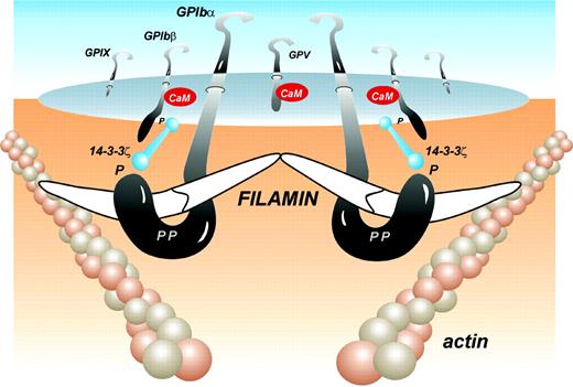
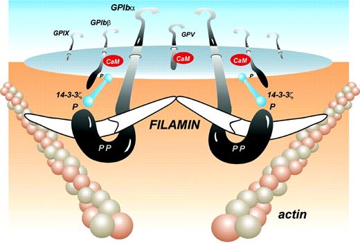
The GPIb-IX-V complex is a primary platelet adhesion receptor mediating the high shear-dependent adhesion of platelets to von Willebrand factor, initiating signals that lead to either hemostasis or thrombosis. Another important function of the GPIb-IX-V complex is in contributing to platelet shape by anchoring, through filamin A, the platelet plasma membrane to a network of submembranous actin filaments, termed the platelet membrane skeleton.1 Bernard-Soulier syndrome platelets that lack the GPIb-IX-V complex are abnormally large. An equivalent phenotype is found in Bernard-Soulier syndrome mice lacking GPIbα, which is rescued by expression of a chimeric protein construct containing the GPIbα cytoplasmic sequence, suggesting a key role for filamin A/GPIb interaction in regulating platelet morphology.2 Filamin A interaction with the GPIb-IX-V complex is also critical for the binding of von Willebrand factor,3 possibly due to a requirement of filamin A for the spatial orientation of the complex on the platelet surface for optimal binding of the A1 domain in multimeric von Willebrand factor. Filamin A is also an important scaffolding protein, with more than 50 known binding partners,4 suggesting that its association with the cytoplasmic face of the GPIb-IX-V complex may play an important role in receptor signaling, supplemental to the known association of GPIb-IX-V with other signaling proteins, such as 14-3-3ζ and calmodulin (see figure).
Filamin A is a homodimeric protein consisting of an approximately 280-kDa subunit, comprising an actin-binding domain at the N-terminus, followed by 24 immunoglobulin-like repeats, with the dimerization site at the ultimate C-terminal domain. Previous studies have localized the GPIb-IX-V/filamin interaction site to the central region of the cytoplasmic tail of GPIbα, approximately residues 556 to 577.3,5 In this issue, Nakamura and colleagues provide convincing evidence that the major GPIbα recognition site in filamin A resides in immunoglobulin repeat 17. They also report the x-ray crystal structure of this repeat, with a peptide encompassing residues 556 to 577 of GPIbα. The filamin A binding site lies within Pro561 and Pro573, with the peptide binding to a groove formed between 2 β-strands (the C and D) of the filamin A immunoglobulin repeat. The structure is consistent with previous observations that mutation of 2 amino acid residues of GPIbα, Phe568 and Trp570,5 abolishes filamin A binding, in that these 2 residues form major hydrophobic contacts with the filamin A binding groove. Remarkably, the interaction of the peptide with repeat 17 is of relatively low affinity, approximately 11 μM. Despite this, GPIb-IX-V can be stably purified with filamin A, implying that higher-order interactions between the GPIb-IX-V complex (minimally 2 GPIb-IX molecules bridged by GPV) and dimeric filamin A must dramatically increase the avidity of binding (see figure). While additional experimentation will be required to confirm this, the detailed structural information in this paper on the key residues in GPIbα and filamin A involved in their interaction will provide a useful road map for future studies aimed at dissecting the functional role of filamin A in platelet morphology and in the regulation of the mechano-receptor properties of the GPIb-IX-V complex as a major platelet adhesion/signaling receptor. ▪
Structure of the GPIb-IX-V complex with filamin A, based on the results of Nakamura et al in this issue. Other potential signaling proteins associated with the cytoplasmic face of the GPIb-IX-V complex, 14-3-3ζ and calmodulin, are also shown along with known phosphorylation sites (P).
Structure of the GPIb-IX-V complex with filamin A, based on the results of Nakamura et al in this issue. Other potential signaling proteins associated with the cytoplasmic face of the GPIb-IX-V complex, 14-3-3ζ and calmodulin, are also shown along with known phosphorylation sites (P).


