Signal transduction mechanisms associated with neutrophil activation by platelet factor 4 (PF4; CXCL4) are as yet poorly characterized. In a recent report, we showed that PF4-induced neutrophil functions (such as adhesion and secondary granule exocytosis) involve the activation of Src-kinases. By analyzing intracellular signals leading to adherence, we here demonstrate by several lines of evidence that in addition to Src-kinases, PF4 signaling involves the monomeric GTPase Ras, the tyrosine kinase Syk, and the MAP kinase JNK. Furthermore, on stimulation, GTPases Rac2 and RhoA were activated, and each was translocated to a different membrane compartment. As shown by inhibitor studies, Rac2 and JNK are located downstream of Syk and Ras. Most intriguingly, the latter 2 elements appear to control the activity of Rac2 and JNK independently of each other at different phases of the activation process. Although a first phase of Rac2 and JNK activation of up to 5 minutes is initiated by Ras, the second phase (5-30 minutes) depends predominantly on the activity of Syk. In summary, we describe that coordinated activity of Syk, Ras, and JNK mediates neutrophil adhesion to endothelial cells and that PF4 induces sequential activation of these elements.
Introduction
Platelet factor 4 (PF4; CXCL4) belongs to the family of CXC chemokines and shares 30% to 60% sequence identity and typical structural properties with other CXC chemokines, including the neutrophil-activating peptide-2 (NAP-2; CXCL7), interleukin-8 (IL-8; CXCL8), and interferon (IFN)-inducible protein 10 (IP-10; CXCL10).1,2 However, with regard to its receptors, signal transduction, and biologic functions, the role of PF4 within the family of chemokines is exceptional. Unlike other chemokines, which affect only a limited set of target cells, PF4 was reported to be active on different cell types including basophils, T cells, NK cells, and monocytes.3-7 In addition, PF4 supports the survival of hematopoietic stem cells and of progenitor cells,8 and it inhibits endothelial cell proliferation and angiogenesis in vitro.9-13 Recently, we described that PF4, in the absence of further costimuli, induces the differentiation of monocytes into HLA-DR-negative macrophages; in combination with IL-4, a specific type of antigen-presenting cell (APC) is developed.6,14
In neutrophils, highly purified PF4 does not induce typical chemokine responses, such as chemotaxis, superoxide anion generation, or degranulation.15 However, PF4 stimulates neutrophils to undergo extremely firm adhesion to unstimulated endothelial cells.16 In the presence of an appropriate costimulus, such as the proinflammatory cytokine TNF, PF4 induces the exocytosis of secondary granule contents.15,16
Although chemokines typically bind to 7-transmembrane domain G protein-coupled receptors, binding sites for PF4 are less well defined. In a recent report, Lasagni et al11 described an alternatively spliced variant of CXCR3, also referred to as CXCR3-B, as a functional receptor for PF4 on endothelial cells. However, our own investigations revealed that CXCR3-B is not expressed on monocytes or neutrophils7 and that PF4 binding to the latter cells is mediated by a distinct receptor, recently identified as chondroitin sulfate proteoglycan.17,18 Interestingly, the binding of PF4 to CXCR3-B on endothelial cells and to proteoglycans on neutrophils is not accompanied by changes in intracellular calcium concentrations.11,15
In the past few years, great progress has been made in elucidating the roles of the different tyrosine kinases in the regulation of neutrophil function in general.19 However, taking into account the unusual receptors and the biologic functions of PF4 on neutrophils, it becomes evident why progress in understanding the underlying signaling processes have been made only recently. In a previous work,20 we have shown that PF4 activates the Src-kinases Lyn, Hck, and Fgr in neutrophils and that the coordinated activation of all 3 kinases is required for the induction of neutrophil adhesion and for exocytosis. Consequently, in the present study, we analyze PF4-mediated signal transduction pathways occurring dependently and independently of Src-kinase activation. Through several lines of evidence, we could demonstrate that PF4-induced adherence to endothelial cells (ECs) required the activation of Syk, Ras (or Ras-related guanosine triphosphate-binding proteins [GTPases]), and c-Jun N-terminal kinase (JNK) mitogen-activated protein (MAP) kinases. Furthermore, we could identify JNK activation, which has not been observed thus far in human suspended neutrophils, as an essential and selective event controlled by Ras and Syk leading to neutrophil adhesion.
Materials and methods
Materials
Human natural PF4 was purified in our laboratory from release supernatants of thrombin-stimulated platelets in a 3-step procedure, as previously described.15,17 The final PF4 preparation exceeded 99% purity and contained no detectable protein contaminants.
Polyclonal rabbit antiserum raised against Rac2 (C-11) and monoclonal antibodies directed against Syk (4D10), RhoA (26C4), phospho-JNK (G-7; Thr183/Tyr185), phospho-c-Jun (KM-1; Ser63), JNK1 (C-17), and phosphotyrosine (PY99) were purchased from Santa Cruz Biotechnology (Heidelberg, Germany). Horseradish peroxidase (HRP)-conjugated goat anti-mouse IgG and HRP-conjugated goat anti-rabbit IgG were from Dianova (Hamburg, Germany). Alexa 680-conjugated goat anti-mouse IgG was obtained from MoBiTec (Göttingen, Germany), and IRDye800-conjugated goat anti-rabbit IgG was from Biotrend (Köln, Germany).
Rac- and Rho-activation assays and PP1 (Src-kinase inhibitor) were obtained from Biomol (Hamburg, Germany). Inhibitors directed against Syk (piceatannol), Ras farnesyltransferase (manumycin A), JNK (SP600125), Rho-associated protein kinase (Y-27632), mowiol 4-88, and n-octyl-β-d-glucopyranoside were purchased from Calbiochem (Schwalbach, Germany). Myelin basic protein (MBP) and chondroitin sulfate A were from Sigma (Deisenhofen, Germany), and c-Jun fusion protein beads were from Cell Signaling (Beverly, MA). Protease inhibitors Complete and Pefabloc SC were obtained from Roche (Mannheim, Germany), whereas phosphatase inhibitor okadaic acid was from Alexis (Grünberg, Germany).
Preparation and stimulation of neutrophils
Neutrophils were routinely isolated from citrated blood of healthy single donors by dextran sedimentation (Plasmasteril; Fresenius, Oberursel, Germany) followed by Ficoll-Hypaque (Pharmacia; Freiburg, Germany) density centrifugation, as described previously.21 More than 98% of the cells were viable, as assessed by trypan blue exclusion, and the percentage of neutrophils exceeded 95% in all experiments, as assessed by hematoxylin staining.
Cells were preincubated for 20 minutes at 37°C in Dulbecco phosphate-buffered saline (D-PBS) in the presence or absence of various inhibitors, as indicated in the Figures 1 and 6 legends, and were supplemented with CaCl2 and MgCl2 to a final concentration of 0.9 mM and 0.5 mM, respectively. Subsequently, the cells were exposed for up to 30 minutes at 37°C to PF4. In some experiments, PF4 was preincubated for 30 minutes at 37°C in the presence of chondroitin sulfate A at concentrations indicated in the legend to Figure S1. Stimulation was performed under gentle agitation in a thermo-shaker (Eppendorf; Hamburg, Germany) and was terminated by rapid centrifugation. Cell pellets were lysed, and protein concentrations were determined by the method of Bradford.22 Approval for these studies was obtained from the institutional review board at the University of Lübeck (Lübeck, Germany), and informed consent was provided according to the Declaration of Helsinki.
Preparation and culture of human endothelial cells
Human endothelial cells (ECs) were isolated from umbilical cord veins by collagenase treatment and were cultured in dishes coated with fibronectin, as described previously.23,24 The cells were maintained in M199 (Biochrom, Berlin, Germany) supplemented with 1% penicillin/streptomycin, 1% l-glutamine (both from Biochrom), 5% fetal calf serum (FCS), 30 μg/mL EC growth factor (both from Roche), and 20 μg/mL heparin (Sigma). Cells were subcultured after trypsinization (0.5% trypsin solution supplemented with 0.2% EDTA; Biochrom) and used throughout passages 2 to 4.
Adhesion assay
Determination of neutrophil adhesion to ECs was performed as described in detail elsewhere.16 Briefly, neutrophils were preincubated for 20 minutes at 37°C in the presence or absence of various inhibitors, as indicated in the Figure 1 legend, and subsequently 2 × 10⁵ neutrophils were allowed to adhere to ECs for 20 minutes at 37°C in the presence of 2 μM PF4. After removing nonadherent cells from the ECs, the amount of remaining cells was determined by measurement of neutrophil-specific endogenous β-glucuronidase enzymatic activity.25 Cell numbers were calculated by means of a standard of lysed cells run in parallel.
Rac- and Rho-activation assays
Immunoprecipitation of Rac2-GTP and RhoA-GTP was performed by using specific activation kits from Biomol. In brief, neutrophils were stimulated for the time periods indicated and immediately were lysed in MLB (Mg-containing lysis buffer; 25 mM HEPES, pH 7.5, 150 mM NaCl, 1% NP-40, 10 mM MgCl2, 1 mM EDTA, 10% glycerol, 2 mM Na3VO4, 2 mM NaF, 500 nM okadaic acid, 4 mM Pefabloc, and 1 × Complete). After 10 minutes on ice, the lysates were cleared by centrifugation (10 minutes, 10 000g, 4°C). Postnuclear lysates containing 750 μg total protein were incubated with PAK-1 PBD agarose or Rhotekin RBD agarose, respectively, for 60 minutes at 4°C under agitation. Beads were then washed 3 times in MLB and resuspended in 60 μL sample buffer. Before electrophoresis, all samples were boiled for 5 minutes. Precipitated Rac2 or RhoA were detected by Western blot analysis, as described in “Western blot analysis.”
Syk immunoprecipitation and in vitro protein kinase assay
Activation of Syk was determined by an in vitro phosphorylation assay using myelin basic protein (MBP) as exogenous substrate. Cells were stimulated for the indicated time periods and subsequently lysed in MLB (see “Rac- and Rho-activation assays“). After extraction for 10 minutes at 4°C, lysates were cleared by centrifugation (10 minutes, 10 000g, 4°C). Samples of precleared cell lysate containing 500 μg total protein were incubated with 2 μg anti-Syk antibody, followed by precipitation with protein A-agarose. After repeated washing, beads were resuspended in kinase buffer (20 mM Tris, pH 7.4, 10 mM MgCl2, 10 mM MnCl2, 1 mM DTT) containing 3 μg MBP, and the kinase reaction was started by the addition of 10 μM adenosine triphosphate (ATP). The reaction was stopped after 15 minutes at 30°C by the addition of 4-fold concentrated sample buffer. Before electrophoresis all samples were boiled for 5 minutes. Phosphorylated Syk and MBP were detected by Western blot analysis as described in “Western blot analysis.”
JNK phosphorylation and enzyme activity assay
Neutrophils were stimulated for the time periods indicated and were lysed immediately in JNK lysis buffer (20 mM Tris, pH 7.5, 150 mM NaCl, 1% Triton X-100, 1 mM EDTA, 1 mM EGTA, 2.5 mM sodium pyrophosphate, 1 mM β-glycerophosphate, 2 mM Na3VO4, 2 mM NaF, 500 nM okadaic acid, 4 mM Pefabloc, and 1 × Complete). After 10 minutes on ice, the lysates were cleared by centrifugation (10 minutes, 10 000g, 4°C). Phosphorylated JNK was detected in cell lysates by Western blot analysis, as described in “Western blot analysis.” JNK enzyme activity was examined using c-Jun fusion protein as exogenous substrate. In brief, neutrophil lysates were incubated with c-Jun fusion protein beads to pull down active/phosphorylated JNK. After repeated washings, c-Jun fusion protein beads were resuspended in kinase buffer (25 mM Tris, pH 7.5, 5 mM β-glycerophosphate, 2 mM DTT, 2 mM Na3VO4, 10 mM MgCl2) supplemented with 100 μM ATP using the c-Jun fusion protein as substrate. Reactions were terminated after 30 minutes at 30°C by the addition of 4-fold concentrated sample buffer, and the samples were boiled for 5 minutes before electrophoresis. Phosphorylated c-Jun was detected by Western blot analysis as described in “Western blot analysis.”
Isolation of detergent-insoluble membrane domains
Neutrophils were stimulated for the time periods indicated and subsequently were lysed in hypotonic lysis buffer (42 mM KCl, 10 mM HEPES, pH 7.4, 5 mM MgCl2) supplemented with inhibitors (2 mM Na3VO4, 2 mM NaF, 500 nM okadaic acid, 4 mM Pefabloc, and 1 × Complete). Lysates were incubated for 30 minutes on ice, followed by centrifugation (10 minutes, 250g, 4°C) to remove nuclei and intact cells. The supernatants were additionally centrifuged at 150 000g for 30 minutes at 4°C (TLA-100 ultracentrifuge; Beckmann Instruments, München, Germany) to separate cytoplasm and membrane fractions. Fractions derived from cytoplasm were diluted in 3-fold concentrated sample buffer. Membrane fractions were further dissolved for 60 minutes at 4°C in TNE buffer (10 mM Tris, pH 7.5, 150 mM NaCl, 5 mM EDTA) containing 1% Triton X-100 and inhibitors and then were adjusted to 42.5% sucrose in TNE buffer. Aliquots of 200 μL were transferred into centrifuge tubes, and 800 μL of 30% sucrose followed by 200 μL of 5% sucrose in TNE buffer were layered on top of the samples. After equilibrium centrifugation at 4°C for 19 hours at 200 000g, the gradients were fractionated from the top by volumes of 150 μL, and fractions were diluted in 3-fold concentrated sample buffer. The Triton-insoluble pellet was dissolved directly in 225 μL sample buffer. Before electrophoresis, all samples were boiled for 5 minutes. Rac2 or RhoA were detected by Western blot analysis, as described in “Western blot analysis.”
Western blot analysis
Proteins derived from cell lysates, pull-downs, or sucrose gradient fractions were separated by SDS-PAGE26 using 10% or 12% polyacrylamide gels and blotted onto polyvinylidene fluoride (PVDF) membranes (Roth, Karlsruhe, Germany). Immunodetection was performed as described in detail elsewhere.27 Briefly, membranes were incubated with the respective primary antibodies and HRP-conjugated goat anti-mouse IgG or HRP-conjugated goat anti-rabbit IgG secondary antibodies. Bands were visualized by an enhanced chemiluminescence method (ECL; Roche) according to the manufacturer's recommendations. In some experiments Alexa 680-conjugated goat anti-mouse IgG or IRDye 800-conjugated goat anti-rabbit IgG secondary antibodies were used instead of HRP conjugates. Under these conditions bands were visualized by an Odyssey infrared imaging system (LICOR, Bad Homburg, Germany), and relative density of protein bands was analyzed using Odyssey software 1.2 (background method: median-top/bottom). For reprobing, the membranes were stripped in 62.5 mM Tris, pH 6.7, 100 mM 2-mercaptoethanol, and 2% SDS, for 30 minutes at 50°C, followed by immunodetection with the appropriate antibodies.
Confocal laser scanning microscopy
Freshly isolated neutrophils were stimulated with 4 μM PF4 for 15 minutes at 4°C or 37°C, respectively. Then the cells were washed and fixed in 3% paraformaldehyde for 10 minutes at 4°C, followed by permeabilization with 0.6% n-octyl-β-d-glucopyranoside (5 minutes at room temperature). Proteins of interest were detected with appropriate primary antibodies and Cy5-labeled secondary antibodies (Dianova). After staining, the cells were embedded in mowiol solution (120 mM Tris, pH 8.5, 30% glycerol, 12% mowiol 4-88), transferred onto glass slides, and analyzed by confocal microscopy. Specimens were analyzed with a Leica TCS-SP confocal scanning microscope equipped with an acousto-optical tunable filter and a 63 ×/1.32 numeric aperture Plan-Apochromat oil-immersion objective (Leica Microsystems, Bensheim, Germany). Images were acquired with Leica TCSNT software and were further processed and assembled using CorelDraw 9 (Corel, Unterschleißheim, Germany).
Results
PF4-mediated neutrophil adhesion involves Syk, monomeric GTPases, and JNK MAP kinases
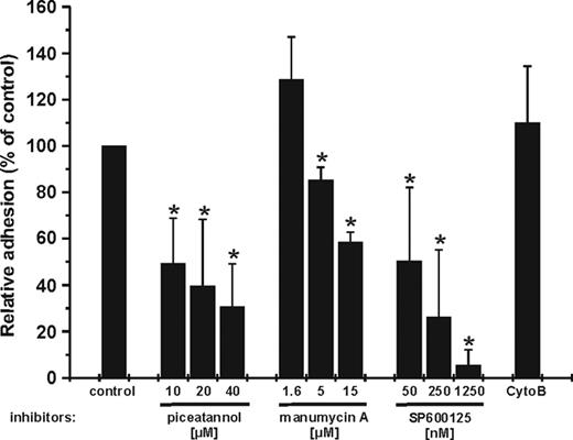
In a previous study, we have shown that neutrophil adhesion induced by PF4 proceeds independently of phosphatidylinositol 3-kinase (PI 3-kinase), p38, or Erk MAP kinases. In contrast, pretreatment of the cells with Src-kinase inhibitor PP1 resulted in a dose-dependent and significant reduction of PF4-induced adherence.20 To identify further the downstream signaling elements involved, we analyzed the effect of different inhibitors on PF4-induced neutrophil adhesion. In a first approach, neutrophils were preincubated in the presence or absence of increasing concentrations of inhibitors directed against Syk (piceatannol), Ras farnesyltransferase (manumycin A), JNK (SP600125), or a constant dosage of cytochalasin B (CytoB; 5 mg/mL), and they were allowed to adhere to cultured endothelial cells (ECs) in the presence of PF4. Preincubation of the cells with piceatannol, manumycin A, or SP600125 each resulted in a dose-dependent and significant reduction of PF4-mediated adhesion to ECs (Figure 1), whereas inhibition of actin polymerization by cytochalasin B was without effect. Interestingly, exocytosis of secondary granule contents in PF4/TNF-costimulated neutrophils was insensitive to JNK inhibitors, whereas blocking of Syk or Ras farnesyltransferase resulted in a dose-dependent reduction of this cellular response (data not shown). These data provide the first evidence that the activation of Syk, Ras, and JNK is involved in neutrophil adhesion, whereas actin polymerization is not involved in this function.
Syk is activated by PF4 in neutrophils
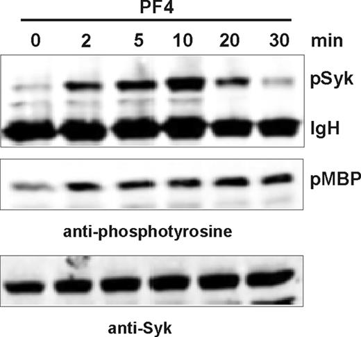
Given that piceatannol reduces neutrophil adhesion, we analyzed directly whether PF4 activates Syk in neutrophils. After PF4 stimulation for the time periods indicated, Syk was immunoprecipitated from neutrophil lysates and evaluated for kinase activity by analysis of Syk autophosphorylation and by its capacity to phosphorylate myelin basic protein (MBP) as a model substrate. Detection of phosphorylated proteins was performed by Western blot analysis. As shown in Figure 2, PF4 induced time-dependent Syk autophosphorylation (pSyk) and MBP (pMBP) phosphorylation with a first effect seen after 2 minutes of stimulation. Although Syk autophosphorylation reached maximal activation after 10 minutes and decreased thereafter, the phosphorylation of MBP continued up to 20 minutes of stimulation and decreased with further incubation. Western blot analysis of Syk in the same lysates confirmed equal protein loading (lower panel). These data clearly show that Syk became activated in PF4-treated neutrophils.
Effect of different inhibitors on PF4-mediated neutrophil adhesion. Freshly isolated neutrophils (1 × 10⁶ cells/mL) were pretreated for 20 minutes with increasing concentrations of piceatannol, manumycin A, or SP600125, a constant dosage of cytochalasin B (CytoB; 5 mg/mL), or without inhibitor and subsequently were incubated with 2 μM PF4 in the presence of a monolayer of cultured endothelial cells. After 20 minutes, nonadherent cells were removed and residual neutrophils were determined. Cell amounts in samples receiving PF4 in the absence of any inhibitor were set at 100%, and data were calculated as the percentage of these controls. Data represent mean ± SD of 3 (manumycin A) or 4 (piceatannol, SP600125, and cytochalasin B) independent experiments, each performed in duplicate. Statistical analysis using one-way ANOVA indicates significant differences (*P < .025) between inhibitor-treated and untreated samples based on the data from 3 or 4 individual experiments.
Effect of different inhibitors on PF4-mediated neutrophil adhesion. Freshly isolated neutrophils (1 × 10⁶ cells/mL) were pretreated for 20 minutes with increasing concentrations of piceatannol, manumycin A, or SP600125, a constant dosage of cytochalasin B (CytoB; 5 mg/mL), or without inhibitor and subsequently were incubated with 2 μM PF4 in the presence of a monolayer of cultured endothelial cells. After 20 minutes, nonadherent cells were removed and residual neutrophils were determined. Cell amounts in samples receiving PF4 in the absence of any inhibitor were set at 100%, and data were calculated as the percentage of these controls. Data represent mean ± SD of 3 (manumycin A) or 4 (piceatannol, SP600125, and cytochalasin B) independent experiments, each performed in duplicate. Statistical analysis using one-way ANOVA indicates significant differences (*P < .025) between inhibitor-treated and untreated samples based on the data from 3 or 4 individual experiments.
PF4 induced time-dependent activation of Syk. Neutrophils were stimulated for the indicated time periods, and Syk was immunoprecipitated from cell lysates. Precipitates were tested for phosphorylated Syk (pSyk; top panel) and Syk enzyme activity in an in vitro phosphorylation assay using MBP as exogenous substrate (middle panel). Phosphorylation of proteins was detected by Western blot analysis using antibodies directed against phosphotyrosine. IgH indicates immunoglobulin heavy chain. The same lysates were probed with anti-Syk antibody to confirm equal protein loading (bottom panel). Bands were visualized by Odyssey infrared imaging system. Data from 1 of 4 representative experiments are given.
PF4 induced time-dependent activation of Syk. Neutrophils were stimulated for the indicated time periods, and Syk was immunoprecipitated from cell lysates. Precipitates were tested for phosphorylated Syk (pSyk; top panel) and Syk enzyme activity in an in vitro phosphorylation assay using MBP as exogenous substrate (middle panel). Phosphorylation of proteins was detected by Western blot analysis using antibodies directed against phosphotyrosine. IgH indicates immunoglobulin heavy chain. The same lysates were probed with anti-Syk antibody to confirm equal protein loading (bottom panel). Bands were visualized by Odyssey infrared imaging system. Data from 1 of 4 representative experiments are given.
PF4 induces activation of small GTPases Rac2 and RhoA
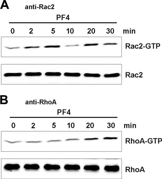
In another set of experiments, we examined the direct activation of Rac2 and RhoA, 2 prominent monomeric GTPases expressed in neutrophils.28 By the use of corresponding activation assays, activated (GTP-bound) GTPases Rac2 and RhoA were precipitated using PAK-1 or Rhotekin binding domain, respectively, and were detected by Western blot analysis with antibodies specific for either GTPase. Surprisingly, stimulation with PF4 provoked a rapid biphasic increase in GTP-bound Rac2, reaching a first maximum after 5 minutes of stimulation (Figure 3A, upper panel). After 10 minutes, amounts of Rac2-GTP decreased to levels comparable to those of unstimulated cells, and a second peak occurred after 20 minutes of stimulation. By contrast, activation of RhoA (Figure 3B, upper panel) displayed monophasic kinetics, with a first effect seen after 10 minutes and increasing amounts of RhoA-GTP up to 30 minutes of stimulation. Aliquots of the same lysates were tested to confirm equal protein loading (Figure 3A-B, lower panels). From these data we conclude that PF4 activates GTPases Rac2 and RhoA, though with different time kinetics. However, in further experiments, we could show that an inhibitor directed against the Rho-associated protein kinase (Y-27632), a downstream signaling element in RhoA signaling, was without effect on PF4-induced adhesion (data not shown). These results argue against a direct involvement of RhoA in PF4-mediated neutrophil adhesion.
PF4 induces protein translocations of Rac2 and RhoA to different membrane fractions
Targeting small GTPases to the plasma membrane is a prerequisite for their activation. Recent advances in membrane biology have led to the identification of glycosphingolipid- and cholesterol-rich plasma membrane microdomains, or lipid rafts, that are characterized by their insolubility in nonionic detergents and low buoyant density when isolated by sucrose gradient centrifugation.29,30 Because lipid rafts have been described to act as a signaling platform (for reviews, see Brown and London31 and Hoessli et al32 ), where signaling molecules accumulate in close proximity, we were interested in determining whether molecules described as involved in adhesion responses and activated by PF4 could also be found in this membrane fraction. In these experiments, the neutrophils were sequentially lysed in a hypotonic buffer, followed by lysis in TNE buffer containing Triton X-100. The latter lysate was then separated by sucrose density centrifugation. An equivalent of 5 × 10⁶ cells from the membrane fractions or 5 × 10⁵ cells from the cytosolic fraction was separated by SDS-PAGE, transferred onto PVDF membranes, and stained with appropriate primary antibodies, as indicated, and bands were visualized with the ECL system.
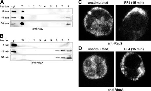
On activation with PF4, GTPases Rac2 and RhoA became translocated from the cytosol to distinct membrane fractions (Figure 4). Although Rac2 was found mainly in the Triton-insoluble (TI) membrane fraction (Figure 4A), RhoA became translocated to Triton-soluble membrane fractions 6 to 8 (Figure 4B). Neither Rac2 nor RhoA was detectable in fractions containing lipid rafts (fractions 2-4).
Interestingly, differences in the subcellular distributions of Rac2 and RhoA after PF4 stimulation could also be observed by microscopic analysis performed in parallel. Neutrophils were stimulated with PF4 (4 μM) or were left untreated, and distribution of GTPases was analyzed by confocal microscopy. In unstimulated cells, Rac2 and RhoA were located predominantly in the cytosol of neutrophils. On stimulation with PF4, Rac2 was translocated to a limited region of the membrane without formation of aggregate-like structures (Figure 4C), whereas RhoA formed small aggregates over the entire plasma membrane (Figure 4D). These data clearly demonstrated that PF4 activated monomeric GTPases Rac2 and RhoA. However, both GTPases displayed individual time kinetics and were translocated to clearly distinct membrane compartments.
PF4 mediates activation of JNK in suspended neutrophils
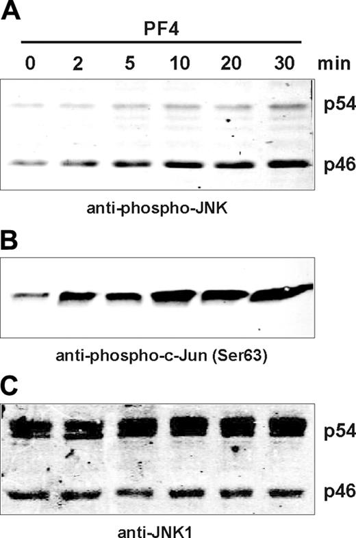
Several recent reports implicate a connection between activation of monomeric GTPases and JNK MAP kinases.33-35 Furthermore, we have shown here that neutrophil adherence induced by PF4 was drastically inhibited in cells pretreated with JNK inhibitor SP600125 (Figure 1). Consequently, we investigated whether stimulation with PF4 could directly induce the phosphorylation and activation of JNK. Neutrophils were treated with PF4 for up to 30 minutes, and lysates were analyzed for phosphorylated JNK by Western blot analysis using antibodies specific for dual-phosphorylated JNK. As shown in Figure 5A, the phosphorylation of JNK could be detected in neutrophil lysates after 2 minutes of stimulation. Interestingly, PF4 preferentially induced the phosphorylation of the p46 isoform, whereas the phosphorylation of the p54 isoform appeared to be much weaker. In parallel, we examined JNK enzyme activity in the same lysates using c-Jun fusion protein to pull down active JNK. In subsequent kinase assay, the c-Jun fusion protein functioned as a substrate for activated JNK, and the phosphorylation of c-Jun fusion protein was determined by Western blot analysis. As expected, the phosphorylation of c-Jun (Figure 5B) correlated with that of JNK (Figure 5A), with a first effect seen after 2 minutes of stimulation (Figure 5B). Western blot analysis of JNK1 in the same lysates confirmed equal protein loading (Figure 5C). These data clearly demonstrate that PF4 induced the phosphorylation and activation of JNK MAP kinases in human suspended neutrophils.
Effect of PF4 on the activation of small GTPases Rac2 and RhoA. Neutrophils were stimulated for the indicated time periods, and activated (GTP-bound) GTPases Rac2 (A) and RhoA (B) were precipitated using PAK-1 binding domain or Rhotekin binding domain, respectively, and were detected by Western blot analysis using anti-Rac2 (A) or anti-RhoA (B) antibodies. Bands were visualized by Odyssey infrared imaging system. An aliquot of the same lysates was tested to confirm equal protein loading (bottom blots of each panel). Data from 1 of 3 representative experiments are given.
Effect of PF4 on the activation of small GTPases Rac2 and RhoA. Neutrophils were stimulated for the indicated time periods, and activated (GTP-bound) GTPases Rac2 (A) and RhoA (B) were precipitated using PAK-1 binding domain or Rhotekin binding domain, respectively, and were detected by Western blot analysis using anti-Rac2 (A) or anti-RhoA (B) antibodies. Bands were visualized by Odyssey infrared imaging system. An aliquot of the same lysates was tested to confirm equal protein loading (bottom blots of each panel). Data from 1 of 3 representative experiments are given.
PF4 induces protein translocation of small GTPases to the plasma membrane. (A-B) Neutrophils were stimulated with PF4 (4 μM) for the time periods indicated in the figure. Cells were lysed, and lysates were separated to obtain cytosol (cyt), Triton-insoluble membrane fraction (TI), raft-containing fractions (fractions 2-4), and non-raft membrane fractions (fractions 6-8). Fractions were separated on 10% SDS-PAGE, and Rac2 (A) and RhoA (B) were detected by Western blot analysis using specific antibodies. Bands were visualized by ECL. Data are representative of 3 independent experiments. (C-D) Neutrophils were stimulated with PF4 (4 μM) for 15 minutes or were left untreated, and GTPases were detected by indirect immunofluorescence staining with anti-Rac2 (C) or anti-RhoA (D) antibodies and were analyzed by confocal microscopy. Data from 1 of 3 representative experiments are given.
PF4 induces protein translocation of small GTPases to the plasma membrane. (A-B) Neutrophils were stimulated with PF4 (4 μM) for the time periods indicated in the figure. Cells were lysed, and lysates were separated to obtain cytosol (cyt), Triton-insoluble membrane fraction (TI), raft-containing fractions (fractions 2-4), and non-raft membrane fractions (fractions 6-8). Fractions were separated on 10% SDS-PAGE, and Rac2 (A) and RhoA (B) were detected by Western blot analysis using specific antibodies. Bands were visualized by ECL. Data are representative of 3 independent experiments. (C-D) Neutrophils were stimulated with PF4 (4 μM) for 15 minutes or were left untreated, and GTPases were detected by indirect immunofluorescence staining with anti-Rac2 (C) or anti-RhoA (D) antibodies and were analyzed by confocal microscopy. Data from 1 of 3 representative experiments are given.
PF4 induces the phosphorylation and activation of JNK. Neutrophils were stimulated with PF4 (4 μM) for the time periods indicated in the figure. Cell lysates were prepared, and proteins were separated by SDS-PAGE. JNK phosphorylation was detected by Western blot analysis using anti-phospho-JNK antibodies (A). Alternatively, cells were lysed, and enzymatically active JNK was pulled down using c-Jun fusion protein beads. Kinase activity was determined by the phosphorylation of c-Jun fusion protein and visualized by Western blot analysis using an antibody directed against Ser63-phosphorylated c-Jun (B). An aliquot of the same lysates was tested with anti-JNK1 antibodies to confirm equal protein loading (C). Bands were visualized by the Odyssey infrared imaging system. Data from 1 of 4 representative experiments are given.
PF4 induces the phosphorylation and activation of JNK. Neutrophils were stimulated with PF4 (4 μM) for the time periods indicated in the figure. Cell lysates were prepared, and proteins were separated by SDS-PAGE. JNK phosphorylation was detected by Western blot analysis using anti-phospho-JNK antibodies (A). Alternatively, cells were lysed, and enzymatically active JNK was pulled down using c-Jun fusion protein beads. Kinase activity was determined by the phosphorylation of c-Jun fusion protein and visualized by Western blot analysis using an antibody directed against Ser63-phosphorylated c-Jun (B). An aliquot of the same lysates was tested with anti-JNK1 antibodies to confirm equal protein loading (C). Bands were visualized by the Odyssey infrared imaging system. Data from 1 of 4 representative experiments are given.
Syk and Ras are located upstream of Rac2 and JNK
To determine whether Src-kinases, Syk, or Ras are participants in the pathway leading to Rac2 or JNK activation in neutrophils, we next examined whether activation of the latter elements could be modulated by specific inhibitors. Neutrophils were preincubated in the presence or absence of inhibitors directed against Src-kinases (PP1), Ras farnesyltransferase (manumycin A), or Syk (piceatannol) and subsequently were stimulated with PF4. Because Rac2 activation displayed biphasic kinetics that peaked after 5 and 20 minutes of stimulation (Figure 3A), these time points were chosen for detailed analysis. Rac activation and JNK activity were determined by Western blot analysis, as described, and bands were visualized and quantified with the Odyssey infrared imaging system (Figure 6, left panels). Furthermore, data derived from quantitative analysis of relative densities of protein bands at 5 and 20 minutes of 5 independent experiments are given (Figure 6, right panels). Compared with controls stimulated for 5 minutes with PF4 in the absence of inhibitors, treatment with manumycin A caused significant reduction of Rac2 (greater than 95% inhibition; Figure 6A) and of JNK (greater than 75% inhibition; Figure 6B) activity. However, at 20 minutes of stimulation, the effects of manumycin A were far less prominent, resulting in an inhibition of Rac2 and JNK of approximately 29% and 48%, respectively. By contrast, the effects of the Syk inhibitor piceatannol were more prominent at the later time point of stimulation. Although Rac2 activity after 5 minutes was reduced by 48% and c-Jun phosphorylation was reduced by 21%, at 20 minutes of PF4 treatment more than 72% and 41% of Rac2 and JNK activity, respectively, were lost in the presence of the inhibitor (Figure 6). Interestingly, the inhibition of Src-kinases by PP1 after 5 minutes of stimulation resulted in a weak but reproducible increase in the activity of both signaling enzymes, whereas at 20 minutes of PF4 challenge, partial reduction of their activity was observed (44% and 22% for Rac2 and JNK, respectively).
From these data we conclude that rapid activation of Rac2 and JNK during the first minutes of stimulation by PF4 requires the activation of Ras, whereas sustained activity of these enzymes at later time points was dependent on Syk. Although Src-kinases played a fundamental role in the induction of PF4-mediated function in neutrophils, their impact on the activation of Rac2 and JNK appeared to be less prominent.
Syk and Ras are located upstream of Rac2 and JNK. Neutrophils were pretreated for 20 minutes with Src-kinase inhibitor PP1 (50 μM), Ras farnesyltransferase inhibitor manumycin A (15 μM), or Syk inhibitor piceatannol (40 μM) or without inhibitor and subsequently were stimulated with 4 μM PF4 for 5 or 20 minutes. Cells were lysed, and activation of Rac2 (A) or JNK (B) was determined as described in Figures 3 and 5B, respectively. Quantification of relative density of protein bands was performed using Odyssey software 1.2 (background method: median, top/bottom). Band density in untreated cells was set as 100%, and the densities of protein bands in inhibitor-treated cells were calculated as percentage of untreated cells. Data represent mean ± SD of 5 independent experiments. Statistical analysis using one-way ANOVA indicates significant differences (*P < .045) between inhibitor-treated and untreated samples based on the data from 5 individual experiments.
Syk and Ras are located upstream of Rac2 and JNK. Neutrophils were pretreated for 20 minutes with Src-kinase inhibitor PP1 (50 μM), Ras farnesyltransferase inhibitor manumycin A (15 μM), or Syk inhibitor piceatannol (40 μM) or without inhibitor and subsequently were stimulated with 4 μM PF4 for 5 or 20 minutes. Cells were lysed, and activation of Rac2 (A) or JNK (B) was determined as described in Figures 3 and 5B, respectively. Quantification of relative density of protein bands was performed using Odyssey software 1.2 (background method: median, top/bottom). Band density in untreated cells was set as 100%, and the densities of protein bands in inhibitor-treated cells were calculated as percentage of untreated cells. Data represent mean ± SD of 5 independent experiments. Statistical analysis using one-way ANOVA indicates significant differences (*P < .045) between inhibitor-treated and untreated samples based on the data from 5 individual experiments.
Effect of soluble chondroitin sulfate A (sCSA) on PF4-mediated neutrophil adhesion and signaling
In previous reports we described the PF4 receptor as a chondroitin sulfate proteoglycan.17,18 To provide the first evidence that chondroitin sulfate proteoglycans may also be involved in PF4 signaling, the inhibitory effect of sCSA on PF4-mediated neutrophil adhesion and activation of Rac2, Syk, and JNK was investigated. Preincubation of PF4 with sCSA at concentrations of 20 μg/mL abrogated neutrophil adhesion and inhibited the activation of Rac2, Syk, and JNK (Figure S1, available on the Blood website; see the Supplemental Figure link at the top of the online article). These results further support the involvement of chondroitin sulfate proteoglycans in PF4-mediated neutrophil activation.
Discussion
The signal transduction mechanisms associated with neutrophil activation by PF4 (CXCL4) are as yet poorly characterized. One reason for this might be that though it is structurally a CXC-chemokine, PF4 does not bind to a 7-transmembrane-spanning G protein-coupled receptor on neutrophils or monocytes but does bind to its own receptor, recently identified as chondroitin sulfate proteoglycan.17,18 In the present study, we could clearly demonstrate that in addition to Src kinases (Hck, Fgr),20 Syk, Ras, and JNK are involved in the induction of neutrophil adhesion by PF4. Furthermore, neutrophil function and signal transduction were abrogated in the presence of sCSA (Figure S1), indicating that PF4 binding to chondroitin sulfate proteoglycans is an essential step in neutrophil activation induced by the chemokine.
The regulation of cellular responses to specific stimuli occurs in part through selective activation of MAP kinase signaling cascades. In a previous report, we have shown that PF4 activates neither p38 nor Erk MAP kinases.20 Here, however, we demonstrate that PF4 stimulates the phosphorylation and activation of a third member of this kinase family, c-Jun N-terminal kinase (JNK; Figure 5). The activation of the latter transduction element has, at least in human suspended neutrophils, not been described thus far. Although one report describes the phosphorylation of JNK in murine bone marrow neutrophils in response to fMLP,36 a number of investigators indeed report that JNK does not become activated in human neutrophils after exposure to such diverse stimuli as cytokines, growth factors, phorbol esters, chemotactic factors, or even potent JNK activators such as UV irradiation or chemical stress.37-42 Recently, Avdi et al43 and Arndt et al44 stated that neutrophil adhesion is a crucial prerequisite for the activation of JNK. Although neither cell-cell/homotypic adherence nor cell-substratum adherence is sufficient to induce JNK activation, the phosphorylation of JNK could be partially blocked by the use of anti-CD11b antibodies.43 By contrast, our data show not only that PF4 induces the activation of JNK in suspended cells but that active JNK is required for neutrophil adherence to endothelial cells (Figure 1). It should be mentioned that in contrast to other chemokines, PF4-mediated adherence does not depend on CD11b/CD18 but involves the activation of CD11a/CD18 (LFA-1).16 Therefore, activation of this—at least in neutrophils—rarely used adhesion molecule may be directly controlled by JNK.
Ten different human JNK isoforms, found as either 46- or 54-kDa proteins, have been described, arising from the alternative splicing of the genes MAPK8 (JNK1), MAPK9 (JNK2), and MAPK10 (JNK3). Although the p54 isoform(s) are clearly expressed in neutrophils, PF4 induces preferentially the phosphorylation of p46 isoform(s) (Figure 5). Interestingly, Avdi et al43 could show a selective activation of JNK p54 in adherent neutrophils stimulated with TNF, whereas in mouse macrophages TNF induced preferentially the activation of JNK p46.45 However, whether the specific phosphorylation of either isoform is associated with distinct cellular functions has yet to be determined.
The fact that an inhibitor to JNKs selectively blocked PF4-induced adhesion but was without effect on neutrophil exocytosis (data not shown) locates this kinase as distal to receptor activation. By analyzing potential upstream signaling elements involved in the control of JNKs, we could show a PF4-mediated activation of Syk and of several members of the family of monomeric GTPases. According to our data, we found that PF4 activates 2 members of the Rho family, RhoA and Rac2, by 2 clearly distinct patterns. Although the first increase in RhoA activity was observed after 10 minutes and was increased further up to 30 minutes of stimulation, Rac2 activation was significantly faster, with elevated activity first visible after 2 minutes of stimulation. Moreover, Rac2 activation occurred in a biphasic mode, showing a first peak after 5 minutes and a second one after 20 minutes of PF4 treatment (Figure 3). Further differences became visible on analysis of stimulus-induced protein translocation of both GTPases. After stimulation, RhoA was found distributed in small aggregates over the entire plasma membrane, whereas Rac2 distribution was restricted to a single defined area of the membrane. Moreover, though RhoA complexes could be dissolved in Triton X-100, Rac2 fractions remained insoluble in this detergent (Figure 4). The latter phenomenon is typical for proteins interacting with the cytoskeleton. The roles of both GTPases in PF4 signaling at their specific membrane sites are not yet clear. In further experiments, we could show that an inhibitor directed against the Rho-associated protein kinase (Y-27632) was without effect on PF4-induced neutrophil adhesion (data not shown). Although these results argue against a direct involvement of RhoA in PF4-mediated neutrophil adhesion, the function of RhoA may be too complex to be elucidated by a simple inhibitor experiment under static conditions. Imhof and Aurrand-Lions46 report that different RhoA-interacting molecules are essential for the tuning of integrin-mediated adhesion, affecting the affinity and mobility of the integrins and their interaction with the cytoskeleton. Further experiments are under way to define the role of RhoA in integrin-mediated neutrophil adhesion.
In human neutrophils, Rac2, p47phox, and p67phox are parts of the oxygen radical-forming NADPH-oxidase system, which is translocated to the plasma membrane on agonist stimulation.47 However, while active on monocytes,7 PF4 is unable to induce the relevant generation of superoxide anions in neutrophils (F.P., unpublished observations, July 2002).
Rac2 and other Ras-related monomeric GTPases are also involved in many cellular processes, such as reorganization of the cytoskeleton, membrane trafficking, transcriptional activation, and cell adhesion (for reviews, see Van Aelst and D'Souza-Schorey48 and Kaibuchi et al49 ). Most interestingly, JNK phosphorylation is consistently decreased in fMLP-treated bone marrow neutrophils from Rac2-deficient mice, providing the first evidence that Rac2 may be at least one signaling element involved in the control of JNK.36 This view is strengthened indirectly by our findings that inhibitors that block JNK activity also affect Rac2. Unfortunately, because of their limited lifespan, neutrophils cannot be transfected, and more specific inhibitors directed against monomeric GTPases (eg, exoenzyme C3 from Clostridium botulinum or lethal toxin Clostridium sordellii) cannot be used. The Ras farnesyltransferase inhibitor manumycin A and the Syk inhibitor piceatannol both reduce PF4-induced adhesion (Figure 1) and Rac2/JNK activation (Figure 6). However, Ras and Syk appeared to trigger their common downstream elements independently of each other. Although manumycin A suppressed predominantly the early activation (5-minute stimulation) of Rac2 and JNK, the effect of piceatannol was more prominent at the later stage (20 minutes) of stimulation (Figure 6). Interestingly, Collins et al50 also described Ras-dependent and -independent JNK activation. Thus, contingent on the cell line the authors used, the expression of dominant-negative Ras (N17Ras) inhibited JNK activation in NIH3T3, COS1 cells, and HeLa cells but did not affect JNK activity in HEK293 cells. According to our data, such Ras-independent activation of JNK could be transmitted by Syk. Although maximal Syk activity became visible after 10 minutes of treatment with PF4, increased Syk phosphorylation and enzyme activity was observed already after 2 minutes of stimulation (Figure 2). This may explain the minor effect of piceatannol on Rac2 and JNK at the early time point of 5 minutes. Interestingly, Syk was described as an essential element in integrin signaling in murine neutrophils and was shown to associate with CD18 during adhesion and cell spreading.51 Here we show that in human cells Syk can also be activated by PF4 in suspended neutrophils and that Syk is directly involved in the induction of cell adhesion to the endothelium. The activation of JNK proceeds through the activation of MKK4 and MKK7 (for a review, see Pearson et al52 ). Currently we are analyzing whether Ras and Syk may use different MKKs leading to JNK activation.
Although Ras, Syk, JNK, and, most likely, Rac2 are central transducers of PF4 signaling leading to neutrophil adhesion, other pathways undoubtedly exist. This is evident from the fact that inhibitors of Src-kinases completely abrogate PF4-induced adhesion20 but affect only minimally the Rac2/JNK-pathway (Figure 6). Thus, our primary assumption of Src-kinases as principal regulators of all PF4-signaling processes most proximal to receptor activation must be adapted to our new findings.
In summary, we show for the first time that in human neutrophils PF4 activates at least 5 additional crucial signaling elements besides Src-kinases, namely Syk, JNK, and the GTPases Ras, RhoA, and Rac2. Syk and Ras were identified as 2 independent regulators of Rac2 and JNK, controlling the latter 2 elements during different time periods of stimulation. Further studies are directed to identifying signaling elements and mechanisms that transduce signals from PF4 receptors toward Ras/Syk activation.
Prepublished online as Blood First Edition Paper, November 1, 2005; DOI 10.1182/blood-2005-06-2501.
Supported in part by Deutsche Forschungsgemeinschaft, Sonderforschungsbereich 415, Projekt B6.
B.K. designed and performed the research and wrote the paper. E.B. contributed vital reagents or analytical tools and wrote the paper. M.E. analyzed the data and proofread the manuscript. F.P. designed and performed the research and wrote the paper.
The online version of this article contains a data supplement.
The publication costs of this article were defrayed in part by page charge payment. Therefore, and solely to indicate this fact, this article is hereby marked “advertisement” in accordance with 18 U.S.C. section 1734.
We thank Diana Heinrich for technical assistance and Christine Engellenner and Alette Hettfleisch for PF4 preparation.

