Abstract
Class 1 phosphoinositide 3-kinases (PI3Ks), consisting of PI3Kα, β, γ, and δ, are a family of intracellular signaling molecules that play important roles in cell-mediated immune responses. In thymocytes, however, their role is less clear, although PI3Kγ is postulated to partially contribute to pre-TCR-dependent differentiation. We now report that PI3Kδ, in conjunction with PI3Kγ, is required for thymocyte survival and ultimately for T-cell production. Surprisingly, genetic deletion of the p110δ and p110γ catalytic subunits resulted in a dramatic reduction in thymus size, cellularity, and lack of corticomedullary differentiation. Total thymocyte counts in these animals were 27-fold lower than in wild-type (WT) controls because of a diminished number of CD4+CD8+ double-positive (DP) cells and were associated with T-cell depletion in blood and in secondary lymphoid organs. Moreover, this alteration in the DP population was intrinsic to thymocytes, because the reconstitution of p110γδ-/- animals with WT fetal liver cells restored the proportions of all thymocyte populations to those in WT controls. The observed defects were related to massive apoptosis in the DP population; TCRB expression, pre-TCR selection, and generation of DP cells appeared relatively unperturbed. Thus, class 1 PI3Ks work in concert to protect developing thymocytes from apoptosis. (Blood. 2006;107:2415-2422)
Introduction
Thymocyte development relies on a series of intracellular signaling events that regulate cell differentiation, proliferation, and survival. This process can be followed based on the presence or absence of cell surface markers such as CD4, CD8, CD25, and CD44.1-3 Early thymocyte progenitors lack CD4 and CD8 expression and are termed double-negative (DN) cells. The DN stage is subdivided into 4 categories. The DN1 stage is characterized by surface expression of CD44 (CD25-CD44+). Maturation of this earliest thymocyte subset then proceeds from the DN2 stage (CD25+CD44+) to the DN3 stage (CD25+CD44-) and finally to the DN4 stage (CD25-CD44-). The first regulatory checkpoint in thymocyte development, termed β-selection, occurs at the DN3 stage. This involves TCRβ gene rearrangement and expression, which permits the subsequent formation of the pre-TCR complex.4,5 Thymocytes unable to generate a functionally rearranged TCRβ gene die by apoptosis.6,7 Subsequently, signals provided by the pre-TCR and local microenvironment result in the proliferation and differentiation of DN thymocytes to the CD4+CD8+ DP stage. A small subset of these cells ultimately bear a mature TCRαβ-CD3 complex and then further differentiate into CD4+ or CD8+ single-positive (SP) T cells.
In addition to TCRB selection, thymocyte development is also shaped by the induction or inhibition of apoptosis. Although many different molecules can regulate this process, the proto-oncogene Bcl-2 appears to have a protective effect with regard to thymocyte survival.8,9 This is supported by the observation that thymocytes in mice expressing a Bcl2 transgene are less prone to dexamethasone-induced cell death.10,11 Moreover, a strong correlation exists between Bcl-2 expression and sensitivity of specific thymocyte populations to apoptotic signals induced not only through stimulation of the TCR and coregulatory molecules, such as CD28, but also by cAMP and corticosteroids.12 For instance, most CD4+CD8+ DP thymocytes do not express Bcl-2, which may contribute to their relatively short lifespan of 3 to 4 days and to their increased sensitivity to various apoptotic stimuli, unlike their CD4+ and CD8+ SP counterparts.13-15 Thus, diminished Bcl-2 expression in DP cells appears to be the result of specific down-regulation, rendering these cells more amenable to thymic selection.
Class 1 PI3Ks can also provide survival signals.16,17 Structurally, they exist as heterodimeric complexes, consisting of a p110 catalytic (classified as α, β, γ, or δ) and a p50, p55, p85, or p101 regulatory subunit.18,19 These enzymes can be further divided into 2 subclasses (1a and 1b) based on their mechanism of activation. Class 1a contains p110α, p110β, and p110δ, each of which associates with a p85 regulatory protein and is activated directly or indirectly on engagement of several cell surface receptors, including TCR.18-20 In contrast, class 1b consists solely of p110γ, which associates with the p101 adaptor molecule and is stimulated by G protein-coupled receptors. In either case, both subclasses transmit signals by generating a common second messenger known as phosphatidylinositol (3,4,5) trisphosphate (PIP3), which remains tethered to the lipid bilayer of the cell membrane. This results in the recruitment of the intracellular effector molecules PDK-1 and Akt/PBK that bind PIP3 through pleckstrin homology (PH) domains. Phosphorylation of Akt/PBK by PDK-1 results in its activation, which then affects cell survival by direct targeting of the proapoptotic proteins BAD and FoxO or by indirect influence on the transcriptional response to apoptotic stimuli.21,22 To date, limited information exists regarding the role of PI3K in thymocyte survival.
Evidence is mounting that class 1 PI3K may participate in thymocyte differentiation. For instance, mice lacking p110γ have reduced thymus size and cellularity and altered percentages of DN and DP thymocytes.23 Further characterization of this defect suggests partial impairment in pre-TCR-dependent DN-to-DP transition that does not affect T-cell numbers in blood or secondary lymphoid organs.24 Moreover, no abnormalities were reported in TCR-mediated Ca2+ flux, tyrosine phosphorylation, or activation of tyrosine kinases in T cells, results not confirmed in thymocytes. T-cell sensitivity to typical apoptotic stimuli, such as γ irradiation or dexamethasone, also remained unaltered, although proliferation and IL-2 secretion were impaired. In contrast to p110γ-/- mice, the catalytic inactivation of p110δ did not perturb thymus size, cellularity, or thymocyte development but did impair antigen-receptor signaling and proliferation of T cells in vitro.25 Similar observations were reported for genetic deletion of the p85 regulatory subunit, which affects the activity of all class 1a PI3Ks.26,27 Thus, it appears that PI3Kδ is not required for thymic development. This may be the consequence of a lack of function, given that it is not known whether p110δ is expressed in developing thymocytes, or of residual PI3K activity due to other class 1a isoforms or perhaps by p110γ.
Recently, we demonstrated that class 1a and 1b PI3Ks work in concert to regulate specific cellular processes. In particular, a deficiency in p110γ and p110δ catalytic subunits in venular endothelium had an additive effect in terms of the ability of this cell type to recruit neutrophils in response to cytokine stimulation.28 Thus, we set forth to determine whether the partial effects in thymic development and thymocyte differentiation associated with a deficiency of p110γ alone may be a consequence of overlapping activity between class 1a and 1b PI3Ks. This was accomplished using in vitro and in vivo experimental approaches that evaluated the effects that genetic deletions of p110γ, p110δ, or both have on regulatory checkpoints involved in T-cell development. Reported observations broaden our understanding of the vital contribution of class 1 PI3K to this process.
Materials and methods
Mice, cell counts, antibodies, and flow cytometry
Mice (p110δ-/- and p110γ-/-) on a mixed B6/129 background were described previously.23,29 Animals were bred to generate a deficiency in both p110 catalytic subunits (p110γδ-/-) and were handled in accordance with the policies administered by the National Institutes of Health and the Institutional Animal Care and Use Committee. Cell counts were measured on a Hemavet 850FS system (CDC Technologies, Oxford, CT), and standard procedures were followed for staining cells with the following antibody conjugates for flow cytometry (BD Biosciences, San Jose, CA)30 : phycoerythrin (PE) anti-CD4 (clone H129.19), fluorescein (FITC), PE, cytochrome c (CyC), or biotin anti-CD8α, FITC CD3ϵ, CyC anti-B220, and Thy 1.2. Biotinylated antibodies were detected with either streptavidin-PE or streptavidin-CyC. Subsets of DN thymocytes were analyzed based on expression of CD25 and CD44 after gating out cells that stained with a cocktail of biotinylated antibodies to CD4, CD8, B220, Mac-1, and Gr-1 followed by streptavidin Cy-Chrome. For intracellular staining of TCRB, cells were first labeled with PE-CD4 and Cy-Chrome-CD8α, then were fixed and permeabilized in 1% saponin, and finally were stained with FITC-labeled anti-Cβ-specific antibody. For identifying apoptotic thymocytes, cell suspensions in DMEM and 10% fetal calf serum (FCS; 2 × 10⁶/mL) were first labeled with PE-CD4 or PE-Cy5 CD8a, washed, and incubated with annexin V-FITC (BD Biosciences) according to the manufacturer's recommendations. A viable lymphocyte gate was first established based on forward and side scatter parameters, and dead cells were excluded by the detection of propidium iodide (PI) uptake in the absence of CD4 or CD8 labeling. For studies evaluating spontaneous apoptosis, purified thymocytes were resuspended in DMEM, 10% FCS, and 2 mM glutamine (25 × 105 cells/mL), and 200 μL was placed in 96-well plates (5% CO2, 37°C). Cells were harvested at 24 hours to determine the extent of apoptosis, as described. All samples were analyzed on a FACSCalibur flow cytometer (BD Biosciences) using CellQuest or FlowJo software. Data are displayed as histograms or dot blots with logarithmic scale. Each plot represents analysis of 2 × 10⁵ or more events collected as list mode files.
Fetal liver reconstitution
Timed pregnant wild-type (WT) littermates were killed on day 14.5 after coitus, and single-cell suspensions of fetal livers were prepared.28 Briefly, 1.5 × 10⁶ cells in PBS were injected intravenously (tail vein) into lethally irradiated 6-week-old p110γδ-/- mice (950 rads [9.5 Gy] single dose, 6 hours before injection). At 6 to 8 weeks after transplantation, complete blood cell counts were taken to confirm engraftment before using mice in experiments.
Tissue histology
Thymi, spleens, and lymph nodes harvested from 4-week-old mice were either formalin-fixed and paraffin embedded or snap frozen at -80°C in liquid nitrogen. Hematoxylin-eosin staining was applied on fixed material for morphologic analysis. Immunohistochemistry was performed according to an indirect immunoperoxidase technique using the following primary antibodies: B220 (Valter Occhiena, Milan, Italy; 1:10), CD3 (Valter Occhiena; 1:10), CD4-biotinylated (Southern Biotechnology, Birmingham, AL; 1:200), CD8 (Valter Occhiena; 1:10), cytokeratin 5 (anti-K5, rabbit polyclonal; Covance, Princeton, NJ; 1:50), and cytokeratin 8 (anti-K8; Progen Biotechnik, Heidelberg, Germany; 1:20). Specimens were visualized using an Olympus BX60 optical microscope, and images were acquired with a DP70 digital camera (Olympus). Image analysis was performed using analySIS (Soft Imaging System, Münster, Germany).
Calcium flux assay
Thymocytes or lymphocytes were preloaded with Fluo-4 AM (Molecular Probes, Eugene, OR) at 5 μg/mL for 30 minutes at 37°C, labeled with anti-CD4-APC conjugate (BD Biosciences) to permit gating on this T-cell subset during analysis, and finally washed and resuspended (2 × 10⁶/mL) in DMEM and 10% FCS. After a baseline was established at quiescence, Ca2+ flux was induced by the addition in tandem of anti-CD3e (hamster antimouse antibody; BD Biosciences) and the anti-hamster IgG polyclonal antibody (Jackson ImmunoResearch, West Grove, PA) for cross-linking. The resultant flux in Ca2+ was measured for 5 minutes by flow cytometry, and total flux was established by the addition of ionomycin (0.5 μg/mL). Percentage overall change in Ca2+ flux is reported as (Ca2+ fluxpeak - Ca2+ fluxbaseline/Ca2+fluxionomycin - Ca2+ fluxbaseline) × 100.
Western blot analysis
Protein extracts from thymus homogenates (30 μg protein per lane) were electrophoresed in polyacrylamide gels (Invitrogen Life Technologies, Carlsbad, CA), transferred to a PVDF membrane (Immobilon-P; Millipore, Billerica, MA) and incubated overnight (4°C) with antibodies to p110α, p110β, p110γ, p110δ, or p85α (Santa Cruz Biotechnology, Santa Cruz, CA) and then with horseradish peroxidase-conjugated secondary antibodies. Bound antibody was detected by chemiluminescence according to the manufacturer's instructions (Amersham Biosciences, Piscataway, NJ). Membranes were stripped and reblotted with antiactin antibody (Sigma-Aldrich, St Louis, MO) to verify equal loading of protein.
Akt/PBK activation
To assess the requirement for p110δ in TCR-induced phosphorylation of Akt/PBK, single-cell suspensions of thymocytes (1 × 10⁸/mL) from PI3Kγ-deficient animals were incubated with the p110δ-specific inhibitor IC87114 (10 μM) or with vehicle control (DMSO) for 30 minutes before TCR cross-linking, as described for the Ca2+ flux assay. Aliquots (100 μL) were collected at 0, 10, 30, and 60 minutes after TCR cross-linking, briefly centrifuged to pellet, and subsequently lysed with ice-cold M-Per (Pierce, Rockford, IL) (according to the manufacturer's recommendations) that contained a cocktail of phosphatase and protease inhibitors.28 Lysates were clarified by centrifugation (12 000g for 15 minutes at 4°C), and total and phosphorylated Akt/PBK were determined by Western blot analysis.
In vivo BrdU labeling
BrdU incorporation analyses were performed using a BrdU labeling kit (BD Biosciences). In brief, mice received intraperitoneal injections with 150 μL BrdU solution (10 mg/mL), and BrdU incorporation was analyzed 20 hours after injection. Thymocyte suspensions were first surface stained with anti-CD4-PE and anti-CD8-CyC antibodies, fixed, and permeabilized in BD Cytofix/Cytoperm buffer, then washed and refixed. To expose incorporated BrdU, cells were treated with DNase solution, washed, stained with anti-BrdU-FITC antibodies, and analyzed by flow cytometry.
Organ culture of E14.5 thymus lobes
Thymus lobes were obtained from mouse embryos, with embryonic day 0 (E0) considered the day of vaginal plug detection. Fetal thymus organ cultures were used to compare the effects of pharmacologic blockade of p110δ activity on thymocyte development in WT, p110δ-/-, and p110γ-/- mice. Briefly, 3 to 4 intact thymi were placed on bare filter inserts (transwell, 3-μm pore size; Corning Costar, Cambridge, MA) and then were inserted into wells containing DMEM, 10% FCS supplemented with either p110δ-specific inhibitor IC87114 (10 μM) or vehicle control (DMSO), and incubated for 1 week at 37°C in 5% CO2. Thymocyte differentiation was evaluated by flow cytometry.
Statistical analysis
Student t test was used for statistical comparisons. Statistical significance was set at P less than .05.
Results
Abnormal thymus size and structure in p110γδ-/- mice
The absence of p110δ and p110γ catalytic subunits in 4-week-old mice resulted in a significant reduction in thymus size compared with either age-matched WT littermate controls (Figure 1Ai-ii) or singly deficient animals (Figure S1, available on the Blood website; see the Supplemental Figures link at the top of the online article). Consequently, total cell counts in p110γδ-/- thymi were significantly reduced compared with WT control (approximately 27-fold) or p110γ-deficient (approximately 10-fold) animals. No defect in thymus size or total cell count, however, was observed for mice deficient in p110δ. Strikingly, thymic sections from p110γδ-/- mice revealed a unique phenotype, that is, a lack of corticomedullary differentiation (Figure 1Aiv-v). This was confirmed by the disorganized pattern of K5+ medullary epithelial cells (ECs), a finding consistent with disorders in T-cell development (Figure 1Aviii).31 Moreover, this defect in corticomedullary differentiation was corrected on the reconstitution of p110γδ-/- animals with WT fetal liver cells (FLCs), as the results of thymic histologic examination were relatively normal (Figure 1Aix). Thymus size and cellularity were also restored to those observed for p110γ-/- mice, which is consistent with previous reports that the activity of this class 1b PI3K is required for thymic growth (Figure 1Aiii).24 Together, these results suggest a previously unrecognized interplay between class 1a and 1b PI3Ks in maintaining thymic organization and cellularity.
Role of class 1 PI3Ks in supporting thymic architecture and cellularity. Representative micrographs depicting thymus size and hematoxylin and eosin-stained sections from WT control (i,iv) and p110γδ-/- (ii,v) mice and from p110γδ-/- animals reconstituted with WT fetal liver cells (iii,vi). Delineation of the thymic medulla in these animals (vii-ix) was performed by immunoperoxidase detection of Keratin5+ epithelial cells counterstained with Meyer hematoxylin. Cortical and medullary regions in the thymus of p110γδ-/- mice are indistinguishable, unlike those of WT and reconstituted animals. (Objective, magnification 40 × 4 ×/numerical aperture [NA] 0.16) in panels iv to vi (scale bar, 500 μm) and 200 × (objective, 20 ×/0.7 NA) in panels vii to ix (scale bar, 100 μm). TC indicates thymic cortex; TM, thymic medulla. Data are representative of at least 3 animals for each genotype depicted.
Role of class 1 PI3Ks in supporting thymic architecture and cellularity. Representative micrographs depicting thymus size and hematoxylin and eosin-stained sections from WT control (i,iv) and p110γδ-/- (ii,v) mice and from p110γδ-/- animals reconstituted with WT fetal liver cells (iii,vi). Delineation of the thymic medulla in these animals (vii-ix) was performed by immunoperoxidase detection of Keratin5+ epithelial cells counterstained with Meyer hematoxylin. Cortical and medullary regions in the thymus of p110γδ-/- mice are indistinguishable, unlike those of WT and reconstituted animals. (Objective, magnification 40 × 4 ×/numerical aperture [NA] 0.16) in panels iv to vi (scale bar, 500 μm) and 200 × (objective, 20 ×/0.7 NA) in panels vii to ix (scale bar, 100 μm). TC indicates thymic cortex; TM, thymic medulla. Data are representative of at least 3 animals for each genotype depicted.
Depletion of DP thymocytes but intact TCRB expression define p110γδ-/- thymi
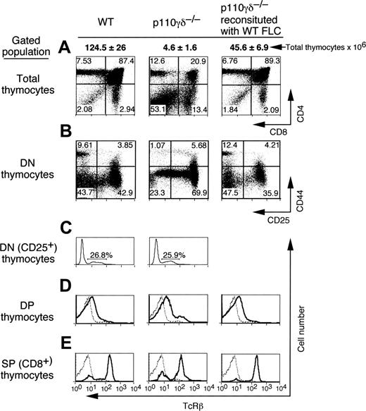
To determine the thymocyte population(s) most affected by the absence of PI3Kδ and PI3Kγ, flow cytometry analyses were performed to detect markers associated with thymocyte differentiation. Although the total number of CD4+ and CD8+ SP and DP cells were reduced overall, the absence of catalytic subunits had the greatest effect on the number of DP cells, typically the largest population of thymocytes in WT mice (Figure 2A). In contrast, DN cells were the preponderant population in p110γδ-/- thymi, as occurs, for instance, in RAG2-/- mice (Figure S2). In the latter, TCRB selection cannot occur at the DN3 stage, resulting in thymocyte death by apoptosis. Although a percentage of the DN3 population (CD44-CD25+) had increased in thymi of p110γδ-/- mice, these cells were still capable of differentiating to the DN4 stage (CD44-CD25-) (Figure 2B). We noted, however, that the populations of DN3 and DN4 thymocytes developing in p110γδ-/- mice appeared to be phenotypically different from those of WT mice. Specifically, there appeared to be a continuum of DN3 to DN4 cells expressing gradually lower levels of CD25+ T cells. Although there was some variation in the percentages of DN1 cells (1.07%-8.82%), we did observe a modest but reproducible increase in the percentages (but not the total numbers) of immature CD8+ SP thymocytes bearing low-level surface TCRB (Figure 2D). These cells are the direct precursors of DP thymocytes. Importantly, the proportion of DN3 cells in p110γδ-/- thymi that expressed TCRB protein was comparable to that of WT controls, as demonstrated by intracellular staining (Figure 2C). Thus, unlike RAG-deficient mice, the depletion of DP cells lacking p110 catalytic subunits does not appear to have resulted from a failure to undergo TCRB selection. The few remaining DP cells, however, still were capable of differentiating into TCRBhigh SP T cells, suggesting that positive selection may be intact (Figure 2D-E). In contrast, the reconstitution of lethally irradiated p110γδ-/- mice with WT FLC restored the proportions of DN, DP, and SP populations to those observed for WT littermates, suggesting that the combined activities of PI3Kδ and PI3Kγ in cells other than thymocytes are not critical for their overall development. Of note, this dramatic alteration in DP and DN thymocyte populations was not observed in p110δ- or p110γ-deficient animals (Figure S1).
Role of PI3Kδ and PI3Kγ in thymocyte development. Flow cytometry analysis of expression of CD4 and CD8 on total thymocyte population (A), CD25 and CD44 on DN thymocytes (B), intracellular TCRB in CD25+ DN thymocytes (C), and TCRB on the surfaces of DP (D) and CD8+ (E) thymocytes. Percentage of gated cells in a particular quadrant is indicated. Data are representative of 3 independent experiments. Total thymocyte counts are in bold (mean ± SE; n = 5).
Role of PI3Kδ and PI3Kγ in thymocyte development. Flow cytometry analysis of expression of CD4 and CD8 on total thymocyte population (A), CD25 and CD44 on DN thymocytes (B), intracellular TCRB in CD25+ DN thymocytes (C), and TCRB on the surfaces of DP (D) and CD8+ (E) thymocytes. Percentage of gated cells in a particular quadrant is indicated. Data are representative of 3 independent experiments. Total thymocyte counts are in bold (mean ± SE; n = 5).
Absence of PI3Kγ and PI3Kδ activity results in depletion of DP thymocytes in vitro
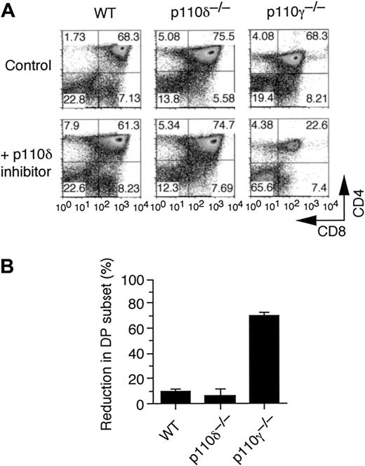
To confirm our in vivo observations and thus demonstrate that a deficiency in PI3Kδ contributed to the reduction in the DP thymocyte population, day 14.5 fetal thymi were harvested from WT, p110δ-/-, and p110γ-/- mice and were cultured in the presence of either p110δ-selective inhibitor IC87114 or vehicle control. Blockade of p110δ activity, in combination with genetic deletion of its gamma counterpart, resulted in a 69.2% ± 2.7% (mean ± SE) reduction in the population of CD4+CD8+ DP thymocytes (Figure 3A-B). Identical treatment of thymic cultures derived from p110δ-/- or WT control mice yielded a 10% or lower decrease in DP cells. Thus, blockade of p110δ function in p110γ-/- mice in lieu of its genetic deletion resulted in a similar alteration in the proportion of DP cells, as observed in p110γδ-/- animals (Figure 2B). Surprisingly, no significant alterations in the percentages of DN or DP populations were detected in p110γ-/- fetal thymi, suggesting that this class 1b PI3K does not have a major effect on thymocyte development under in vitro culture conditions. Moreover, the use of fetal thymic organ cultures excludes the possibility of glucocorticoid-induced thymocyte apoptosis as the primary mechanism for the observed reduction in cell numbers in vivo.32
Increased apoptosis in p110γδ-/- DP thymocytes
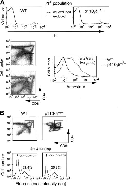
It is conceivable that the observed reduction in the DP thymocyte population in p110γδ-/- mice may result from either an increase in cell death or an overall decrease in the generation of this subset of cells. To determine whether this reduced cellularity might have reflected the former, we evaluated this population of cells for evidence of enhanced apoptosis. Flow cytometry analysis of PI-negative DP thymocytes revealed a 42% ± 6.1% increase in annexin V staining compared with WT littermates (Figure 4A). Moreover, DP thymocytes from p110γδ-/- mice showed decreased survival in in vitro cultures compared with WT or DP cells lacking p110γ or p110δ alone (data not shown). On the other hand, in vivo labeling of thymocytes with BrdU revealed no differences in the rate of generation of p110γδ-/- or WT DP 20 hours after the BrdU pulse (25.4 ± 5.7 vs 23.3 ± 0.2, respectively), indicating that PI3Kγ and PI3Kδ activity is not essential for the generation of DP thymocytes (Figure 4B).33 Rather, these results suggest that one major function of class 1 PI3Ks is to protect DP thymocytes from enhanced cell death, which, in turn, has a direct effect on thymic cellularity.
Contribution of p110γ and p110δ activity in thymocyte development in vitro. (A) Representative flow cytometry profiles of fetal thymic organ cultures harvested from day 14.5 WT, p110δ-/-, and p110γ-/- embryos that were treated with either vehicle control or p110δ-specific inhibitor IC87114 (10 μM) for 1 week. (B) Percentage reduction in DP thymocyte population after treatment with IC87114 compared with control treatment (mean ± SE; n = 3).
Contribution of p110γ and p110δ activity in thymocyte development in vitro. (A) Representative flow cytometry profiles of fetal thymic organ cultures harvested from day 14.5 WT, p110δ-/-, and p110γ-/- embryos that were treated with either vehicle control or p110δ-specific inhibitor IC87114 (10 μM) for 1 week. (B) Percentage reduction in DP thymocyte population after treatment with IC87114 compared with control treatment (mean ± SE; n = 3).
Akt/PBK phosphorylation and Ca2+ flux in the absence of PI3Kδ and PI3Kγ function
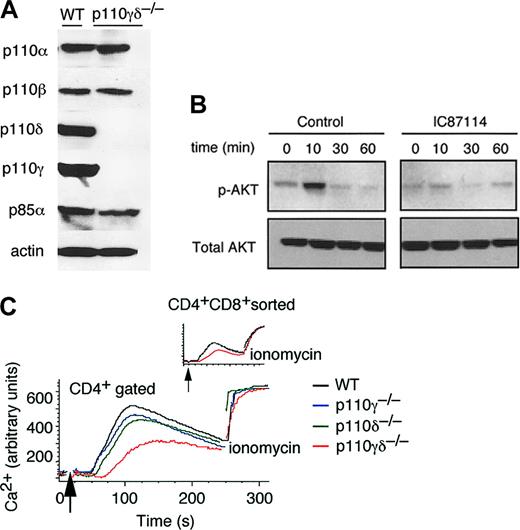
To date, no evidence exists demonstrating that p110δ is expressed or is functionally active in thymocytes. Indeed, Western blot analysis revealed the presence of this catalytic subunit and other class 1a and class 1b isoforms in thymocytes harvested from WT control mice (Figure 5A). Importantly, the expression pattern of p110α and p110β remained unchanged in thymocytes harvested from p110γδ-/- mice, with the exception of a small reduction in levels of the p85α regulatory subunit. The latter, however, is consistent with that previously reported for B cells obtained from mice lacking p110δ alone.29 To demonstrate that p110δ is functional in thymocytes, TCR-induced phosphorylation of the PI3K target Akt/PKB was used as an indirect measure of its activity. To isolate PI3Kδ activity, thymocytes from p110γ-/- mice were harvested and pretreated with vehicle control or with the p110δ-specific inhibitor IC87114 before TCR cross-linking. Our results indicate that PI3Kδ does contribute to antigen receptor-induced activation of Akt/PKB in thymocytes because the phosphorylated form of this protein kinase was not detected in p110γ-/- cells treated with IC87114 under our assay conditions (Figure 5B). Optimal TCR-induced Ca2+ flux required the activity of both class 1 PI3K isoforms (Figure 5C). Given that the proportion of cells capable of responding to TCR cross-linking in doubly-deficient thymi was different from that of its WT counterpart because of a larger proportion of DN cells in the former, we also evaluated Ca2+ flux in DP cells sorted from p110γδ-/- mice. Results indicate the persistence of this attenuated response, implicating both PI3Kδ and PI3Kγ as important mediators of antigen receptor signals in DP thymocytes (Figure 5C, inset).
DP thymocytes lacking p110γ and p110δ are prone to apoptosis. (A) Representative flow cytometry profiles of annexin V staining of the PI- population of DP thymocytes live-gated from WT control and p110γδ-/- mice (n = 3). PI staining of the live-gated population of thymocytes was performed first to identify and thus exclude necrotic cells as defined by forward- and side-scatter parameters. Gates in the CD4+ and CD8+ panels indicate the DP thymocyte population gated for analysis of annexin V staining (histogram, which was exclusive of the PI+ staining, as stated). (B) Density plots of DP thymocytes harvested from WT control and p110γδ-/- mice after treatment with BrdU. Representative histograms depict the percentages of DP cells that stained with BrdU (n = 3 for each group).
DP thymocytes lacking p110γ and p110δ are prone to apoptosis. (A) Representative flow cytometry profiles of annexin V staining of the PI- population of DP thymocytes live-gated from WT control and p110γδ-/- mice (n = 3). PI staining of the live-gated population of thymocytes was performed first to identify and thus exclude necrotic cells as defined by forward- and side-scatter parameters. Gates in the CD4+ and CD8+ panels indicate the DP thymocyte population gated for analysis of annexin V staining (histogram, which was exclusive of the PI+ staining, as stated). (B) Density plots of DP thymocytes harvested from WT control and p110γδ-/- mice after treatment with BrdU. Representative histograms depict the percentages of DP cells that stained with BrdU (n = 3 for each group).
Evaluation for p110δ protein and activity in thymocytes. (A) Representative immunoblots of class 1a and 1b p110 subunits expressed in thymocytes harvested from WT control and p110γδ-/- mice. Western blot of β-actin illustrates equal loading of proteins. (B) Detection of Akt/PKB in Western blots of total lysates from p110γ-/- thymocytes treated with vehicle control or the p110δ-specific inhibitor IC87114 (10 μM) before TCR cross-linking. (C) Ca2+ flux in CD4+-gated thymocytes in WT control, p110γ-/-, p110δ-/-, and p110γδ-/- mice in response to TCR cross-linking. Ca2+ flux in CD4+CD8+-sorted thymocytes from WT control and p110γδ-/- animals is shown for comparison (inset). Data are representative of 3 to 4 separate experiments.
Evaluation for p110δ protein and activity in thymocytes. (A) Representative immunoblots of class 1a and 1b p110 subunits expressed in thymocytes harvested from WT control and p110γδ-/- mice. Western blot of β-actin illustrates equal loading of proteins. (B) Detection of Akt/PKB in Western blots of total lysates from p110γ-/- thymocytes treated with vehicle control or the p110δ-specific inhibitor IC87114 (10 μM) before TCR cross-linking. (C) Ca2+ flux in CD4+-gated thymocytes in WT control, p110γ-/-, p110δ-/-, and p110γδ-/- mice in response to TCR cross-linking. Ca2+ flux in CD4+CD8+-sorted thymocytes from WT control and p110γδ-/- animals is shown for comparison (inset). Data are representative of 3 to 4 separate experiments.
Effect of PI3Kδ and PI3Kγ deficiency on T-cell numbers and Ca2+ mobilization
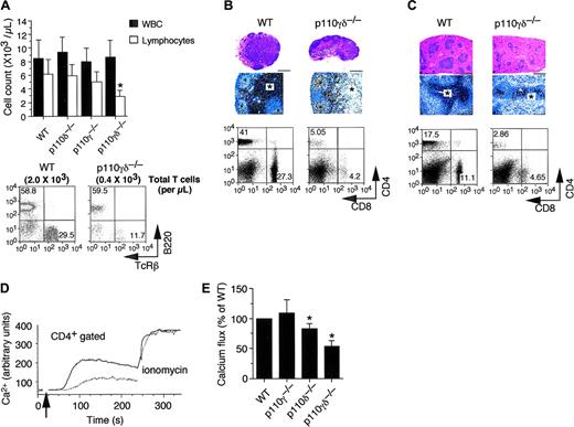
The abnormalities observed in T-cell numbers and TCR-signaling associated with a deficiency in p110γ and p110δ catalytic subunits was not limited to the thymus but persisted in secondary lymphoid organs. In particular, a defect in DP cell development appears to have a direct effect on extrathymic T-cell populations. Although the white blood cell count was similar among all genetic phenotypes tested, the total lymphocyte count was significantly reduced in p110γδ-/- mice compared with WT littermates (2.9 ± 1.1 K/μLvs 6.2 ± 2.1 K/μL, respectively; Figure 6A). Moreover, this corresponded to a 5-fold reduction in total number of circulating TCRB+ cells in the former. Similarly, T-cell populations in peripheral lymph nodes and spleen were diminished, as determined by immunohistology (Figure 6B-C). No such dramatic reduction of T cells was observed in secondary lymphoid organs in p110γ- or p110δ-deficient mice (data not shown). TCR-induced Ca2+ flux in mature T cells also relied on the activity of class 1 PI3Ks, mirroring the defect observed in thymocytes. For example, we observed a greater than 45% reduction in Ca2+ flux in CD4+ T cells from p110γδ-/- animals compared with WT littermates (Figure 6D-E). No defect was observed for p110γ-deficient cells, results consistent with those of a previous study.23 Moreover, only a modest reduction (approximately 15%) was noted for CD4+ T cells from p110δ-/- mice. These results suggest that PI3Kγ and PI3Kδ must work in concert to ensure effective signaling through this antigen receptor in mature T cells.
Effect of p110δ and p110γ deletion on extrathymic T cells. (A) Cell counts and flow cytomtery analysis of surface expression of TCRB were performed on whole blood and isolated PBMCs, respectively, whereas CD4 and CD8 expression was evaluated on total cells harvested from peripheral lymph nodes (B) and spleens (C) of WT control and p110γδ-/-. Histologic examination of hematoxylin and eosin-stained lymph node (B) and splenic (C) sections (objective magnifications each 4 ×). Delineation of the T-cell population by immunoperoxidase detection of CD3+ counterstained with Meyer hematoxylin was also performed (100 ×; scale bar, 100 μm). Ca2+ flux in CD4+-gated T cells from WT control (D-E), p110γ-/- (E), p110δ-/- (E), and p110γδ-/- (D-E) mice in response to TCR cross-linking. Values depicted represent the mean ± SE for 3 independent experiments performed in duplicate or triplicate. *Statistical significance compared with WT control (P < .05).
Effect of p110δ and p110γ deletion on extrathymic T cells. (A) Cell counts and flow cytomtery analysis of surface expression of TCRB were performed on whole blood and isolated PBMCs, respectively, whereas CD4 and CD8 expression was evaluated on total cells harvested from peripheral lymph nodes (B) and spleens (C) of WT control and p110γδ-/-. Histologic examination of hematoxylin and eosin-stained lymph node (B) and splenic (C) sections (objective magnifications each 4 ×). Delineation of the T-cell population by immunoperoxidase detection of CD3+ counterstained with Meyer hematoxylin was also performed (100 ×; scale bar, 100 μm). Ca2+ flux in CD4+-gated T cells from WT control (D-E), p110γ-/- (E), p110δ-/- (E), and p110γδ-/- (D-E) mice in response to TCR cross-linking. Values depicted represent the mean ± SE for 3 independent experiments performed in duplicate or triplicate. *Statistical significance compared with WT control (P < .05).
Discussion
Class 1 PI3Ks are essential for supporting innate and adaptive immune responses. By contrast, previous studies suggest they play a more limited role in thymocyte development and differentiation. We describe here a novel defect in thymocyte development in mice that is dependent on the activities of 2 distinct subclasses of PI3Ks. Genetic deletion of p110δ, in conjunction with its gamma counterpart, had a dramatic and unanticipated effect on thymus size, cellularity, and architecture. In particular, the combined absence of these 2 catalytic subunits resulted in a more than 4-fold reduction in the percentage and a 10- to 30-fold reduction in total numbers of cortical CD4+CD8+ DP thymocytes compared with WT littermates. Depletion of DP cells in p110γδ-/- thymi was accompanied by a corresponding compensatory increase in percentages, but not total numbers, of DN thymocytes and a paucity in the number of mature CD4+ and CD8+ SP T cells found in blood and secondary lymphoid organs. Thus, the reduction in DP thymocytes is of importance as relates to T-lymphocyte production because there may be insufficient quantities of this subset in p110γδ-/- thymi to yield normal numbers of mature SP cells compared with WT animals (1.0 × 10⁶ ± 0.3 vs 109.6 × 10⁶ ± 22.6 DP cells, respectively).
Mechanistically, we propose that the combined activity of PI3Kδ and PI3Kγ is critical to the survival of DP thymocytes in vivo. Indeed, given the inherent susceptibility of DP thymocytes to programmed cell death, presumably because of the down-regulation of the antiapoptotic Bcl-2 protein at this stage of development, this population would be particularly vulnerable to the loss of survival signals generated by class 1 PI3Ks. In this context, an antiapoptotic role has been indicated by the immunologic consequences of constitutive PI3K signaling that occurs in the absence of the tumor-suppressor gene PTEN, a phosphatase that converts PIP3 to PIP2. Selective deletion of PTEN in murine T cells not only results in uncontrolled proliferation of this lymphocyte subset, it leads to autoimmunity that is thought to be a consequence of impaired programmed cell death in the thymus.34 Thus, our ability to demonstrate that class 1 PI3Ks do indeed participate in PIP3 generation in thymocytes was central to this hypothesis (Figure 5B). Further evidence in support of our claim is provided by annexin V staining. A significant percentage of DP cells in p110γδ-/- thymi were annexin V-positive, a marker indicative of apoptosis, unlike that of WT and single null animals. Moreover, our ability to reproduce this in vivo abnormality in thymocyte development by exogenously blocking the activity of PI3Kδ in cultured fetal thymi harvested from E14 p110γ-/- embryos suggests an inherent defect in thymocyte signaling. Thus, we exclude a role for external factors such as a potential elevation in glucocorticoid levels in p110γδ-/- animals in this process.
Although the activity of PI3Kδ and PI3Kγ is involved in maintaining DP thymocyte survival, it is conceivable that they could participate in TCRβ-selection. During normal development, TCRβ chain gene rearrangement and expression reaches completion at the DN3 stage, permitting the formation of the pre-TCR complex. As a result, DN3 thymocytes can activate several signaling pathways, including lck/fyn and ZAP-70/Syk tyrosine kinases, SLP-76 and LAT linker proteins, Vav-family GEFs, and PLCγ1 phospholipase, that collectively mediate the transition of these cells to the CD4+CD8+ DP stage.35-39 Consequently, mice lacking structural or signaling components of the pre-TCR complex exhibit a developmental block at the DN3 stage. In this context, PI3K activity has been implicated in Vav and PLCγ activation and Ca2+ flux through direct (PIP3 binding to PH domains) and indirect (induction of Tec-family kinases) mechanisms.40,41 Indeed p110γδ-/- thymocytes show impaired TCR-mediated Ca2+ flux in vitro. Thus, a deficiency in p110δ and p110γ could result in the perturbation of DN to DP checkpoint through defective pre-TCR signaling. Our data, however, do not appear to support this mechanism because equal proportions of p110γδ-/- compared with WT DN3 thymocytes express TCRβ intracellularly. Moreover, pre-TCR complex-mediated events such as proliferative expansion, loss of CD25+ expression (transition to the DN4 stage), and acquisition of CD8+ and CD4+ coreceptors (transition to DP stage) were readily visible in thymocytes from p110γδ-/- mice. Thus, the resultant phenotype is clearly distinct from that associated with known defects in TCRB selection, such as RAG deficiency (Figure S2). Importantly, animals lacking both PI3Kδ and PI3Kγ can still generate DP thymocytes at rates similar to those in WT mice, as indicated by BrdU-incorporation experiments. Despite this finding, we noted that the populations of DN3 and DN4 thymocytes in p110γδ-/- mice were phenotypically different from those of WT mice because there appeared to be a continuum of DN3 to DN4 cells expressing gradually lower levels of CD25+ T cells in the former. Although at present we do not completely understand the mechanism for this abnormality, the most plausible explanation is that the gradual loss of CD25+ cells simply mirrors Bcl-2 down-regulation and the subsequent necessity for class 1 PI3K-dependent survival signals.
Although we provided evidence that the combined activities of PI3Kδ and PI3Kγ are essential for thymocyte development, it appears that either subclass is sufficient to maintain T-cell production. This potential redundancy in function may ensure that adequate levels of PIP3 are maintained to protect cells from proapoptotic stimuli. How these 2 PI3K subclasses, which are activated through distinct pathways, are linked through receptors (such as the TCR) that promote the development and survival of immature DP thymocytes remains to be determined. That said, it has been demonstrated that ligation of an ITAM-bearing receptor on cells, such as FcγRI, can result in the activation of class 1a and class 1b PI3Ks.42 Moreover, it was speculated that the activation of p110γ, which typically occurs through G protein-coupled receptors, may involve the Tec family of tyrosine kinases, which have the capacity to physically interact with PIP3 and heterotrimeric G-protein subunits.43 Such a scenario may hold true for T cells, because PI3Ks and Tec kinases are intricately linked in TCR-mediated signaling. For example, Tec kinases are required for the regulation of PLCγ activity and Ca2+ signaling, an event that involves PI3Kδ.25 Thus, it is conceivable that in response to PI3Kδ activation or other class 1a isoforms, a Tec tyrosine kinase family member will be become localized at the plasma membrane through interactions with PIP3, which in turn may recruit a heterotrimeric G-protein that could activate p110γ and thus enhance PIP3 production. Further investigation is warranted to determine the molecular mechanism(s) that link the activities of these PI3K subclasses in T cells.
Prepublished online as Blood First Edition Paper, November 22, 2005; DOI 10.1182/blood-2005-08-3300.
Supported by National Institutes of Health grants HL75805-01A2 (T.G.D.) and HLA106107702 (W.S.) and by the American Lung Association (T.G.D.).
J.D. and K.P. are employed by ICOS Corp, whose potential product (IC87114) was studied in the present work.
The online version of this article contains a data supplement.
The publication costs of this article were defrayed in part by page charge payment. Therefore, and solely to indicate this fact, this article is hereby marked “advertisement” in accordance with 18 U.S.C. section 1734.
We thank Hairu Zhou and Kui Tan for genotyping and Francesa Gentili for histology.

![Figure 1. Role of class 1 PI3Ks in supporting thymic architecture and cellularity. Representative micrographs depicting thymus size and hematoxylin and eosin-stained sections from WT control (i,iv) and p110γδ-/- (ii,v) mice and from p110γδ-/- animals reconstituted with WT fetal liver cells (iii,vi). Delineation of the thymic medulla in these animals (vii-ix) was performed by immunoperoxidase detection of Keratin5+ epithelial cells counterstained with Meyer hematoxylin. Cortical and medullary regions in the thymus of p110γδ-/- mice are indistinguishable, unlike those of WT and reconstituted animals. (Objective, magnification 40 × 4 ×/numerical aperture [NA] 0.16) in panels iv to vi (scale bar, 500 μm) and 200 × (objective, 20 ×/0.7 NA) in panels vii to ix (scale bar, 100 μm). TC indicates thymic cortex; TM, thymic medulla. Data are representative of at least 3 animals for each genotype depicted.](https://ash.silverchair-cdn.com/ash/content_public/journal/blood/107/6/10.1182_blood-2005-08-3300/4/m_zh80060692830001.jpeg?Expires=1769487749&Signature=H5hT0-ANd37b5hkEt9ULOP56h1nLhqV-Y5J1JH56VTk23rgt8hontbfEN-5ACq01AFlgdlZsF4wrfvTuaLwjzT7dLCN6PHkKgEw9PCX9ZGkTAcCiB~oPMrrlnyftX-OXREwCsI2SCjym-b1cBGAz~mfQQj3kd6T~4SPdDJzi58WMZWS5vVYtwaU2hEdQ79TwdJWVKjNBqYb9mHxAyy~la3omWFjpYa03xT4CQj2XKz3Y3-~je~I7ntG63iP782JmRr~78XBJrFux8qbfa8Hm46AbaDf~63VgRHOSgxMJrxMlGHqDTPLf0PFI8qHXfMAl6T7dQmROPiizPXsjykm0JQ__&Key-Pair-Id=APKAIE5G5CRDK6RD3PGA)
