Abstract
Dendritic cells (DCs) from patients with cancer are functionally defective, but the molecular mechanisms underlying these defects are poorly understood. In this study, we used the murine 5TGM1 myeloma model to examine the effects and mechanisms of tumor-derived factors on the differentiation and function of DCs. Myeloma cells or tumor culture conditioning medium (TCCM) were shown to inhibit the differentiation and function of BM-derived DCs (BMDCs), as evidenced by the down-regulated expression of DC-related surface molecules, decreased IL-12, and compromised capacity of the cells to activate allospecific T cells. Moreover, TCCM-treated BMDCs were inferior to normal BMDCs at priming tumor-specific immune responses in vivo. Neutralizing antibodies against IL-6, IL-10, and TGF-β partially abrogated the effects. TCCM treatment activated p38 mitogen-activated protein kinase (MAPK) and Janus kinase (JNK) but inhibited extracellular regulated kinase (ERK). Inhibiting p38 MAPK restored the phenotype, cytokine secretion, and function of TCCM-treated BMDCs. BMDCs from cultures with TCCM and p38 inhibitor was as efficacious as normal BMDCs at inducing tumor-specific antibody, type 1 T cell, and cytotoxic T lymphocyte (CTL) responses and at prolonging mouse survival. Thus, our results suggested that tumor-induced p38 MAPK activation and ERK inhibition in DCs may be a new mechanism for tumor evasion and that regulating these pathways during DC differentiation provides new strategies for generating potent DC vaccines for immunotherapy in patients with cancer. (Blood. 2006;107:2432-2439)
Introduction
Dendritic cell (DC)-based immunotherapy holds great promise for treating malignancies,1-3 including multiple myeloma.2,4 However, preliminary reports of DC vaccines in human trials have demonstrated minor clinical responses.1,2 The lack of effectiveness of DC vaccines in tumor patients may be associated at least in part with defects in DCs.5-8 Accumulating evidence shows that DCs generated ex vivo from their progenitor cells in tumor patients or tumor-bearing animals are functionally abnormal.5-8 Thus, a better understanding of the molecular mechanisms underlying the impairment of DC functions by tumor-derived factors and restoration of functions of DCs from tumor patients will be important for the application of DC-based immunotherapy in multiple myeloma and other malignancies.
The 5T murine model of myeloma, originally described by Radl et al9 in an inbred substrain of C57 black mice (C57BL/KaLwRij substrain), offers a unique opportunity for in vivo studies of myeloma biology, drug treatment, and tumor immunology. Several of the 5T myeloma lines closely mimic myeloma in humans, with monoclonal gammopathy, marrow replacement, focal osteolytic bone lesions, hind limb paralysis, and occasionally hypercalcemia.9,10 With the use of this murine myeloma model, the aim of this study was to examine whether and how tumor cells and their derived factors affected the differentiation and generation of DCs and whether it was possible to restore cell function. Our results showed that culture of murine BM cells with myeloma cells, both in a Transwell system and by direct contact, and with tumor culture conditioning medium (TCCM) impaired the differentiation and generation of BM-derived DCs (BMDCs) and that myeloma-derived cytokines, such as IL-6, IL-10, and TGF-β, were partially responsible. Mitogen-activated protein kinase (MAPK) p38, which was activated in the cultured BM cells by treatment with myeloma cells or TCCM, played an important and detrimental role in the differentiation of BMDCs. Inhibiting p38 MAPK activity in BM cells cultured in the presence of TCCM restored the generation of functional BMDCs.
Materials and methods
Mice, cell lines, and reagents
BALB/c and C57BL/KaLwRij mice were purchased from the Jackson Laboratory (Bar Harbor, ME) and Harlan CPB (Zeist, The Netherlands), respectively. The murine myeloma cell line 5TGM111,12 was kindly provided by Dr G.R. Mundy at the University of Texas Health Science at San Antonio. Murine myeloma cell lines MCP-11 and MOPC-315 were purchased from ATCC (Rockville, MD).
p38 MAPK inhibitors SB203580 and SB202190, p38 MAPK inhibitor 3, and JNK inhibitor 2 were purchased from EMD Biosciences (San Diego, CA). These inhibitors were dissolved in DMSO (Sigma, St Louis, MO), and the final concentration of DMSO in cultures was 0.05%. IL-6, IL-10, VEGF, MCP-1, MCP-5, RANTES, TGF-β1, and all their neutralizing or blocking antibodies were purchased from R&D Systems (Minneapolis, MN).
Preparation of TCCM
5TGM1 cells were cultured in IMDM complete medium; 24 hours later, supernatants were harvested, filtered, and concentrated 10-fold using an Amicon Ultra Filter (Millipore, Bedford, MA). Concentrated TCCM was divided into aliquots and stored at -80°C until use. Unless otherwise noted, all TCCM used in the experiments was from 5TGM1 cells. Medium control, prepared from freshly prepared IMDM complete medium in a manner similar to TCCM preparation, and TCCM from murine myeloma cell lines MCP-11 and MOPC-315 were used in the experiments.
Generation of BMDCs and treatment with myeloma cells
BMDCs were generated as described previously.13 BM cells were flushed from tibias and femurs of C57BL/KaLwRij mice and were cultured in RPMI 1640 medium supplemented with 10% heat-inactivated fetal bovine serum with the addition of 20 ng/mL GM-CSF (R&D Systems). At day 4 (d4), 90% of the medium was taken out and was replaced with fresh medium containing 10 ng/mL GM-CSF. At d8, cultures were replaced with fresh medium containing TNF-α (10 ng/mL) and IL-1β (10 ng/mL) (R&D Systems) for 48 hours to mature the cells. Portions of the cells were taken out on d8 and d10 for analysis.
To examine the effects of myeloma cells or their derived factors on the differentiation of BMDCs, TCCM was added to the cell cultures (10% TCCM to 90% fresh medium) on d0 and on d4, when the medium was changed. No additional TCCM was added on d8. In other experiments, BM cells were cocultured with 5TGM1 (8 × 10⁴ cells/mL) placed on a 0.4-μm transwell insert (Nunc, Naperville, IL) or were cocultured directly with irradiated 5TGM1 cells (8 × 10⁴/mL). Medium changes and maturation induction were similar to those for normal BMDCs.
Flow cytometry analysis
BMDCs or cultured cells were incubated with FITC- or PE-conjugated mAbs against CD11c, CD40, CD80, CD86, and major histocompatibility complex (MHC) class II molecules (BD PharMingen, San Diego, CA) for 30 minutes at 4°C, washed twice, and resuspended in PBS. Analyses of fluorescence staining were performed using a FACScan (Becton Dickinson; San Jose, CA).
Detection of cytokine expression and production
Intracellular IL-12 and IL-10 staining for BMDCs and IFN-γ and IL-4 staining for T cells were performed using a Cytofix/Cytoperm kit (BD Biosciences) according to the manufacturer's instructions. Cells (BMDCs or activated T cells) were stained with FITC-labeled anti-CD11c mAb or CD3 and then with PE-labeled anti-IL-12, -IL-10, -IFN-γ, or -IL-4 (BD PharMingen); this was followed by washing and analysis.
Enzyme-linked immunosorbent assay (ELISA) for IL-12 and TGF-β1 was used to measure the secreted cytokines. Supernatants from BM cell cultures at different time points were collected, and the cytokines in the supernatants were quantified using commercially available ELISA kits (R&D Systems).
Cytokine array analysis (RayBiotech, Norcross, GA) was used to examine a broader spectrum of cytokines secreted by different cells. Supernatants from 24-hour culture of 5TGM1 and 4-day cultures of BM cells with or without addition of TCCM were collected for analysis. Cytokines in the supernatants were analyzed using cytokine array kits according to the manufacturer's instructions. Mean intensities of dots on the membranes, which represented relative protein levels of cytokines, were semiquantified by RayBiotech.
RT-PCR for detecting cytokine mRNA expression
Reverse transcription-polymerase chain reaction (RT-PCR) was used to detect cytokine mRNA expression by BMDCs or cultured cells on day 5 of culture. Total cellular RNA was extracted by Tri-Reagent (Molecular Research Center, Cincinnati, OH). Reverse transcription was performed using a Transcriptor First Strand cDNA Synthesis Kit (Roche, Indianapolis, IN). Taq DNA polymerase was purchased from Roche, and PCR was performed according to instructions in a thermal cycler. Primer sets used for these analyses are listed in Table 1. Each of the primer sets was confirmed by running samples on agarose gels. β-Actin transcript levels were used to normalize the amount of cDNA in each sample. Water controls were always included in each experiment.
Western blot analysis
To examine intracellular signaling, phosphorylated (p) p38 and pERK, pMEK, pJNK, and pIκB-α were detected as previously described.14 Samples were subjected to sodium dodecyl sulfate-polyacrylamide gel electrophoresis (SDS-PAGE), and, after transfer to nitrocellulose membrane and subsequent blocking, the membranes were immunoblotted with respective antibodies (Cell Signaling, Beverly, MA) and were visualized with alkaline phosphatase-conjugated secondary antibodies followed by enhanced chemiluminescence (Bio-Rad Laboratories, Hercules, CA) and autoradiography.
To examine p38 activity in the cells, a p38 functional assay kit (US Biological, Swampscott, MA) was used to study the capacity of p38 to phosphorylate one of its substrates, ATF-2, by Western blotting. Nonphosphorylated p38 was examined and served as control for cell lysate proteins.
In vitro functional test for BMDCs and cultured cells
Splenic T cells from BALB/c mice (H-2d) were isolated and used as alloreactive T cells, as described previously.14 Mature BMDCs, TCCM-treated cells, or TCCM+SB203580-treated cells (all from H-2b C57BL/KaLwRij mice) were irradiated and used as stimulatory cells. T cells (2 × 10⁵/mL) and stimulatory cells were seeded into 96-well U-bottom plates (Corning, Corning, NY) at different (T/DC) ratios. Cultures with T cells alone served as a control. On day 5, cells were pulsed with 1 μCi (0.037 MBq)/well 3[H]-thymidine and, 18 hours later, were harvested, and radioactivity was measured. All tests were performed in triplicate, and the results are expressed as mean cpm.
In vivo functional test for BMDCs and cultured cells
We compared the ability of the cells to immunize C57BL/KaLwRij mice against the idiotype (Id) protein secreted by the 5TGM1 myeloma cells (IgG2b) and to treat established myeloma. To enhance the immunogenicity of Id protein, Id-KLH (EMD Biosciences, La Jolla, CA) conjugate was made using glutaraldehyde (Sigma), as described previously.15 Immature BMDCs, TCCM-treated cells, TCCM+SB203580-treated cells, and SB203580-treated cells from d8 cultures were collected and pulsed with Id-KLH conjugate at a concentration of 50 μg/mL. Four hours later, TNF-α and IL-1β were added to induce the cells to maturation. After 48 hours of culture, Id-KLH-pulsed, mature BMDCs were collected and used as vaccines to immunize the mice.
The experiments included 5 groups of mice, each receiving injections of PBS, Id-KLH-pulsed BMDCs, TCCM-treated cells, TCCM+SB203580-treated cells, or SB203580-treated cells. DC vaccines were injected subcutaneously at weeks 0, 1, and 2 with 1 × 10⁶ cells per injection. Serum samples were collected weekly. At week 3, mice were killed, and spleens were removed. Serum samples and splenocytes were used in the subsequent experiments. Mouse studies were approved by the Institutional Animal Care and Use Committee of the University of Texas M.D. Anderson Cancer Center.
ELISA for antibody detection. Mouse serum was diluted in microtiter plates coated with purified Id or KLH. Antibodies in the serum to Id or KLH were detected by horseradish peroxidase (HRP)-conjugated goat anti-mouse IgG (Caltag Laboratories, Burlingame, CA). To detect antibodies against the Id protein, HRP-conjugated goat anti-mouse IgG was preabsorbed against Id protein to reduce unspecific binding. Absorption was evaluated at an optical density of 490 nm.
Cytotoxicity assay. Standard chromium 51 (51Cr)-release assay was performed to examine the cytotoxicity of T cells.16 In brief, splenocytes of 5 mice from each group were pooled and cultured with irradiated syngeneic stimulators (5TGM1 cells) for 5 days, and T cells were then incubated with 51Cr-labeled 5TGM1 cells (104 cells/well) at different effector-target cell ratios. MPC-11 cells were used as control target cells. After 4 hours of culture, 100 μL supernatant was collected, and radioactivity was measured. Percentage specific lysis was calculated using the following formula: percentage specific lysis = (experimental counts - spontaneous counts)/(maximal counts - spontaneous counts).
Statistical analysis
Student t test was used for the comparison of various experimental groups, and the Kaplan-Meier test was used for the comparison of mouse survival. Significance was set at P below .05.
Results
Phenotype, cytokine expression, and function of BMDCs in cultures with or without myeloma cells or their derived factors
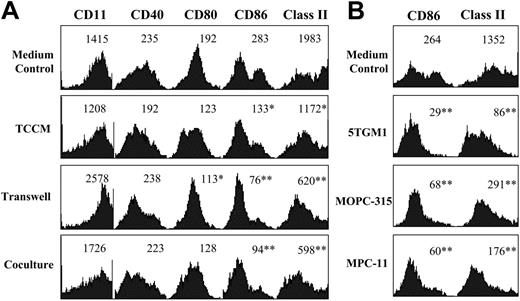
To determine the effects of 5TGM1 myeloma cells on DC phenotype, BM cells were cultured with GM-CSF in the presence or absence of TCCM or with 5TGM1 cells in a transwell system or in direct contact with irradiated 5TGM1 cells. In most of the experiments described, d10 cultured cells (mature BMDCs) were used for analyses. Expression of DC-related surface molecules CD86 and MHC class II were significantly inhibited by the presence of myeloma cells or TCCM (P < .05 to P < .01; Figure 1A), indicating that soluble factors secreted by myeloma cells rather than by direct cell contact impaired the phenotype of cultured BMDCs.
Phenotypes of normal and myeloma-treated BMDCs. (A) Expression of DC-related molecules on normal DCs (medium control) and BMDCs cultured in the presence of TCCM, 5TGM1 cells on Transwell insets, or irradiated 5TGM1 cells in a direct coculture. (B) Expression of CD86 and MHC class II on normal mature BMDCs (medium control) and on cells in cultures with the addition of TCCM from 5TGM1, MOPC-315, or MPC-11 myeloma cells. Cells from d10 cultures were analyzed. Values above histograms represent mean fluorescence intensity. Representative results of 4 experiments are shown. *P < .05; **P < 0.01 (compared with normal controls).
Phenotypes of normal and myeloma-treated BMDCs. (A) Expression of DC-related molecules on normal DCs (medium control) and BMDCs cultured in the presence of TCCM, 5TGM1 cells on Transwell insets, or irradiated 5TGM1 cells in a direct coculture. (B) Expression of CD86 and MHC class II on normal mature BMDCs (medium control) and on cells in cultures with the addition of TCCM from 5TGM1, MOPC-315, or MPC-11 myeloma cells. Cells from d10 cultures were analyzed. Values above histograms represent mean fluorescence intensity. Representative results of 4 experiments are shown. *P < .05; **P < 0.01 (compared with normal controls).
To verify these results, we tested TCCM from 2 other myeloma cell lines, MPC-11 and MOPC-315. Indeed, the addition of 10% TCCM from these 2 cell lines also impaired the phenotype of the cultured cells, as evidenced by the down-regulated expression of CD86 and MHC class II molecules (Figure 1B) and other surface antigens (data not shown).
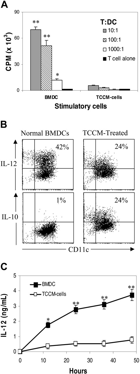
We next determined whether the function of the treated BMDCs was also affected. As shown in Figure 2A, the allostimulatory capacity of TCCM-treated cells, compared with normal BMDCs, was dramatically reduced (P < .01). This phenomenon was also observed with cells cultured with 5TGM1 in transwell or in direct contact or with the addition of TCCM from MPC-11 and MOPC-315 cells (data not shown). The poor allostimulatory capacity of the cells did not appear to contribute to apoptosis because Annexin-V staining revealed no difference among the cultures (data not shown).
Given that the function of DCs is associated with the relative amounts of IL-12 and IL-10 secretion,17 we performed intracellular staining of these cytokines. As shown by the representative experiment depicted in Figure 2B, normal, mature BMDCs contained approximately 42% (42% ± 6.9%; from 3 independent experiments, same as below) IL-12-expressing cells and only 1% (1% ± 1.3%) IL-10-expressing cells. TCCM treatment significantly reduced the percentage of IL-12-expressing cells to approximately 24% (24% ± 3.7%; P < .05) and increased the percentage of IL-10-expressing cells to 24% (24% ± 4.3%; P < .01). To confirm these results, ELISA was used to quantify secreted IL-12 by the cultured cells during DC maturation (d8-d10). As shown in Figure 2C, compared with normally maturing BMDCs in which a time-dependent increase in the secretion of IL-12 was detected, significantly reduced (5- to 8-fold less) secretion of IL-12 was noted in cells cultured with the addition of TCCM (P < .01). These results indicated that myeloma-derived factors impaired the phenotype and cytokine secretion of BMDCs, leading to poor antigen presentation capacity of the cells.
Identification of tumor-derived factors affecting BMDC differentiation
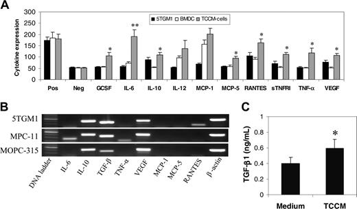
To determine the soluble factors responsible for the observed effects, cytokine array analysis was used to examine cytokines in the culture media of 5TGM1, normal BMDCs, and TCMM-treated BMDCs. As shown in Figure 3A, 5TGM1 cells secreted relatively large amounts of regulated on activation, normal T-cell expressed and secreted (RANTES), IL-10, vascular endothelial growth factor (VEGF), and soluble TNF receptor-1 (sTNFR1) and modest amounts of IL-6 and monocyte chemotactic protein-1 (MCP-1). Because this cytokine array does not include TGF-β, which might be an important negative regulator of DC differentiation,18-22 RT-PCR was used to detect TGF-β mRNA expression in 5TGM1 myeloma cells. As shown in Figure 3B, TGF-β mRNA was detected. Secretion of this cytokine, detected with an ELISA kit for TGF-β1 quantification (R&D Systems), showed that a low level of TGF-β1 protein was present in the culture medium of 5TGM1 cells (P < .05; Figure 3C). The same assays were also used to detect cytokine expression and production in 2 other myeloma cell lines, MPC-11 and MOPC-315, and their conditioning media. As exemplified by the results of RT-PCR, similar levels of IL-6, IL-10, TGF-β, and VEGF mRNA were detected in all 3 myeloma cell lines (Figure 3B).
Allostimulatory capacity and cytokine production profiles of normal and TCCM-treated BMDCs. (A) MLR examining the ability of normal and TCCM-treated BMDCs to activate allospecific T cells. Results shown are the mean ± SD of 3 experiments. (B) Intracellular cytokine staining showing the percentages of IL-12- and IL-10-expressing CD11c+ cells in normal and TCCM-treated BMDCs. Cells from d10 cultures were analyzed. Representative results of 3 experiments are shown. (C) ELISA results demonstrating the secretion of IL-12 by normal maturing BMDCs and TCCM-treated cells during d8 to d10. Supernatants from cell cultures with the addition of TNF-α and IL-1β were collected at 0, 12, 24, 36, and 48 hours after the addition of the cytokines. Concentration of IL-12 in supernatants was quantified by ELISA. Results shown are the mean ± SD of 3 experiments. *P < .05; **P < .01 (compared with normal controls).
Allostimulatory capacity and cytokine production profiles of normal and TCCM-treated BMDCs. (A) MLR examining the ability of normal and TCCM-treated BMDCs to activate allospecific T cells. Results shown are the mean ± SD of 3 experiments. (B) Intracellular cytokine staining showing the percentages of IL-12- and IL-10-expressing CD11c+ cells in normal and TCCM-treated BMDCs. Cells from d10 cultures were analyzed. Representative results of 3 experiments are shown. (C) ELISA results demonstrating the secretion of IL-12 by normal maturing BMDCs and TCCM-treated cells during d8 to d10. Supernatants from cell cultures with the addition of TNF-α and IL-1β were collected at 0, 12, 24, 36, and 48 hours after the addition of the cytokines. Concentration of IL-12 in supernatants was quantified by ELISA. Results shown are the mean ± SD of 3 experiments. *P < .05; **P < .01 (compared with normal controls).
Array analysis revealed that during differentiation, cultured BM cells secreted a low level of IL-6 and modest levels of IL-12, MCP-1, and RANTES (Figure 3A), whereas in cultures with the added TCCM, the levels of G-CSF, IL-6, MCP-5, RANTES, sTNFR1, TNF-α, and VEGF were significantly increased (P < .05 to P < .01). Hence, these cytokines might have contributed to the detrimental effects through autocrine action on the cultured BMDCs.
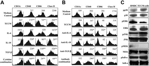
We next determined which cytokines were important for impairing BMDC differentiation. Except for TGF-β, which we added at a final concentration of 5 ng/mL, we first added these cytokines individually or in combination to the cell culture at a final concentration of 10 ng/mL (these may be 2- to 10-fold higher than levels detected in TCCM; for descriptions, see Figures 3 and 6). Cultures with the added 10% TCCM served as a positive control. As shown in Figure 4A, adding IL-6, TGF-β, and, more important, IL-10 or a combination of these 3 cytokines impaired the phenotype of the cultured cells (P < .05 to P < .01), whereas adding RANTES, MCP-1, and VEGF had no effect (data not shown).
Neutralizing antibodies against cytokines IL-6, IL-10, TGF-β, VEGF, RANTES, and MCP-1 (20 μg/mL), singly or in combination, were added to cell cultures containing 10% TCCM. As shown in Figure 4B, neutralizing antibodies against IL-6, TGF-β, and IL-10 in particular partially restored the BMDC phenotype. Similar results were obtained with a combination of all the antibodies. Neutralizing antibodies against other cytokines, such as VEGF, RANTES, and MCP-1, had no effect (data not shown). Thus, these results indicate that the 3 cytokines, especially IL-10, were partially responsible for retarding the differentiation of BMDCs.
Cell signaling pathways induced by TCCM
To examine signaling pathways involved in TCCM-treated cells, cell lysates from d5 cultures were prepared for the analyses. As shown in Figure 4C, increased pp38 and pJNK expression and decreased pERK and pMEK levels were observed in cells treated with TCCM compared with normal cells. No changes in expression were observed with IκB-α or pIκB-α. Protein levels of nonphosphorylated p38, ERK, MEK, and JNK remained stable. These findings suggest that tumor-derived factors and cytokines activated p38 MAPK and JNK pathways and inhibited the MEK/ERK signaling pathway.
Inhibiting p38 MAPK restored the phenotype and function of TCCM-treated BMDCs
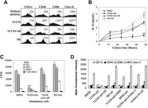
To examine whether p38 MAPK activation suppressed the differentiation of BMDCs, the p38 inhibitor SB203580 was added to the cells at the beginning of the cultures, without further addition of inhibitors at medium changes. As shown in Figure 5A, in cultures without TCCM, inhibiting p38 further enhanced the up-regulation of CD86 and MHC class II expression, whereas in cultures with the added TCCM, the inhibitor not only restored the phenotype (Figure 5A) and the secretion of IL-12 (Figure 5B), it restored the allostimulatory capacity of the cells (Figure 5C). To confirm these results, 2 other selective p38 inhibitors, SB202190 and p38 inhibitor 3, were also used. As did SB203580, these 2 inhibitors restored the phenotype (Figure 5D), IL-12 secretion, and antigen presentation activity (data not shown) of TCCM-treated cells. Inhibition of JNK by JNK inhibitor 2 completely abrogated differentiation and induced apoptosis in cultured cells (data not shown), indicating that low-level activation of the JNK pathway is essential for cell survival and differentiation.
Cytokine profiles of myeloma cells, normal BMDCs, and TCCM-treated BMDCs. (A) Cytokine array analysis showing the relative levels of secreted cytokines in the medium of 5TGM1, normal, and TCCM-treated BMDCs. Positive (Pos) and negative (Neg) controls were included in the array. An arbitrary unit, measured as the density of dots on a membrane for each cytokine in relationship to positive and negative controls, is provided to represent the relative concentration of the cytokines. Representative results of 2 experiments are shown. (B) RT-PCR detecting cytokine mRNA expression in 3 myeloma cell lines. Representative results of 3 experiments are shown. (C) ELISA assay detecting TGF-β1 secretion by 5TGM1 cells. Unconcentrated culture medium of the cell line was used to quantify the secreted cytokine. Results shown are the mean ± SD of 3 experiments. *P < .05; **P < .01 (compared with normal controls).
Cytokine profiles of myeloma cells, normal BMDCs, and TCCM-treated BMDCs. (A) Cytokine array analysis showing the relative levels of secreted cytokines in the medium of 5TGM1, normal, and TCCM-treated BMDCs. Positive (Pos) and negative (Neg) controls were included in the array. An arbitrary unit, measured as the density of dots on a membrane for each cytokine in relationship to positive and negative controls, is provided to represent the relative concentration of the cytokines. Representative results of 2 experiments are shown. (B) RT-PCR detecting cytokine mRNA expression in 3 myeloma cell lines. Representative results of 3 experiments are shown. (C) ELISA assay detecting TGF-β1 secretion by 5TGM1 cells. Unconcentrated culture medium of the cell line was used to quantify the secreted cytokine. Results shown are the mean ± SD of 3 experiments. *P < .05; **P < .01 (compared with normal controls).
We next examined signaling molecules and cytokine secretion profiles in cells treated with p38 inhibitor SB203580. As shown in Figure 6A, inhibiting p38 in TCCM-treated cells restored the protein levels of pERK and pMEK, suggesting that SB203580 abrogated TCCM-mediated inhibition on the MEK/ERK signaling pathway. Because SB203580 binds to the ATP-binding pocket of p38 kinase and inhibits its activity but does not prevent it from phosphorylation,23 the expression of pp38 in TCCM-treated cells was not affected by the inhibitor (Figure 6B). To verify that p38 MAPK activity was indeed inhibited by SB203580, a p38 functional assay was used to test the capacity of pp38 to phosphorylate its substrate activating transcription factor 2 (ATF-2). As shown in Figure 6B, in normal, differentiating BM cells in which a low level of pp38 was detected, low-level phosphorylation of ATF-2 induced by pp38 was observed in the in vitro assay. TCCM treatment increased the levels of pp38 and the phosphorylation of ATF-2. SB203580 treatment of TCCM-treated cells did not affect the expression of pp38, but its capacity to phosphorylate ATF-2 was significantly reduced, thus confirming the inhibition of pp38 activity by treatment.
Cytokine profiles of the cells were examined by RT-PCR and cytokine array analysis. As shown in Figure 6C, though TCCM treatment increased mRNA expression of IL-6, IL-10, and TGF-β in treated cells, adding the inhibitor significantly reduced mRNA expression of these cytokines in TCCM-treated cells. SB203580 alone had no effect. Furthermore, as shown in Figure 6D, using cytokine array analysis to examine a broad spectrum of cytokines, the p38 inhibitor successfully reduced the secretion of cytokines, including IL-6, IL-10, TNF-α, and VEGF in TCCM-treated cells. Secretion of TGF-β1, measured by ELISA, was also significantly reduced in TCCM-treated cells (Figure 6E).
Identifying cytokines and signaling pathways responsible for impairing BMDC differentiation. (A) Expression of DC-related surface markers on normal BMDCs, TCCM-treated BMDCs, or cells in culture with addition of cytokines IL-6, IL-10, or TGF-β individually or in combination. Representative results of 4 experiments are shown. *P < .05; **P < .01 (compared with normal controls). (B) Expression of DC-related surface markers on normal BMDCs, TCCM-treated BMDCs, or cells in cultures with the addition of TCCM and neutralizing antibody, individually or in combination, against IL-6, IL-10, or TGF-β. Representative results of 3 experiments are shown. Cells from d10 cultures were analyzed. Values above histograms represent mean fluorescence intensity. *P < .05 (higher than TCCM-treated cells). (C) TCCM-activated p38 MAPK and inhibited ERK signaling. Western blot analysis showing protein levels of phosphorylated (p) and nonphosphorylated p38, ERK, JNK, MEK, and IκB-α. Lysates from d5 cultures of normal (BMDCs) or with the addition of TCCM (TCCM cells) for 24 hours were prepared for the analyses. Representative results of 3 experiments are shown.
Identifying cytokines and signaling pathways responsible for impairing BMDC differentiation. (A) Expression of DC-related surface markers on normal BMDCs, TCCM-treated BMDCs, or cells in culture with addition of cytokines IL-6, IL-10, or TGF-β individually or in combination. Representative results of 4 experiments are shown. *P < .05; **P < .01 (compared with normal controls). (B) Expression of DC-related surface markers on normal BMDCs, TCCM-treated BMDCs, or cells in cultures with the addition of TCCM and neutralizing antibody, individually or in combination, against IL-6, IL-10, or TGF-β. Representative results of 3 experiments are shown. Cells from d10 cultures were analyzed. Values above histograms represent mean fluorescence intensity. *P < .05 (higher than TCCM-treated cells). (C) TCCM-activated p38 MAPK and inhibited ERK signaling. Western blot analysis showing protein levels of phosphorylated (p) and nonphosphorylated p38, ERK, JNK, MEK, and IκB-α. Lysates from d5 cultures of normal (BMDCs) or with the addition of TCCM (TCCM cells) for 24 hours were prepared for the analyses. Representative results of 3 experiments are shown.
In vivo capacity of normal BMDCs and TCCM-treated cells to immunize against KLH and myeloma-derived Id
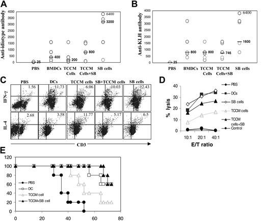
To compare the efficiency of cells to prime tumor-specific immunity in vivo, normal mature BMDCs and other treated cells pulsed with Id-KLH conjugates were used as vaccines to immunize C57BL/KaLwRij mice. As shown in Figure 7, vaccination with TCCM-treated BMDCs induced lower titers of antibodies against Id (Figure 7A) and KLH (Figure 7B) (P < .05) compared with normal cells, whereas vaccination with TCCM- and SB203580-treated cells had an efficacy similar to that of normal BMDCs at inducing antibody responses against these antigens. Vaccination with SB203580-treated BMDCs resulted in significantly higher titers of antibody production compared with normal BMDCs (P < .05).
Inhibiting p38 MAPK restored the phenotype, cytokine production, and function of normal BMDCs and of TCCM-treated, TCCM-, SB203580 (SB)-treated, and SB-treated cells. (A) Phenotypes of the cells. Representative results of 3 experiments are shown. Values above histograms represent mean fluorescence intensity. (B) IL-12 secretion by the cells during a 48-hour period in the presence of TNF-α and IL-1β. Results shown are the mean ± SD of 4 experiments. (C) MLR examining the ability of the cells to activate allospecific T cells. Results shown are the mean ± SD of 3 experiments. (D) Expression (mean fluorescence intensity) of DC-related surface markers on the cells. Also shown are cells treated with TCCM in the presence of other p38 inhibitors, such as SB202190 (SB90) and p38 MAPK inhibitor 3 (inhibitor III). These inhibitors were as potent as SB203580 at restoring the phenotype of TCCM-treated cells. Results shown are the mean ± SD of 3 experiments. Unless otherwise stated, cells from d10 cultures were analyzed. *P < .05; **P < .01 (compared with normal controls).
Inhibiting p38 MAPK restored the phenotype, cytokine production, and function of normal BMDCs and of TCCM-treated, TCCM-, SB203580 (SB)-treated, and SB-treated cells. (A) Phenotypes of the cells. Representative results of 3 experiments are shown. Values above histograms represent mean fluorescence intensity. (B) IL-12 secretion by the cells during a 48-hour period in the presence of TNF-α and IL-1β. Results shown are the mean ± SD of 4 experiments. (C) MLR examining the ability of the cells to activate allospecific T cells. Results shown are the mean ± SD of 3 experiments. (D) Expression (mean fluorescence intensity) of DC-related surface markers on the cells. Also shown are cells treated with TCCM in the presence of other p38 inhibitors, such as SB202190 (SB90) and p38 MAPK inhibitor 3 (inhibitor III). These inhibitors were as potent as SB203580 at restoring the phenotype of TCCM-treated cells. Results shown are the mean ± SD of 3 experiments. Unless otherwise stated, cells from d10 cultures were analyzed. *P < .05; **P < .01 (compared with normal controls).
T-cell immunity against these antigens was also analyzed in vaccinated mice by using pooled and restimulated spleen T cells with irradiated 5TGM1 cells for 5 days. The production of IFN-γ and IL-4 by these T cells was analyzed by intracellular cytokine staining. As shown in Figure 7C, vaccination with TCCM-treated BMDCs induced a significantly lower percentage of IFN-γ- and a higher percentage of IL-4-expressing T cells (P < .05) compared with normal cells, whereas the percentages of the cytokine-expressing cells in mice vaccinated with SB203580-treated or TCCM+SB203580-treated BMDCs were comparable to those of normal cells. Moreover, the cytotoxicity against the 5TGM1 myeloma cells of the T cells was also lower in mice vaccinated with TCCM-treated BMDCs than in those vaccinated with normal BMDCs, SB203580-treated BMDCs, or TCCM+SB203580-treated BMDCs (P < .05; Figure 7D).
Inhibiting p38 MAPK abrogated TCCM-mediated inhibition of MEK/ERK signaling and cytokine secretion. Western blot analysis showing protein levels of phosphorylated (p) and nonphosphorylated (A) ERK and MEK and (B) p38 and pATF-2 in normal (medium [Med]), TCCM-treated (TCCM), and TCCM- and SB203580-treated (TCCM+SB) cells. Lysates from d5 cultures of cells with or without the addition of TCCM and SB for 24 hours were prepared for the analyses. Representative results of 3 experiments are shown. Shown is cytokine expression in normal BMDCs (DMSO) and in SB203580 (SB)-treated, TCCM-treated, TCCM-, and SB-treated cells. (C) RT-PCR detecting cytokine mRNA expression in the cells. Representative results of 3 experiments are shown. (D) Cytokine array analysis showing the relative protein levels of cytokines in the culture medium of 5TGM1 myeloma cells and these cells. Results shown are the mean ± SD of 3 experiments. (E) ELISA detecting secreted TGF-β in TCCM and the culture media of these cells. Results shown are the mean ± SD of 3 experiments. *P < .05; **P < .01 (compared with normal controls).
Inhibiting p38 MAPK abrogated TCCM-mediated inhibition of MEK/ERK signaling and cytokine secretion. Western blot analysis showing protein levels of phosphorylated (p) and nonphosphorylated (A) ERK and MEK and (B) p38 and pATF-2 in normal (medium [Med]), TCCM-treated (TCCM), and TCCM- and SB203580-treated (TCCM+SB) cells. Lysates from d5 cultures of cells with or without the addition of TCCM and SB for 24 hours were prepared for the analyses. Representative results of 3 experiments are shown. Shown is cytokine expression in normal BMDCs (DMSO) and in SB203580 (SB)-treated, TCCM-treated, TCCM-, and SB-treated cells. (C) RT-PCR detecting cytokine mRNA expression in the cells. Representative results of 3 experiments are shown. (D) Cytokine array analysis showing the relative protein levels of cytokines in the culture medium of 5TGM1 myeloma cells and these cells. Results shown are the mean ± SD of 3 experiments. (E) ELISA detecting secreted TGF-β in TCCM and the culture media of these cells. Results shown are the mean ± SD of 3 experiments. *P < .05; **P < .01 (compared with normal controls).
To examine the capacity of these cells to treat established myeloma, mice were first inoculated with 5TGM1 cells. Ten days later, when circulating IgG2b Id was increased by 1.5- to 2-fold, 3 weekly DC vaccinations were given. As shown in Figure 7E, compared with PBS controls, vaccination with all these BMDCs prolonged the survival of the mice (P < .05). However, mice vaccinated with either normal BMDCs or TCCM+SB203580-treated BMDCs had better survival than those receiving TCCM-treated BMDCs (P < .05). Taken together, these results indicate that TCCM-treated cells had an impaired ability to immunize mice against these antigens; inhibiting p38 activity restored their in vivo capacity to induce potent and therapeutic immune responses in vaccinated mice.
Inhibiting p38 MAPK restored the ability of TCCM-treated BMDCs to immunize mice against tumor antigen and KLH and to prolong mouse survival. Immune responses monitored included titers of (A) anti-Id and (B) anti-KLH antibodies, measured by ELISA assays. Bars and values beside them represent the median titers of the antibodies. (C) Intracellular staining of IFN-γ- and IL-4-expressing T cells. Value in each graph represents the percentage of CD3+ T cells expressing IFN-γ or IL-4. (D) Cytotoxicity of spleen T cells from mice (5 per group) vaccinated with normal BMDCs, TCCM-treated (TCCM cells), TCCM- and SB203580 (SB)-treated (TCCM-cells+SB), and SB-treated cells (SB cells) pulsed with Id-KLH conjugates. Mice that had received injections of PBS served as a negative control group. (E) Survival curve of myeloma-bearing mice. Mice (5 per group) were first inoculated with 2 × 10⁶ 5TGM1 myeloma cells. Ten days later, when circulating IgG2b Id protein was increased 1.5- to 2-fold over the background level, mice received 3 weekly DC vaccinations, as indicated. Mice were humanely killed when moribund. Representative results of 3 experiments are shown.
Inhibiting p38 MAPK restored the ability of TCCM-treated BMDCs to immunize mice against tumor antigen and KLH and to prolong mouse survival. Immune responses monitored included titers of (A) anti-Id and (B) anti-KLH antibodies, measured by ELISA assays. Bars and values beside them represent the median titers of the antibodies. (C) Intracellular staining of IFN-γ- and IL-4-expressing T cells. Value in each graph represents the percentage of CD3+ T cells expressing IFN-γ or IL-4. (D) Cytotoxicity of spleen T cells from mice (5 per group) vaccinated with normal BMDCs, TCCM-treated (TCCM cells), TCCM- and SB203580 (SB)-treated (TCCM-cells+SB), and SB-treated cells (SB cells) pulsed with Id-KLH conjugates. Mice that had received injections of PBS served as a negative control group. (E) Survival curve of myeloma-bearing mice. Mice (5 per group) were first inoculated with 2 × 10⁶ 5TGM1 myeloma cells. Ten days later, when circulating IgG2b Id protein was increased 1.5- to 2-fold over the background level, mice received 3 weekly DC vaccinations, as indicated. Mice were humanely killed when moribund. Representative results of 3 experiments are shown.
Discussion
We show in this study that myeloma cells and their TCCM inhibited the differentiation and function of BMDCs. This was supported by the decreased expression of surface DC-related molecules, reduced IL-12, elevated IL-10 secretion, compromised capacity to activate allospecific T cells in vitro, poor ability of the cells to prime tumor-specific immune responses, and a bias toward induction of the type 2 T-cell response in vivo. Myeloma cells secreted many cytokines, and adding IL-6, TGF-β, and, more important, IL-10 to the cultures could partially mimic the effects of TCCM. Signal pathway analysis revealed that p38 MAPK was activated and that MEK/ERK was inhibited by the treatment of TCCM. We also show that inhibiting p38 MAPK by selective inhibitors restored the phenotype, cytokine secretion, and function of TCCM-treated BMDCs. Vaccinating mice with BMDCs obtained from cultures with the addition of TCCM and p38 inhibitor was as efficient as normal BMDCs at inducing tumor-specific antibody, type 1 (IFN-γ) T cells, and CTL responses and at prolonging the survival of myeloma-bearing mice. Three different p38 inhibitors (SB203580, SB202190, and p38 MAPK inhibitor 3) were used, all of which were shown to have similar activities on the differentiation of TCCM-treated DCs, indicating that inhibiting p38 MAPK was indeed responsible for the effect. Thus, our study demonstrates that regulation of these MAPK signaling pathways during DC differentiation may provide new strategies for the generation of potent DC vaccines for immunotherapy in patients with cancer.
Our results indicated that soluble factors were responsible for retarding the differentiation of BMDCs; hence, various assays were used to detect and identify the contributing cytokines. Given that high levels of IL-6, IL-10, VEGF, G-CSF, TNF-α, RANTES, MCP-1, MCP-5, and TGF-β were detected in TCCM or the supernatant of TCCM-treated BMDCs, we showed, using exogenous cytokines and neutralizing antibodies, that IL-6, TGF-β, and especially IL-10 might partially be responsible for the inhibitory activity of TCCM on the differentiation of BMDCs. VEGF, which was previously shown to be responsible for inhibiting the functional maturation of dendritic cells in solid tumors,18,24,25 had no effect. We do not believe these results are conflicting because differences in the cytokines could be attributed to the diverse biology of different cancers, precluding any single tumor type or approach from serving as a dominant paradigm for elucidating the mechanisms of tumor evasion or biology across a broad spectrum of cancers. More studies are, therefore, required to better understand the heterogenicity of cancers and their effects on the immune system.
In our study, we identified p38 MAPK as a novel target for intervention. On interaction with tumor cells or their TCCM, BM cells up-regulated the expression of pp38 and down-regulated pMEK/ERK, suppressing the differentiation of BMDCs. These results suggest that activation of p38 MAPK inhibited the Raf/MEK/ERK signaling pathway, which is important and crucial for cell differentiation.26,27 Indeed, it has been shown that activated p38 is capable of forming a complex with ERK, subsequently sequestering ERK and blocking its phosphorylation.28 Inhibiting p38 MAPK by selective inhibitors such as SB203580 restored the differentiation of BMDCs in the presence of TCCM, possibly through mechanisms of reduced production of IL-6, TGF-β, and especially IL-10 in treated cells and blocked p38 MAPK signaling, thus leading to reactivation of the Raf/MEK/ERK pathway in the cultured cells. As a result, the cells were as potent as their normal counterparts in activating allospecific T cells in vitro and priming tumor-specific immune responses, including the type 1 T-cell and CTL responses in vaccinated mice. Moreover, adding p38 inhibitors to normal DC cultures for the first several days accelerated or improved the differentiation and generation of immature DCs, and vaccination with p38 inhibitor-treated normal BMDCs induced stronger immune responses compared with normal BMDCs (Figure 7). This phenomenon was also observed with human monocyte-derived DCs.29 Therefore, inhibiting p38 MAPK signaling during DC generation and differentiation may provide a new strategy to generate potent DC vaccines for immunotherapy in patients with cancer.
DCs are professional APCs that play a central role in the initiation and modulation of adaptive immune responses.17,30 DC vaccination may also be an important option for the treatment of cancers. However, in the tumor microenvironment, tumor-derived factors such as IL-6, IL-10, VEGF, and TGF-β could impair the differentiation and maturation of DCs and bias the induction of immune response toward a type 2 T-cell response,5,19,20 leading to a state of tolerance against tumors.20,31,32 Clinical trials with DC vaccines have demonstrated only minor responses1,2 that might be associated with defects in DCs obtained from patients with cancer.5-8 Indeed, we found in the present study that vaccination with DCs treated with TCCM induced inferior immune responses with the type 2 (IL-4) phenotype. However, our finding that it is possible to correct the defects in DCs, such as inhibiting p38 MAPK during DC differentiation, is both interesting and important. Such findings warrant further study to examine the applicability of this approach in the preparation of DC vaccines for patients with cancer.
In conclusion, this study demonstrates that tumor cells or their derived cytokines suppressed the differentiation and generation of BMDCs. In contrast with previous reports in which VEGF was identified as the major player in solid tumors,6,18,33 the current study identified myeloma-derived IL-6, TGF-β, and, more important, IL-10 as the cytokines that suppress the differentiation of BMDCs. We also identified p38 MAPK as a novel target for intervention and showed that inhibiting p38 MAPK activity during DC differentiation could restore the phenotype, cytokine secretion, and function of BMDCs. We believe that this study is the first to show that blocking p38 MAPK can improve the quality of DCs obtained from cancers. Further preclinical studies are needed to examine the applicability of this approach to preparing DC vaccines for immunotherapy in patients with cancer.
Prepublished online as Blood First Edition Paper, November 15, 2005; DOI 10.1182/blood-2005-06-2486.
Supported by a start-up fund from the University of Texas M.D. Anderson Cancer Center, by grants R01 CA96569 and CA103978 from the National Cancer Institute, and by Translational Research Grant 6041-03 from the Leukemia and Lymphoma Society.
S.W. and Q.Y. designed the study. S.W., J.Y., J.Q., and M.W. performed the research and analyzed the data. S.W., L.W.K., and Q.Y. wrote the paper.
The publication costs of this article were defrayed in part by page charge payment. Therefore, and solely to indicate this fact, this article is hereby marked “advertisement” in accordance with 18 U.S.C. section 1734.
We thank A. Woo for editorial assistance.

![Figure 6. Inhibiting p38 MAPK abrogated TCCM-mediated inhibition of MEK/ERK signaling and cytokine secretion. Western blot analysis showing protein levels of phosphorylated (p) and nonphosphorylated (A) ERK and MEK and (B) p38 and pATF-2 in normal (medium [Med]), TCCM-treated (TCCM), and TCCM- and SB203580-treated (TCCM+SB) cells. Lysates from d5 cultures of cells with or without the addition of TCCM and SB for 24 hours were prepared for the analyses. Representative results of 3 experiments are shown. Shown is cytokine expression in normal BMDCs (DMSO) and in SB203580 (SB)-treated, TCCM-treated, TCCM-, and SB-treated cells. (C) RT-PCR detecting cytokine mRNA expression in the cells. Representative results of 3 experiments are shown. (D) Cytokine array analysis showing the relative protein levels of cytokines in the culture medium of 5TGM1 myeloma cells and these cells. Results shown are the mean ± SD of 3 experiments. (E) ELISA detecting secreted TGF-β in TCCM and the culture media of these cells. Results shown are the mean ± SD of 3 experiments. *P < .05; **P < .01 (compared with normal controls).](https://ash.silverchair-cdn.com/ash/content_public/journal/blood/107/6/10.1182_blood-2005-06-2486/4/m_zh80060692990006.jpeg?Expires=1769470597&Signature=ONl9jt4RBdsf0yZUeIqe6PgDvy4JFpWJLcCEQuMEZe97j9Vq~DbTxG3mWxB5wm0aj6nJGEmef82kiQO2UAsGeSj-g7qb327PElCtujIZBq~ARkIr6LQP0zrCxl8PHS0u6cwvMOcmIDuJjnfQDVxWcHe2aIdXwWE2DQo8Yf~O8sedO4HfndrgC48DMiHTU2KSbYy3-ZHQTaAZSAIboTlud1QoFiBpyRqbW61bsRgY1PVvGmnpHJCie6gdhjJt7-NyV9I7Pc5nqpghTTx82ppm-zBBqC--gSfcFjpUHqka2CY2aF~aWtxxjuo9v7caQ9VylNbwveIjD5cQncaoFFTAqg__&Key-Pair-Id=APKAIE5G5CRDK6RD3PGA)
