Comment on Grosse et al, page 3350
In this issue of Blood, Grosse and colleagues present a completely novel autoinflammatory syndrome and a completely novel inflammatory mechanism.
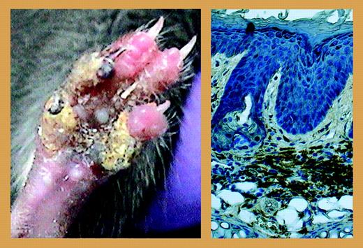
Autoinflammatory diseases are inherited disorders characterized by random episodes of unprovoked inflammation in the absence of high-titer autoantibodies or antigen-specific T cells; among these disorders are familial Mediterranean fever, pyogenic arthritis, pyoderma gangrenosum and acne (PAPA) syndrome, and TNF receptor–associated periodic syndrome (TRAPS).1,2 In this issue of Blood, Grosse and colleagues present a completely novel autoinflammatory syndrome and a completely novel inflammatory mechanism. Created by chemical mutagenesis and identified by phenotypic screen, the authors present a mouse with overall unimpaired survival, but with inflammation of the digits that is so severe, it ultimately leads to osteolysis and destruction (see the left panel of the figure). Most intriguing, the infiltrating cell type is not the neutrophil, but the macrophage (see the right panel of the figure). The underlying point mutation, an isoleucine to asparagine conversion at position 282 of the MAYP/PSTPIP2 gene on chromosome 18, encodes a protein expressed uniquely in macrophages and presumptive macrophage precursors. The authors provide a thorough and thoroughly pleasing evaluation of this new finding, as they demonstrate that the infiltrating macrophages are crucial to initiate and to maintain the inflammatory response. Grosse et al likewise document that the inflammatory disorder is not mediated by lymphocytes and can be transferred to wild-type mice via bone marrow transplantation. The authors go on to present results suggesting that the mutation is detrimental to function at the posttranscriptional level.FIG1
The left panel shows inflammation observed in the homozygous mutant (MaypLp/Lp) mouse, leading to osteolysis and localized necrosis of the digits. The right panel shows extensive infiltrates of F4/80-positive macrophages observed in thickened dermis in ear tissue in the homozygous mutant (MaypLp/Lp) mouse. See the complete figure in the article beginning on page 3350.
The left panel shows inflammation observed in the homozygous mutant (MaypLp/Lp) mouse, leading to osteolysis and localized necrosis of the digits. The right panel shows extensive infiltrates of F4/80-positive macrophages observed in thickened dermis in ear tissue in the homozygous mutant (MaypLp/Lp) mouse. See the complete figure in the article beginning on page 3350.
Of the many questions that arise from this work, two stand out in our minds, and are considered by the authors in their “Discussion.” What precisely is causing bone destruction? Macrophages are not traditionally associated with this role. The authors document little to no granulocyte influx, a point that is even more interesting given the profound degree of granulocyte involvement observed in the joints of individuals affected with PAPA syndrome, which results from a mutation in the related gene, CD2BP1/PSTPIP1. Among many directions, the authors consider osteoclast activation. This is an interesting possibility that is worthy of further consideration.
Does this model represent a natural disease? As of right this minute, none of us can pinpoint a human disease that relates specifically to a mutation in MAYP/PSTPIP2. Of course, that could change at any time, as the dissemination of information often leads the perceptive clinician to think harder about a perplexing patient. Likewise, there is a whole wide world of animal medicine, particularly the problems faced by veterinary clinicians dealing with genetically inbred strains of domestic animals. Could this disease already be out there? Perhaps. But in a much larger sense, this work stands on its own without reference to this question at all. Disease model or not, the authors have identified a novel and intriguing regulatory pathway in the cellular inflammatory network, as it occurs, where it counts, in vivo.
The author appreciates helpful discussions with Hirsh Komarow, MD, Laboratory of Allergic Diseases, National Institute of Allergy and Infectious Diseases, National Institutes of Health in preparing this commentary. ▪


