Abstract
Apoptotic cells were shown to induce dendritic cell immune tolerance. We applied a proteomic approach to identify molecules that are secreted from apoptotic monocytes, and thus may mediate engulfment and immune suppression. Supernatants of monocytes undergoing apoptosis were collected and compared using sodium dodecyl sulfate-polyacrylamide gel electrophoresis (SDS-PAGE), and differentially expressed proteins were identified using tandem mass spectrometry. Thrombospondin-1 (TSP-1) and its cleaved 26-kDa heparin-binding domain (HBD) were identified. We show that TSP-1 is expressed upon induction of monocyte apoptosis in a caspase-dependent pattern and the HBD is cleaved by chymotrypsin-like serine protease. We further show that CD29, CD36, CD47, CD51, and CD91 simultaneously participate in engulfment induction and generation of an immature dendritic cell (iDC) tolerogenic and phagocytic state. We conclude that apoptotic cell TSP-1, and notably its HBD, creates a signalosome in iDCs to improve engulfment and to tolerate engulfed material prior to the interaction with apoptotic cells.
Introduction
In recent years, it has become apparent that upon induction of apoptosis, apoptotic cells play an active role in their own engulfment by signaling professional phagocytes and/or antigen-presenting cells, without triggering an inflammatory or autoimmune response.1-5 This process seems to play an important role in homeostasis, resolution of inflammation, and peripheral tolerance induction.4,6-8
Apoptotic cells have been shown to signal the innate immune system in a variety of ways. “Eat me” signals on apoptotic cells serve as markers for phagocytes to specifically recognize these cells and subsequently ingest them. Such signals can appear on apoptotic cell membranes. Direct signals include alteration in cell surface phospholipid composition,9 changes in cell surface glycoprotein expression, distinct alterations in cell surface charge,10,11 or expression of specific molecules.12 Alternatively, certain serum or phagocyte-derived proteins can opsonize an apoptotic cell surface and signal phagocytes to engulf the opsonized cells.4,13-17 Viable cells actively express “do not eat me” signals by restriction of phosphatydilserine to the inner leaflet of their membrane, or “stay away” signals using CD31 expression.18 Recently, attention has been given not only to apoptotic cell membrane changes and phagocyte receptors, but also to the release of a membrane-derived phospholipid, lysophosphatidylcholine, which acts as a “find me” signal that is important for phagocytic cell recruitment.19 Most of these mechanisms suggest efficient identification and clearance of cells undergoing apoptosis, with noninflammatory and nonautoimmune consequences.
We decided to further explore whether apoptotic cells can actively express and secrete molecules that have a physiological significance for their own engulfment and for the environmental immune suppression. We examined whether apoptosis-induced immune suppression exists in an autologous primary cell system such as apoptotic monocytes and immature dendritic cells (iDCs). Monocytes can survive in tissues as macrophages for long periods, but a substantial portion of them constantly undergoes apoptosis, either in the absence of antiapoptotic factors or following infection or activation. Monocyte apoptosis might also have a special role in cross tolerance and presentation,8,20 and clearance of noninfected dying monocytes should proceed efficiently with immunosuppressive consequences.
In the current study, we hypothesized that apoptotic monocytes express and secrete proteins that have a role in mediating engulfment and immune suppression. We have applied a proteomic approach to identify proteins that are expressed and secreted when monocyte apoptosis is triggered.
We show that, as a part of death preparation, apoptotic monocytes express and secrete thrombospondin-1 (TSP-1), and the cleaved 26-kDa N-terminal heparin-binding domain (HBD) of TSP-1, in a caspase- and serine protease-dependent way. Using inhibitory monoclonal antibodies within a model of iDC-apoptotic cell interactions, we further show that TSP-1 and HBD directly and indirectly mediate both engulfment and immune suppression.
Materials and methods
Media and reagents
The iDC culture medium consisted of RPMI 1640, 1% l-glutamine, 1% penicillin/streptomycin (Biological Industries, Kibbutz Beit-Haemek, Israel), 1% autologous human plasma, and recombinant human cytokines GMCSF and IL-4 (R&D Systems, Minneapolis, MN, or PeproTech, London, United Kingdom). Ficoll-Paque was from Amersham Pharmacia Biotech (Uppsala, Sweden). Mouse anti-human HLA-DR-FITC and CD83-PE were from Becton Dickinson (Franklin Lakes, NJ) and Serotec (Oxford, United Kingdom), respectively; anti-human CD1a FITC, CD86 FITC, CD3-PE, and isotype controls were from Dako Cytomation (Glostrup, Denmark). Latex beads (LB-11), green fluorescent latex beads (L-4655), and LPS were obtained from Sigma-Aldrich (St Louis, MO). Thrombospondin-1 (TSP-1) was obtained from Sigma-Aldrich and Protein Sciences (Meriden, CT). Anti-β1 integrins and 1,1′-dioctadecyl-3,3,3′,3′-tetramethyl-indocarbocyanineperchlorate (DiI) were obtained from Molecular Probes (Eugene, OR). Unless otherwise indicated, all chemicals for mass spectrometry were analytical grade reagents purchased from Sigma-Aldrich. MilliQ water (Millipore, Bedford, MA) was used to prepare all solutions. For mass-spectral analysis and preparation of digests, HPLC-grade methanol and acetonitrile (JT Baker, Phillipsburg, NJ) were used. A sequencing-grade trypsin was from Promega (Madison, WI). N-p-Tosyl-L-phenylalanine chloromethyl ketone (TPCK) was obtained from Sigma-Aldrich. CFSE stock (5 μM) was purchased from Molecular Probes.
Induction and detection of apoptosis
Monocytes were obtained from buffy coats using CD14 magnetic beads (Miltenyi Biotech, Bergisch Gladbach, Germany) according to the manufacturer's instructions. Serum withdrawal apoptosis was used for generation of apoptotic monocytes in serum-free RPMI after monocyte plating at a concentration of 7.5 × 10⁶/mL, in 35-mm diameter dishes with up to 24 hours of incubation at 37°C in a humidified incubator containing 5% CO2. Blood neutrophils were also isolated from buffy coats. Briefly, red blood cells (RBCs) were sedimented using HetaSep 6% dextran solution (StemCell Technologies, Vancouver, BC) and kept at 25°C for 45 minutes. The white blood cell (WBC)-rich upper layer of the suspension was then collected and centrifuged on a density gradient with Ficoll-Paque (Amersham Biosciences, Buckinghamshire, England). Residual erythrocytes were removed by hypotonic lysis. For serum withdrawal apoptosis induction, neutrophils (6 × 10⁶/mL) were incubated in 24-well plates. Fas-induced apoptosis was obtained by exposing cells to monoclonal IgM anti-Fas antibody at 500 ng/mL (clone CH11; Upstate Biotechnology, Lake Placid, NY). Staurosporine (Sigma-Aldrich) was dissolved in dimethyl sulfoxide and was used at 400 ng/mL for apoptosis induction. Apoptosis was detected by double staining with annexin V-FITC and propidium iodide (Nexins Research BV, Hoeven, The Netherlands), as well as by estimating the proportion of hypodiploid fraction following propidium iodide staining of ethanol-fixed cells.21
For apoptosis inhibition, broad-spectrum caspase-inhibitor zVAD-fmk (Bachem, Bubendorf, Switzerland) was used. For serine protease inhibition, TPCK was dissolved in methanol and added in the indicated concentrations to cells at incubation.
Transmission electron microscopy
Freshly isolated and apoptotic monocytes were obtained as described above, fixed with 2.5% glutaraldehyde for 2.5 hours at room temperature, and then washed and resuspended in PBS x1 (Ca++ and Mg++ free). The cells were then sedimented, postfixed in 1% OsO4, rinsed in cacodylate buffer, and dehydrated in an ethanol gradient series. The cells then were treated with propylene oxide for 20 minutes (2 changes) and embedded in a propylene oxide-epoxy resin (Huntsman Advanced Materials, Everborg, Belgium). Thin sections were prepared with an ultramicrotome and examined at an accelerated voltage of 100 KW using the Philips transmission electron microscope (TEM) (CM12; Philips, Amsterdam, The Netherlands) at × 5600 magnification.
Generation of monocyte-derived dendritic cells (DCs) and interaction with apoptotic monocytes
iDCs were generated as described elsewhere.4 For interaction assays, 8 × 10⁵ apoptotic monocytes were labeled with 5 μg/mL DiI, as described,4 and were offered to 2 × 10⁵ iDCs on day 6 of culture (4:1 ratio) for 5 hours at 37°C, in 96-well plates, in 300 μL iDC culture medium. Uptake was read by flow cytometry (FACScan; Becton Dickinson, Franklin Lakes, NJ), as described.4 Briefly, iDCs were separated from monocytes based on CD1a and CD14 staining. FSC/SSC distribution and DiI acquisition by iDCs were measured. Validation of the results was done using interaction index.21
For light-microscopic evaluation (interaction index), serum-withdrawal apoptotic monocytes were added to iDCs at a ratio of 4:1 or 8:1 and incubated for 5 hours. Next, the cells were washed, cytospun using a Shandon cytospin centrifuge, fixed, and stained according to standard cytologic procedures. Fluorescence microscopy was performed using the DeltaVision system (Applied Precision, Issaquah, WA) attached to an inverted Zeiss Axiovert microscope using a 100 ×/1.4 numerical aperture Plan-APOCHROMAT objective (Zeiss, Oberkochen, Germany). Image processing was performed using Photoshop imaging software (version 8.0; Adobe Systems, Mountain View, CA).
Stimulation of iDCs
For DC maturation assays, unlabeled apoptotic monocytes were offered to iDCs for 5 hours as described under “Generation of monocyte-derived dendritic cells (DCs) and interaction with apoptotic monocytes,” after which 1 to 10 ng/mL LPS (Sigma-Aldrich) was added. The expression of maturation-related membrane molecules CD86, HLA-DR, and CD83 was examined 20 hours later.
CFSE labeling of responder cells for mixed lymphocyte reaction (MLR) experiments
These experiments were performed as described in Document S1 (available at the Blood website; see the Supplemental Materials link at the top of the online article).
Proteomic-based identification of secreted proteins from cells undergoing apoptosis
Sample preparation for gel electrophoresis. Apoptotic and viable monocyte culture media were collected and compared using sodium dodecyl sulfate-polyacrylamide gel electrophoresis (SDS-PAGE). Culture media of 3 × 10⁷ zVAD-fmk-treated monocytes and 3 × 10⁷ serum-withdrawal apoptotic monocytes were cleared of cells and undesired cell debris by sequential centrifugations—first at 290g for 5 minutes, then at 14 000g for 5 minutes, and finally at 55 000g for 1 hour—using a Beckman Ti100 centrifuge with a TLS55 rotor (Beckman Coulter, Krefeld, Germany). The resulting supernatant was collected and analyzed. Prior to electrophoresis, proteins were concentrated and desalted using Sep-Pak C-18 cartridges (Waters, Milford, MA). Protein concentration was determined using the Bradford assay (Bio-Rad, Hercules, CA).
SDS-PAGE. Gradient 4% to 20% polyacrylamide-SDS gels and SDS buffer were prepared according to the Laemmli method.22 The molecular mass of the protein bands was determined by means of a Precision Plus Protein Standards Kit (Bio-Rad). Proteins were visualized using a silver-staining kit (Amersham Pharmacia Biotech) or Bio-Safe Coomassie (Bio-Rad), according to the manufacturer's instructions. The gel images were acquired using a Umax Power Look III scanner (Umax Systems, Willich, Germany).
ESI-MS/MS. Nanoelectrospray ionization tandem mass spectrometry (ESI-MS/MS) was carried out at the mass spectrometry facility in the Interdepartmental Unit of Hadassah Medical School at the Hebrew University of Jerusalem. For trypsin digestion, proteins from concentrated medium fractions were separated by SDS-PAGE. The region corresponding to the differential protein was excised and subjected to an in-gel digestion procedure, as previously described by Matsui et al.23 Briefly, the procedure includes washing and drying of gels, reduction and alkylation, rehydration with 10 ng/mL trypsin in 25 mM ammonium bicarbonate buffer solution, incubation for 12 to 16 hours at 37°C, and peptide extraction. In-gel tryptic digests were further desalted using C18 ZipTips (Millipore) and were eluted in 5 μL of an elution buffer containing 60% (vol/vol) acetonitrile in 0.1% (vol/vol) formic acid (JT Baker).
Mass spectrometry was performed using a Micromass Q-Tof system, equipped with a NanoFlow Probe Tip Type F (Micromass UK, Manchester, United Kingdom). The extracted peptide solution was collected in a borosilicate capillary tip (Protana, Odense, Denmark) and subjected to ESI at a flow rate of 10 nL/min. The MS spectra were analyzed with MicroMass Protein Lynx software. Protein identification was conducted using the MS-FIT proteomic tool from the Matrix-Science web server.
Identification of secreted proteins from cells undergoing apoptosis
TSP-1 medium concentrations were determined using TSP-1 enzyme immunoassay (EIA; Chemicon, Temecula, CA). IL-12 concentrations were determined using IL-12 enzyme-linked immunosorbent assay (ELISA; Diaclone, Besançon, France), according to the instructions provided by the manufacturer. Western blotting was preformed using monoclonal antibodies against TSP-1 (Ab-11; Neomarkers, Fremont, CA). Protein extracts from 3 × 10⁷ apoptotic monocyte culture media (separated as described under “Proteomic-based identification of secreted proteins from cells undergoing apoptosis”), and from 3 × 10⁷ lysed monocytes, were loaded on a 4% to 20% gradient SDS-PAGE gel, transferred to a PVDF membrane (Millipore), and blocked using 20% skimmed milk in PBST (PBS x1, 0.05%-0.1% Tween 20). The membrane was incubated with primary antibody for 2 hours at room temperature or overnight at 4°C, and then washed with PBST and incubated for 30 minutes with 1:10 000 HRP-conjugated goat anti-mouse secondary antibody (Amersham Biosciences). Proteins were visualized with EZ-ECL (enhanced chemiluminescence) detection kit (Biological Industries).
cDNA synthesis and reverse transcription-polymerase chain reaction (RT-PCR) amplifications
Total RNA was isolated by using the EZ-RNA isolation kit (Biological Industries) according to the manufacturer's protocol. Single-stranded cDNA was synthesized from 2-μg RNA samples of the apoptotic monocytes using the superscript preamplification system for first-strand cDNA synthesis, according to the manufacturer's instructions. PCR was performed using 5′-GAGTCTGGCGGAGACAACAGC and 5′-TTCCTGCACAAACAGGGTGAT primers for TSP-1 (Sigma-Aldrich). The primers were optimized using a specific cloned DNA as well as the temperature gradient cycler (BioMetra, Goettingen, Germany). Relative gene expression levels were adjusted based on β-actin intensity, using 5′-ATGGTGGGAATGGGTCAGAAG and 5′-CACGCAGCTCATTGTAGAAGG primers (Sigma-Aldrich).
Inhibition assays
iDCs were exposed to inhibiting antibodies and to various TSP-1 receptors or TSP-1 motifs, before the addition of 2 μg/mL TSP-1 and/or apoptotic monocytes. Uptake of FITC-labeled latex beads (Sigma-Aldrich), either with or without the addition of exogenous TSP-1, was used as a control for phagocytosis. Various blocking antibodies were used against TSP-1 receptors or TSP-1 at concentrations of 10 μg/mL: anti-CD47 (Neomarkers), anti-CD51, anti-CD29 (Chemicon), and anti-CD36 (Serotec); and antibodies against TSP-1 type I repeats (clone A4.1; Biomeda, Foster City, CA); and N-terminal domain (Santa Cruz Biotechnology, Santa Cruz, CA).
TSP-1 binding assays
Viable and apoptotic monocytes and iDCs were washed twice with RPMI and incubated for 30 minutes on ice with 10 μg/mL TSP-1. Cells were then rewashed and stained with anti-TSP-1 antibody (Biomeda) by fluorescence-labeled secondary antibody, with either FITC-labeled annexin V or propidium iodide (PI).
Statistics
Statistical comparisons of mean data were performed using one-way analysis of variance (ANOVA) and the Student t test with Bonferroni correction for multiple comparisons. The Student t test was also used to compare uptake, and to compare the expression of surface molecules on DCs.
Results
Monocyte serum withdrawal apoptosis yields a homogeneous apoptotic population with minimal level of necrosis
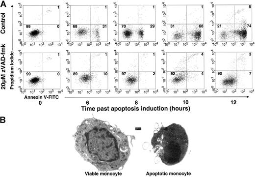
We studied serum withdrawal-, Fas-, and staurosporine-induced apoptosis pathways in peripheral blood monocytes and found that serum withdrawal is the most homogenous and reproducible method for apoptosis induction. This method results in more than 70% of monocytes at their early apoptotic phase with a minimal proportion of necrotic cells, as evident from the annexin V and PI staining (Figure 1A). Since maximal apoptosis and minimal necrosis were observed at 10 to 12 hours following serum withdrawal, we used these conditions in all the following experiments, unless indicated otherwise. Apoptosis was also validated using transmission electron microscopy (Figure 1B).
Phagocytosis of apoptotic monocytes induces immune paralysis in iDCs
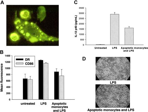
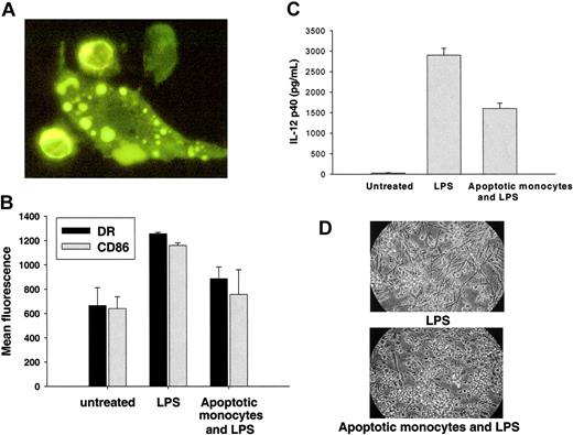
Next, we wanted to verify that autologous apoptotic monocyte binding or engulfment has an immunosuppressive effect on iDCs. To estimate the ability of iDCs to phagocytose apoptotic monocytes, DiI-stained apoptotic monocytes were offered to iDCs for 5 hours. DiI acquisition by the iDCs was quantified using flow cytometry4,24 and showed a linear correlation to the number of interacting apoptotic monocytes (Figure S1). Interaction and internalization were also observed with light microscopy and verified using fluorescent microscopy (Figure 2A). Additional validation was performed using the interaction index, and showed an index of 170 ± 108 for 4:1 and 267 ± 143 for 8:1 (P < .001) (“Generation of monocyte-derived dendritic cells (DCs) and interaction with apoptotic monocytes”; and Shoshan et al21 ).
Exposure to LPS is widely used as a model for triggering DC maturation, which in turn leads to up-regulation of MHC class II and costimulatory molecules. We therefore tested the ability of interacting autologous apoptotic monocytes to inhibit LPS-induced maturation of DCs. iDCs that were pretreated with autologous apoptotic monocytes exhibited immune paralysis in response to LPS (1-10 ng/mL), with a decrease in expression of both cell surface molecules CD86 and MHC-II (Figure 2B) as well as in IL-12 production (Figure 2C). Further, a morphologic appearance that is distinct from that of classical mature DCs (mDCs) was observed (Figure 2D). Taken together, these findings demonstrate that interaction with apoptotic monocytes has an immunosuppressive effect on iDCs.
Monocyte serum withdrawal apoptosis. (A) Time course of monocyte serum withdrawal apoptosis in the presence and absence of zVAD-fmk was assayed by flow cytometry. After 12 hours, more than 70% of monocytes were shown to be annexin V positive and propidium iodide (PI) negative, and therefore in the early stage of apoptosis. Secondary necrotic cells were 5% or less of cells, as indicated by annexin V-positive, PI-positive cells. The specificity of the apoptotic process was further shown by marked apoptosis inhibition in the presence of 20 μM of the pan-caspase inhibitor zVAD-fmk. The dot plots represent viable cells (bottom left quadrant), early apoptotic cells (bottom right quadrant), and secondary necrotic cells (top right quadrant). The percentage of viable, early apoptotic, and secondary necrotic cells is indicated within the respective quadrant. Data are representative of 6 different experiments. (B) Viable and apoptotic monocyte morphology at transmission electron microscopy (TEM). Viable and apoptotic monocytes were prepared for TEM as described in “Transmission electron microscopy.” Apoptotic cells show the typical morphology of condensed cytoplasm and chromatin with cellular membrane blebbing.
Monocyte serum withdrawal apoptosis. (A) Time course of monocyte serum withdrawal apoptosis in the presence and absence of zVAD-fmk was assayed by flow cytometry. After 12 hours, more than 70% of monocytes were shown to be annexin V positive and propidium iodide (PI) negative, and therefore in the early stage of apoptosis. Secondary necrotic cells were 5% or less of cells, as indicated by annexin V-positive, PI-positive cells. The specificity of the apoptotic process was further shown by marked apoptosis inhibition in the presence of 20 μM of the pan-caspase inhibitor zVAD-fmk. The dot plots represent viable cells (bottom left quadrant), early apoptotic cells (bottom right quadrant), and secondary necrotic cells (top right quadrant). The percentage of viable, early apoptotic, and secondary necrotic cells is indicated within the respective quadrant. Data are representative of 6 different experiments. (B) Viable and apoptotic monocyte morphology at transmission electron microscopy (TEM). Viable and apoptotic monocytes were prepared for TEM as described in “Transmission electron microscopy.” Apoptotic cells show the typical morphology of condensed cytoplasm and chromatin with cellular membrane blebbing.
Immunosuppression has generally been attributed to apoptotic cell binding or engulfment.2,4,25,26 However, as the apoptotic culture media by itself mediated immunosuppression (not shown), we were interested to find out whether proteins secreted from apoptotic monocytes mediate such an effect.
Phagocytosis of apoptotic monocytes induces immune paralysis in iDCs. (A) Apoptotic monocytes interacting with iDCs. Two DiI-stained apoptotic monocytes are surrounded by iDC pseudopods, and engulfed apoptotic fragments are seen within the dendritic cell (fluorescence microscopy, magnification × 100; further described under “Generation of monocyte-derived dendritic cells (DCs) and interaction with apoptotic monocytes”). For the micrograph in panel A, DiI-stained apoptotic cells were dried on a glass slide with Fluoromount-G (Southern Biotech), and then visualized under a Zeiss Axiovert 200 microscope equipped with a Plan-APOCHROMAT 100×/1.40 objective lens (Zeiss, Oberkochen, Germany). The image was captured with a SensiCam (PCO, Kelheim, Germany) and acquired via ImagePro Plus 4.5 software (Media Cybernetics, Silver Spring, MD). Final processing was performed with Adobe Photoshop 8.0 software (Adobe Systems, San Jose, CA). (B) Apoptotic monocyte engulfment down-regulates the expression of maturation-related molecules on DCs. Apoptotic monocytes were offered, or not offered, to iDCs at a ratio of 4:1. Five hours later, iDCs were exposed to 5 ng/mL LPS. HLA-DR-FITC and CD86 FITC expression was measured by flow cytometry. DR and CD86 are expressed at baseline levels in iDCs (mean fluorescence: 918 and 640, respectively) and are up-regulated following exposure to LPS (mean fluorescence: 1256 and 1159, respectively). Up-regulation by LPS is inhibited by exposure to apoptotic monocytes for both DR (mean fluorescence: 886, P < .001) and CD86 (mean fluorescence: 756, P < .01).Values represent the mean ± SEM of 3 experiments. (C) Apoptotic monocyte engulfment decreases IL-12 p40 production by DCs exposed to LPS. Immature DCs secrete IL-12 in response to LPS. Following interaction with apoptotic cells, down-regulation of IL-12 secretion is observed. Data represent the mean ± SD of 3 experiments. (D) Apoptotic monocytes prevent appearance of mature DC morphology in response to LPS. iDCs change morphology, becoming elongated and more “dendritic,” as they transform into mDCs in response to LPS. Following interaction with apoptotic cells, iDC morphology is retained despite exposure to LPS (light microscopy magnification, × 10). For the micrograph in panel D, cells were mounted in RPMI culture medium for visualization under a Nikon Eclipse TS100 microscope equipped with a Nikon Ph2 ADL 10×/0.25 objective lens (Nikon, Melville, NY). The image was then captured with a Nikon Coolpix 995 camera and processed with Adobe Photoshop 8.0 software.
Phagocytosis of apoptotic monocytes induces immune paralysis in iDCs. (A) Apoptotic monocytes interacting with iDCs. Two DiI-stained apoptotic monocytes are surrounded by iDC pseudopods, and engulfed apoptotic fragments are seen within the dendritic cell (fluorescence microscopy, magnification × 100; further described under “Generation of monocyte-derived dendritic cells (DCs) and interaction with apoptotic monocytes”). For the micrograph in panel A, DiI-stained apoptotic cells were dried on a glass slide with Fluoromount-G (Southern Biotech), and then visualized under a Zeiss Axiovert 200 microscope equipped with a Plan-APOCHROMAT 100×/1.40 objective lens (Zeiss, Oberkochen, Germany). The image was captured with a SensiCam (PCO, Kelheim, Germany) and acquired via ImagePro Plus 4.5 software (Media Cybernetics, Silver Spring, MD). Final processing was performed with Adobe Photoshop 8.0 software (Adobe Systems, San Jose, CA). (B) Apoptotic monocyte engulfment down-regulates the expression of maturation-related molecules on DCs. Apoptotic monocytes were offered, or not offered, to iDCs at a ratio of 4:1. Five hours later, iDCs were exposed to 5 ng/mL LPS. HLA-DR-FITC and CD86 FITC expression was measured by flow cytometry. DR and CD86 are expressed at baseline levels in iDCs (mean fluorescence: 918 and 640, respectively) and are up-regulated following exposure to LPS (mean fluorescence: 1256 and 1159, respectively). Up-regulation by LPS is inhibited by exposure to apoptotic monocytes for both DR (mean fluorescence: 886, P < .001) and CD86 (mean fluorescence: 756, P < .01).Values represent the mean ± SEM of 3 experiments. (C) Apoptotic monocyte engulfment decreases IL-12 p40 production by DCs exposed to LPS. Immature DCs secrete IL-12 in response to LPS. Following interaction with apoptotic cells, down-regulation of IL-12 secretion is observed. Data represent the mean ± SD of 3 experiments. (D) Apoptotic monocytes prevent appearance of mature DC morphology in response to LPS. iDCs change morphology, becoming elongated and more “dendritic,” as they transform into mDCs in response to LPS. Following interaction with apoptotic cells, iDC morphology is retained despite exposure to LPS (light microscopy magnification, × 10). For the micrograph in panel D, cells were mounted in RPMI culture medium for visualization under a Nikon Eclipse TS100 microscope equipped with a Nikon Ph2 ADL 10×/0.25 objective lens (Nikon, Melville, NY). The image was then captured with a Nikon Coolpix 995 camera and processed with Adobe Photoshop 8.0 software.
Apoptotic monocytes secrete thrombospondin-1 (TSP-1)
The secreted proteome of serum withdrawal apoptotic monocytes, in the absence and presence of the pan-caspase inhibitor zVAD-fmk (see Figure 1 for apoptotic phenotype), was collected and compared by SDS-PAGE and Coomassie staining (Figure S2A). Differentially expressed proteins were then further analyzed by MS. One of the proteins identified was TSP-1, which was found both in its full length and its cleaved 26-kDa, N-terminal heparin-binding domain (HBD, Figure S2B).
TSP-1, a homotrimeric glycoprotein of approximately 145 kDa/subunit,27 was first described as a platelet alpha-granule protein that is released upon activation.28 TSP-1 has been found to mediate numerous cell-matrix and cell-cell activities through a variety of receptors (reviewed in Adams29 ) and to act as a mediator of apoptotic cell engulfment.30,31 It has been previously suggested that TSP-1 is secreted by macrophages20 and dendritic cells,32 as well as by fibroblasts31 and other cell types.29 TSP-1 HBD was previously shown to mediate cell-cell adhesion, but despite its recognition as a possible separate biologically active protein, HBD's physiological role has not yet been established.33 As we separately identified the 228-amino acid N-terminal fragment of TSP-1 (Figure S2B), we hypothesized that the TSP-1 and, specifically, the TSP-1 N-terminal domain are likely candidates for mediating apoptotic monocyte engulfment and DC immunosuppression.
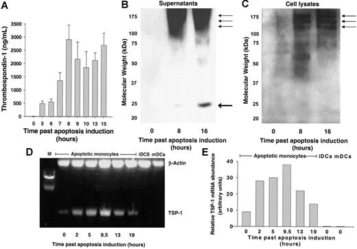
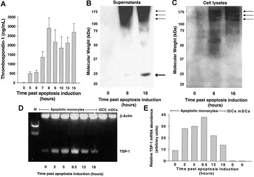
We chose conditions that yield a minimal proportion of necrotic cells during our apoptosis induction assays (Figure 1). Thus, we assumed that TSP-1 observed in the apoptotic medium did not leak from the cytosol due to loss of membrane integrity, but was secreted during induced apoptosis. To confirm the correlation between apoptosis progression and TSP-1 secretion, we examined TSP-1 levels in the apoptotic monocyte medium by EIA using a polyclonal TSP-1 Ab. As shown in Figure 3A, TSP-1 concentration in apoptotic monocyte culture medium was shown to rise to a maximum level of approximately 2 μg/mL as apoptosis progressed. No TSP-1 was detected in a lysate of viable monocytes.
As both full-length and cleaved HBDs were identified by MS, we wanted to confirm the secretion of both TSP-1 and HBD by apoptotic monocytes using Western blot analysis. As shown in Figure 3B, Western blot analysis of the apoptotic medium showed both full-length TSP-1 and 26-kDa TSP-1 fragments, whereas Western blot of apoptotic cell lysate (Figure 3C) showed only the full-length protein. Viable monocytes (Figure 3B-C), iDCs, and mDCs (not shown) did not contain or secrete TSP-1 in any significant levels on Western blot.
Monocyte serum withdrawal apoptosis induces de novo TSP-1 synthesis
We examined whether TSP-1 is secreted from readily available pools due to the apoptotic process or is synthesized de novo upon apoptosis induction. As shown in Figure 3A, a gradual increase in supernatant content of TSP-1 correlated with the apoptotic process. Furthermore, lysates of viable monocytes did not contain TSP-1, indicating that preapoptotic cells did not contain cellular TSP-1. We then tested TSP-1 mRNA transcription, as shown in Figure 3D-E. TSP-1 mRNA levels rose upon induction of apoptosis and peaked after 10 hours. Thus, despite being in the process of cell death, apoptotic monocytes actively synthesized mRNA for de novo generation of TSP-1. In contrast, TSP-1 mRNA levels were much lower in viable monocytes.
TSP-1 is transcribed, translated, and secreted upon monocyte apoptosis. (A) TSP-1 secretion by apoptotic monocytes correlates to early apoptosis. TSP-1 concentrations in apoptotic monocyte culture media were measured by TSP-1 EIA at several time points after apoptosis induction. TSP-1 levels peaked 8 hours following induction of apoptosis, corresponding to early apoptosis, as shown in Figure 1. Data are mean ± SD of 3 experiments. (B-C) The N-terminal heparin-binding domain of TSP-1 appears exclusively in the extracellular milieu of apoptotic monocytes. (B) Smear at average 130 kDa represents differentially degraded TSP-1 monomers (arrows) as verified by mass spectrometry. A degraded 26-kDa fragment (thick arrow) is also seen. TSP-1 monomers appear as 3 bands at 130 kDa or more, as verified by mass spectrometry (arrows). TSP-1 is seen only in apoptotic monocytes. (C) TSP-1 differentially degraded/glycosylated monomers appear as 3 bands at 130 kDa or more, and all perform full peptide coverage at mass-spectral analysis (arrows). TSP-1 is evidently seen only in apoptotic monocytes. Protein from 3 × 10⁷ cells, from culture media of viable (0 hours, B) or apoptotic monocytes (8 and 16 hours, B), and from whole cell lysed viable (0 hours, C) or apoptotic monocytes (8 and 16 hours, C) was loaded and separated by SDS-PAGE, as described under “Proteomic-based identification of secreted proteins from cells undergoing apoptosis.” Proteins were transferred to the PVDF membrane and exposed to mouse anti-human triclonal TSP-1 antibody and then to goat anti-mouse HRP. ECL results are presented. (D-E) TSP-1 mRNA is transcribed upon monocyte apoptosis. Total mRNA was extracted from 107 monocytes at various time intervals following serum withdrawal apoptosis induction, and then reverse-transcribed and enhanced by polymerase chain reaction. (D) TSP-1 mRNA and β-actin mRNA were enhanced by PCR using specific primers, and their relative abundance was measured by ethidium bromide photospectrometry. Viable monocytes (0 hours) had very low levels of TSP-1. The level increased as apoptosis progressed. TSP-1 mRNA was not detected in viable iDCs or mDCs. (E) Relative abundance of TSP-1 mRNA adjusted to β-actin mRNA level, as measured by densitometry. TSP-1 mRNA levels peaked at 9.5 hours, in agreement with a state of early monocyte apoptosis.
TSP-1 is transcribed, translated, and secreted upon monocyte apoptosis. (A) TSP-1 secretion by apoptotic monocytes correlates to early apoptosis. TSP-1 concentrations in apoptotic monocyte culture media were measured by TSP-1 EIA at several time points after apoptosis induction. TSP-1 levels peaked 8 hours following induction of apoptosis, corresponding to early apoptosis, as shown in Figure 1. Data are mean ± SD of 3 experiments. (B-C) The N-terminal heparin-binding domain of TSP-1 appears exclusively in the extracellular milieu of apoptotic monocytes. (B) Smear at average 130 kDa represents differentially degraded TSP-1 monomers (arrows) as verified by mass spectrometry. A degraded 26-kDa fragment (thick arrow) is also seen. TSP-1 monomers appear as 3 bands at 130 kDa or more, as verified by mass spectrometry (arrows). TSP-1 is seen only in apoptotic monocytes. (C) TSP-1 differentially degraded/glycosylated monomers appear as 3 bands at 130 kDa or more, and all perform full peptide coverage at mass-spectral analysis (arrows). TSP-1 is evidently seen only in apoptotic monocytes. Protein from 3 × 10⁷ cells, from culture media of viable (0 hours, B) or apoptotic monocytes (8 and 16 hours, B), and from whole cell lysed viable (0 hours, C) or apoptotic monocytes (8 and 16 hours, C) was loaded and separated by SDS-PAGE, as described under “Proteomic-based identification of secreted proteins from cells undergoing apoptosis.” Proteins were transferred to the PVDF membrane and exposed to mouse anti-human triclonal TSP-1 antibody and then to goat anti-mouse HRP. ECL results are presented. (D-E) TSP-1 mRNA is transcribed upon monocyte apoptosis. Total mRNA was extracted from 107 monocytes at various time intervals following serum withdrawal apoptosis induction, and then reverse-transcribed and enhanced by polymerase chain reaction. (D) TSP-1 mRNA and β-actin mRNA were enhanced by PCR using specific primers, and their relative abundance was measured by ethidium bromide photospectrometry. Viable monocytes (0 hours) had very low levels of TSP-1. The level increased as apoptosis progressed. TSP-1 mRNA was not detected in viable iDCs or mDCs. (E) Relative abundance of TSP-1 mRNA adjusted to β-actin mRNA level, as measured by densitometry. TSP-1 mRNA levels peaked at 9.5 hours, in agreement with a state of early monocyte apoptosis.
TSP-1 is expressed in other modes of monocyte apoptosis and in neutrophil apoptosis
In order to examine if TSP-1 is expressed in other modes of monocyte apoptosis, we induced apoptosis using anti-Fas monoclonal antibody or staurosporine. Both anti-Fas and staurosporine induced TSP-1 expression and secretion (Figure S3). We then set out to examine the specificity of TSP-1 expression and secretion to cell type. We examined TSP-1 levels in the apoptotic neutrophil medium by EIA, and were able to see increasing TSP-1 levels upon apoptosis progression (Figure S3) similar to levels detected with apoptotic monocytes. Thus, TSP-1 expression is not confined to apoptotic monocytes and may represent a more general phenomenon of leukocyte and other cell apoptosis. The 26-kDa fragment was also seen in both other modes of monocyte apoptosis, as well as in neutrophil apoptosis (not shown).
Expression of 26-kDa HBD of TSP-1 is caspase and serine protease dependent
TSP-1 expression was markedly reduced in the presence of zVAD-fmk, indicating that TSP-1 expression is caspase dependent. However, the cleavage site of HBD does not show an aspartic acid motif that could indicate caspase involvement in cleavage. Several proteases other than caspases, including serine proteases, have been implicated in various activities during apoptosis.34-36 Indeed, the HBD cleavage site suggests involvement of serine proteases, specifically of a chymotrypsin-like protease.37-39 To examine the possibility that a chymotrypsin-like protease was involved in generation of the 26-kDa TSP-1 cleavage product, we added the chymotrypsin inhibitor N-p-Tosyl-L-phenylalanine chloromethyl ketone (TPCK). EIA and Western blot analysis indicated that TPCK inhibited cleavage of the 26-kDa HBD in apoptotic monocytes at 1- to 10-μM concentrations (Figure S4A and S4B, respectively), and was toxic (80% of cells were depleted) at 100 μM. Thus, upon induction of apoptosis, chymotrypsin-like serine protease is activated and cleaves TSP-1, yielding the 26-kDa HBD fragment.
TSP-1 improves engulfment and induces immunosuppression in iDCs
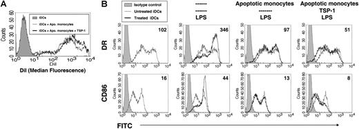
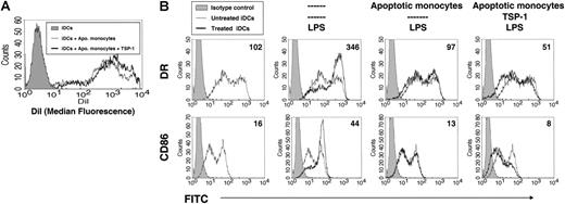
We then set out to examine whether TSP-1 can account for the observed apoptotic cell engulfment, immunosuppression, or both. iDCs were incubated with washed apoptotic monocytes, either with or without the addition of exogenous TSP-1. The TSP-1 concentration chosen was the same level observed following 10 hours of monocyte apoptosis (2 μg/mL; Figure 3A). Adding TSP-1 to the interacting apoptotic monocytes and iDCs improved iDC uptake of apoptotic monocytes by 60% on average, with a mean fluorescence of 601 in the absence of exogenous TSP-1, and 960 in the presence of TSP-1 (Figure 4A). TSP-1 also inhibited DC plasma membrane expression of maturation-related molecules CD86 and MHC class II (Figure 4B) by 47.4% and 38.5%, respectively. IL-12 production was also decreased by 51% (not shown).
TSP-1 extensively binds iDCs, but scarcely binds apoptotic monocytes
TSP-1 was previously suggested to function as a bridging molecule between apoptotic cells and macrophages, thereby increasing apoptotic cell phagocytosis.30 We therefore hypothesized a similar role for TSP-1 in our model, and examined whether TSP-1 binds both apoptotic monocytes and iDCs. Purified TSP-1 was added to washed apoptotic monocytes and iDCs, and cell surface-bound TSP-1 was measured by flow cytometry using anti-TSP-1 monoclonal antibody directed against the type I repeats (clone A4.1). Surprisingly, in comparison with binding of viable monocytes, only scant additional binding to annexin V-positive monocytes was observed (Figure 5A). This mild increase in binding was confined to PI-positive cells (Figure 5B). In contrast, a 3-fold increase in binding of TSP-1 to iDCs was observed (Figure 5C).
TSP-1 enhances apoptotic monocyte interaction with iDCs, and inhibits DC maturation. (A) TSP-1 enhances apoptotic monocyte interaction with iDCs. Apoptotic monocytes were stained with DiI and offered to iDCs, at a 4:1 ratio. The iDCs acquired apoptotic cell-derived DiI. A control group of iDCs was not exposed to apoptotic monocytes. Addition of 2 μg/mL exogenous TSP-1 significantly increased (60%, P < .001), with median fluorescence from 601 (in the absence of TSP-1) to 960 (in the presence of TSP-1). Apo monocytes indicates apoptotic monocytes. Results are representative of 5 experiments. (B) iDC maturation is inhibited by TSP-1 in the presence of apoptotic monocytes. Expression of CD86 and HLA-DR on iDCs following exposure to 5 ng/mL LPS. In the presence of apoptotic monocytes, marked down-regulation of CD86 and DR was documented (P < .001). Addition of 2 μg/mL TSP-1 even further inhibited DR and CD86 expression (P < .001). Anti-DR-FITC (top panel) and anti-CD86-FITC (bottom panel) are presented. Numbers indicating the median fluorescence are presented in each histogram. Isotype controls are shown as gray-filled curves.
TSP-1 enhances apoptotic monocyte interaction with iDCs, and inhibits DC maturation. (A) TSP-1 enhances apoptotic monocyte interaction with iDCs. Apoptotic monocytes were stained with DiI and offered to iDCs, at a 4:1 ratio. The iDCs acquired apoptotic cell-derived DiI. A control group of iDCs was not exposed to apoptotic monocytes. Addition of 2 μg/mL exogenous TSP-1 significantly increased (60%, P < .001), with median fluorescence from 601 (in the absence of TSP-1) to 960 (in the presence of TSP-1). Apo monocytes indicates apoptotic monocytes. Results are representative of 5 experiments. (B) iDC maturation is inhibited by TSP-1 in the presence of apoptotic monocytes. Expression of CD86 and HLA-DR on iDCs following exposure to 5 ng/mL LPS. In the presence of apoptotic monocytes, marked down-regulation of CD86 and DR was documented (P < .001). Addition of 2 μg/mL TSP-1 even further inhibited DR and CD86 expression (P < .001). Anti-DR-FITC (top panel) and anti-CD86-FITC (bottom panel) are presented. Numbers indicating the median fluorescence are presented in each histogram. Isotype controls are shown as gray-filled curves.
TSP-1 scarcely binds to late apoptotic monocytes but extensively binds to iDCs. Viable and apoptotic monocytes and iDCs were washed twice with RPMI and incubated for 30 minutes on ice with 10 μg/mL TSP-1. Cells were then rewashed and stained with anti-TSP-1 antibody (Biomeda) followed by fluorescence-labeled secondary antibody, and with either FITC-labeled annexin V or propidium iodide (PI). (A) TSP-1 scarcely binds apoptotic monocytes. Double staining with anti-TSP-1-PE and annexin V-FITC is shown. Only 28% (18/64) of annexin V-FITC-positive monocytes bound TSP-1. Numbers indicate percentage of cells included in the respective quadrants. (B) TSP-1 binds late, rather than early, to apoptotic monocytes. Double staining with anti-TSP-1-FITC and PI is shown. Almost all monocytes that bind anti-TSP-1 are PI-positive late apoptotic cells. This is true also for the small fraction of PI-positive cells that is included in viable cells. (C) TSP-1 binds extensively to iDCs. TSP-1-bound iDCs had a mean fluorescence of 26, whereas viable and apoptotic monocytes show mean fluorescence of 9.14 and 9.73, respectively (P < .001). Data shown are representative of 3 experiments. Isotype control is shown as a gray-filled curve. Less than 5% of iDCs were annexin V positive and less than 1% were PI positive, excluding binding to iDCs due to apoptosis.
TSP-1 scarcely binds to late apoptotic monocytes but extensively binds to iDCs. Viable and apoptotic monocytes and iDCs were washed twice with RPMI and incubated for 30 minutes on ice with 10 μg/mL TSP-1. Cells were then rewashed and stained with anti-TSP-1 antibody (Biomeda) followed by fluorescence-labeled secondary antibody, and with either FITC-labeled annexin V or propidium iodide (PI). (A) TSP-1 scarcely binds apoptotic monocytes. Double staining with anti-TSP-1-PE and annexin V-FITC is shown. Only 28% (18/64) of annexin V-FITC-positive monocytes bound TSP-1. Numbers indicate percentage of cells included in the respective quadrants. (B) TSP-1 binds late, rather than early, to apoptotic monocytes. Double staining with anti-TSP-1-FITC and PI is shown. Almost all monocytes that bind anti-TSP-1 are PI-positive late apoptotic cells. This is true also for the small fraction of PI-positive cells that is included in viable cells. (C) TSP-1 binds extensively to iDCs. TSP-1-bound iDCs had a mean fluorescence of 26, whereas viable and apoptotic monocytes show mean fluorescence of 9.14 and 9.73, respectively (P < .001). Data shown are representative of 3 experiments. Isotype control is shown as a gray-filled curve. Less than 5% of iDCs were annexin V positive and less than 1% were PI positive, excluding binding to iDCs due to apoptosis.
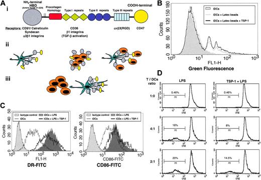
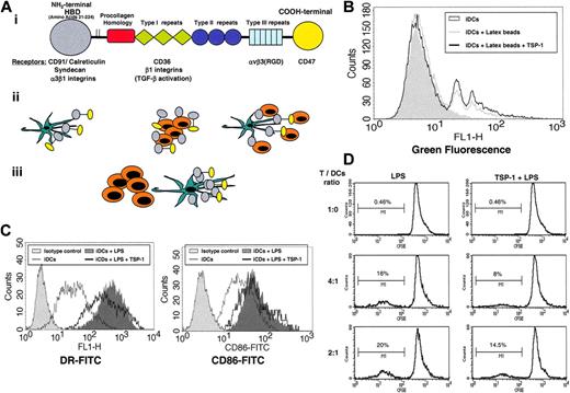
We concluded that the proposed classical bridging role of TSP-1 as a factor increasing phagocytosis of apoptotic cells may not be the only mechanism responsible for the increase in engulfment that was observed. We then were interested to know whether TSP-1 may mediate its function by directly affecting iDCs and not necessarily during the actual uptake of apoptotic cells (Figure 6A).
TSP-1, by itself, enhances apoptotic monocyte engulfment by iDCs, and inhibits iDC maturation and T-cell activation. (A) Proposed mechanisms of TSP-1's effect on iDCs. (Ai) TSP-1 structural and functional domains that are relevant to apoptotic cell clearance. The relevant interacting receptors for each domain are indicated. HBD indicates heparin binding domain. (Aii) Mechanisms of TSP-1 as a bridging molecule. TSP-1 may be expressed by phagocytes (Aii, left) or by apoptotic cells (Aii, middle) that also can secrete the N-terminal domain. Whether the source is the engulfing cell or the apoptotic cell, TSP-1 or the N-terminal domain serves as a bridge between apoptotic cells and phagocytes (Aii, right). (Aiii)Alternatively (no bridge), TSP-1 or the N-terminal domain may bind to iDCs and induce ameliorated phagocytosis and immune suppression, even in the absence of attached apoptotic cells. (B) TSP-1 enhances latex bead engulfment by iDCs. Green fluorescent latex beads were offered to iDCs at a 16:1 ratio, in the presence or absence of 2 μg/mLTSP-1. The gray-filled curve represents iDCs that were not exposed to green fluorescent latex beads. Of the TSP-1-treated iDCs, 22.6% ± 0.1% engulfed at least 1 latex bead, compared with 15.6% ± 1.8% in the absence of TSP-1, indicating approximately 45% augmentation of phagocytic capacity following TSP-1 exposure (P < .001). Data are representative of 3 experiments. (C) TSP-1 inhibits iDC maturation. iDCs were treated with 5 ng/mL LPS in the absence or presence of 1 μg/mL TSP-1, or remained untreated. Mean fluorescence decreased from 453 to 283 (38%, P < .001, representative of 3 experiments) for DR and from 53 to 21 (39%, P < .001, representative of 3 experiments) for CD86. The bright-filled curve represents isotype control. (D) TSP-1 inhibits T-cell activation. CFSE-labeled T cells were cocultured in different ratios with LPS-treated DCs (left) or DCs that were exposed to TSP1 for 5 hours before LPS treatment (right). Exposure to TSP1 prior to LPS treatment inhibited DC-induced T-cell activation by 27% at 2:1 T-cell/DC ratio and by 50% at 4:1 T-cell/DC ratio.
TSP-1, by itself, enhances apoptotic monocyte engulfment by iDCs, and inhibits iDC maturation and T-cell activation. (A) Proposed mechanisms of TSP-1's effect on iDCs. (Ai) TSP-1 structural and functional domains that are relevant to apoptotic cell clearance. The relevant interacting receptors for each domain are indicated. HBD indicates heparin binding domain. (Aii) Mechanisms of TSP-1 as a bridging molecule. TSP-1 may be expressed by phagocytes (Aii, left) or by apoptotic cells (Aii, middle) that also can secrete the N-terminal domain. Whether the source is the engulfing cell or the apoptotic cell, TSP-1 or the N-terminal domain serves as a bridge between apoptotic cells and phagocytes (Aii, right). (Aiii)Alternatively (no bridge), TSP-1 or the N-terminal domain may bind to iDCs and induce ameliorated phagocytosis and immune suppression, even in the absence of attached apoptotic cells. (B) TSP-1 enhances latex bead engulfment by iDCs. Green fluorescent latex beads were offered to iDCs at a 16:1 ratio, in the presence or absence of 2 μg/mLTSP-1. The gray-filled curve represents iDCs that were not exposed to green fluorescent latex beads. Of the TSP-1-treated iDCs, 22.6% ± 0.1% engulfed at least 1 latex bead, compared with 15.6% ± 1.8% in the absence of TSP-1, indicating approximately 45% augmentation of phagocytic capacity following TSP-1 exposure (P < .001). Data are representative of 3 experiments. (C) TSP-1 inhibits iDC maturation. iDCs were treated with 5 ng/mL LPS in the absence or presence of 1 μg/mL TSP-1, or remained untreated. Mean fluorescence decreased from 453 to 283 (38%, P < .001, representative of 3 experiments) for DR and from 53 to 21 (39%, P < .001, representative of 3 experiments) for CD86. The bright-filled curve represents isotype control. (D) TSP-1 inhibits T-cell activation. CFSE-labeled T cells were cocultured in different ratios with LPS-treated DCs (left) or DCs that were exposed to TSP1 for 5 hours before LPS treatment (right). Exposure to TSP1 prior to LPS treatment inhibited DC-induced T-cell activation by 27% at 2:1 T-cell/DC ratio and by 50% at 4:1 T-cell/DC ratio.
TSP-1 actions of engulfment and immunosuppression are mediated even in the absence of apoptotic monocytes
Next, we checked whether TSP-1 alone can account for iDC enhancement of engulfment and the observed tolerogenic profile, or whether apoptotic monocytes must also be present. TSP-1-treated iDCs were incubated with green fluorescent latex beads as bait. As seen in Figure 6B, iDC engulfment of the latex beads increased by 35.7% ± 3.5% over control in the presence of TSP-1. Thus, an increased engulfing capacity that is nonspecific to apoptotic cells was observed, possibly due to bridging effect or due to direct effect on iDCs. Furthermore, TSP-1-treated DCs displayed an immunoparalyzed phenotype (Figure 6C), even in the absence of apoptotic monocyte engulfment. Adding only TSP-1 to iDCs decreased expression of iDC maturation molecule levels by 38% ± 10.5% (P < .001) and 39% ± 6% (P < .001) for MHC-II and CD86, respectively. In order to see whether DC immunoparalyzation was indeed effective, we conducted MLR experiments in which T-cell proliferation was clearly inhibited by 30% to 50% (Figure 6D).
We conclude that TSP-1 increases iDC engulfing capability either by binding to iDCs or by binding to both latex beads and IDCs, and has a direct immunosuppressive effect resulting in generation of tolerogenic DCs, even in the absence of apoptotic cells.
Induction of engulfment and immunosuppression requires cooperation between multiple receptors and is mediated mainly by the heparin-binding domain (HBD)
In order to explore the mechanisms underlying these observations, we sought to elucidate the exact sites for TSP-1 binding that mediate these effects. We focused on known TSP-1 receptors. For this, we carried out inhibition assays by exposing iDCs to inhibiting antibodies directed to several TSP-1 receptors or domains, both in the presence and the absence of apoptotic monocytes. While exposure of iDCs to αCD36 or αCD91 inhibiting antibodies prior to interaction significantly decreased apoptotic monocyte engulfment, exposure to an antibody against the N-terminal domain (HBD) decreased engulfment even further, almost to the basal level of TSP-1-mediated engulfment. In contrast, addition of either anti-β1 integrins or anti-CD47 had no significant influence on engulfment, and addition of anti-CD51 only slightly inhibited apoptotic monocyte uptake (Figure 7A).
As for inhibition of the immunosuppressive effect, addition of anti-N-terminal domain, anti-β1 integrins, and anti-CD36 antibodies had the most striking effect on maturation molecule expression. In most cases, there was no significant difference whether or not apoptotic monocytes were added (Figure 7B-C). There was a similar effect by most inhibiting antibodies to avoid down-regulation of DR and CD86 expression by TSP-1, but the impact was more pronounced with antibodies directed against the N-terminal domain of TSP-1.
As CD36 and CD91 may mediate binding or engulfment via a non-TSP-1-dependent mechanism, experiments were also conducted in the absence of TSP-1, and in the presence of anti-CD36 and anti-CD91 inhibitory antibodies. No significant inhibition was found in these conditions (not shown), suggesting that the major inhibitory effect was related to TSP-1.
Thus, TSP-1 has multiple domains that induce phagocytic and tolerogenic states in iDCs. HBD, interacting with iDCs, is a critical domain in regard to both engulfment and immunosuppression. As shown here, this domain is expressed by apoptotic monocytes and neutrophils, both to mediate their own engulfment and to negatively signal iDCs.
Discussion
In the current study, we have used an unbiased proteomic approach to identify apoptosis-related secreted proteins that may participate in engulfment mediation and immune suppression. We were able to identify TSP-1, whose secretion has previously been ascribed mainly to the engulfing cell, as a protein that is synthesized de novo upon apoptosis induction in monocytes.
TSP-1 is a calcium-binding protein that participates in cellular responses to growth factors, cytokines, and injury.29,40 It regulates cell proliferation, migration, and apoptosis in a variety of physiological and pathological settings, including wound healing, inflammation, angiogenesis, and neoplasia. TSP-1 binds to a wide variety of integrin and nonintegrin cell surface receptors (Figure 6A). The binding sites for these receptors on TSP-1 are dispersed throughout the molecule, with most domains binding multiple receptors. In some cases, TSP-1 binds to multiple receptors concurrently, and recent data indicate that there is cross talk between receptor systems. Thus, TSP-1 may direct the clustering of receptors to specialized membrane domains for adhesion and signal transduction.
The N-terminal (heparin-binding) domain and CD36 mediate engulfment capacity for apoptotic monocytes, and multiple domains mediate the immunosuppressive effect. iDCs were exposed to several blocking antibodies directed against various potential TSP-1 receptors, washed, and then offered DiI-stained apoptotic monocytes in the presence of 2 μg/mL TSP-1. (A) TSP-1-dependent apoptotic monocyte engulfment is mediated mainly through the HBD. Striking (90%) inhibition of apoptotic monocyte uptake is seen upon inhibition of binding through the HBD (P < .001). Significant uptake inhibition is seen upon inhibition of CD36 (64%, P < .001), whereas slight uptake inhibition is observed upon neutralizing CD51 (24%, P < .05). No significant engulfment inhibition is seen when CD47 and β1-integrin domains (CD29) are inhibited. Inhibition is presented as a percentage of TSP-1-dependent engulfment. Data are mean ± SEM of 3 experiments. (B-C) TSP-1-induced maturation inhibition involves multiple domains. Expression of DR (B) and CD86 (C) was indicative of the level of DC maturation. iDCs were treated with inhibiting antibodies as described under “Inhibition assays,” and were then exposed to TSP-1 with or without the addition of apoptotic monocytes. LPS (5 ng/mL) was added 5 hours later, and expression of DR and CD86 was examined 20 hours later. As shown, the main effect was achieved by blocking HBD (P < .001), but other binding sites, such as CD36, CD29 (β1 integrins), CD51, and CD47, also had important immunosuppressive effects (P < .001 for each site). The relative inhibition effects on maturation are expressed as the relative changes in DR and CD86 median fluorescence of DCs, compared with DCs treated with isotype control and exposed to LPS. Data are mean ± SEM of 4 experiments.
The N-terminal (heparin-binding) domain and CD36 mediate engulfment capacity for apoptotic monocytes, and multiple domains mediate the immunosuppressive effect. iDCs were exposed to several blocking antibodies directed against various potential TSP-1 receptors, washed, and then offered DiI-stained apoptotic monocytes in the presence of 2 μg/mL TSP-1. (A) TSP-1-dependent apoptotic monocyte engulfment is mediated mainly through the HBD. Striking (90%) inhibition of apoptotic monocyte uptake is seen upon inhibition of binding through the HBD (P < .001). Significant uptake inhibition is seen upon inhibition of CD36 (64%, P < .001), whereas slight uptake inhibition is observed upon neutralizing CD51 (24%, P < .05). No significant engulfment inhibition is seen when CD47 and β1-integrin domains (CD29) are inhibited. Inhibition is presented as a percentage of TSP-1-dependent engulfment. Data are mean ± SEM of 3 experiments. (B-C) TSP-1-induced maturation inhibition involves multiple domains. Expression of DR (B) and CD86 (C) was indicative of the level of DC maturation. iDCs were treated with inhibiting antibodies as described under “Inhibition assays,” and were then exposed to TSP-1 with or without the addition of apoptotic monocytes. LPS (5 ng/mL) was added 5 hours later, and expression of DR and CD86 was examined 20 hours later. As shown, the main effect was achieved by blocking HBD (P < .001), but other binding sites, such as CD36, CD29 (β1 integrins), CD51, and CD47, also had important immunosuppressive effects (P < .001 for each site). The relative inhibition effects on maturation are expressed as the relative changes in DR and CD86 median fluorescence of DCs, compared with DCs treated with isotype control and exposed to LPS. Data are mean ± SEM of 4 experiments.
Although it has long been appreciated that TSP-1 plays a role in mediating apoptotic cell engulfment, its source was attributed mainly to the engulfing cell and its role was attributed mainly to facilitating phagocytosis as a bridging molecule. We were able to show for the first time that, upon apoptosis, monocytes transcribe and translate TSP-1 de novo and that TSP-1 has a direct facilitating effect on engulfment and immunosuppression.
How may TSP-1 receptor binding ameliorate apoptotic cell engulfment? Of interest, TSP-1 was shown to induce Fak-dependent signaling to stimulate focal adhesion disassembly and cell migration. Furthermore, RhoA inactivation is required for this response.41 RhoA inactivation may occur via mechanisms similar to those described for apoptotic cell engulfment in C elegans42 with CD91 homology to mammals.
Until now, immunosuppressive characteristics were attributed only to TSP-1 interaction with CD47,32 which in turn interacts with the C-terminal domain. In the current study, we were able to determine that the N-terminal region, the HBD, serves as a potent ligand for engulfment and immunosuppression. The N-terminus of TSP-1 and TSP-2 has sequence similarity to the pentraxin superfamily, by virtue of its globular structure. The pentraxin family has been associated with apoptotic cell clearance. Furthermore, the heparin-binding domain has been shown to interact with calreticulin and CD91, which are related to apoptotic cell clearance in both mammals43 and C elegans.44,45 Although TSP-1 is not found in C elegans, the HBD motif is found in 4 proteins in C elegans (InterPro46 ). The heparin-binding domain supports cellular adhesion47,48 and chemotaxis.49,50
What advantages could a 26-kDa HBD fragment have over the full-length TSP-1? It is possible that HBD is cleaved rather late in the apoptotic process, in order to ameliorate the clearance of long-standing apoptotic cells. Another possibility is that HBD has different properties and roles than TSP-1, such as proangiogenic activity that may be important in the context of cellular adaptation to injury.33 Alternatively, experiments using blocking antibodies suggest that signalosome created by HBD may differ from this of whole TSP-1. For example, Saumet et al51 showed that domains outside the HBD, namely the type III repeats/C-terminal domain, trigger caspase-independent cell death.
Given the findings of this and earlier studies, we would like to propose a mechanism of action involving de novo TSP-1 synthesis by apoptotic monocytes and secretion of either intact TSP-1 or its cleaved 26-kDa HBD. Upon synthesis or secretion, TSP-1 or the HBD binds mainly to iDCs or engulfing cells. TSP-1 triggers the formation of a signalosome that increases phagocytic capacity and mediates inhibitory signals, resulting in a tolerogenic iDC phenotype, and may form a bridging molecule. Apoptotic cells then bind either via TSP-1 and its relevant receptors—CD91/calreticulin (LRP), CD36, and αvβ3—or via other specific engulfing apoptotic cell receptors such as Mer, CD11b/CD18 (if complement opsonization occurs), αvβ5, or αvβ3. The theoretic phosphatydilserine receptor is another possibility.52,53 Most important, this tolerogenic phenotype is effective for T-cell suppression (Figure 6D) and can be acquired by iDCs, even in the absence of interaction with apoptotic cells, indicating the crucial role that TSP-1, and particularly the 26-kDa heparin-binding domain fragment of TSP-1, plays in creation of a tolerizing state. We were not able to document TSP-1 generation by iDCs at the mRNA or protein level, as suggested by Doyen et al,32 but this is another possible mechanism (Figure 6A).
The mechanism described here also suggests the formation of multiprotein complexes on cell surfaces and the clustering of receptors that initiate signal transduction, such as the T-cell receptor signalosome.54 Apoptotic cell and iDC interactions show a dynamic structure with expanding complexity. However, as demonstrated here, the vast majority of the consequences of these interactions can be mediated by a single protein. This can allow expression of CD91's phagocytic property, but not its proinflammatory property.55 This mechanism is not only important in homeostasis, but may also be a major mechanism for turning down inflammation and avoiding autoimmunity.
Finally, this study demonstrates, for the first time, an innate altruistic community property within cells. Despite being doomed, apoptotic monocytes actively express and secrete a molecule that is important for the sake of the continuation of life in the organism.
Prepublished online as Blood First Edition Paper, August 1, 2006; DOI 10.1182/blood-2006-03-013334.
Supported in part by grants from the Israel Science Foundation (D.M.) and Hadassah-Hebrew University (D.M.), and by the Sudarsky Center for Computational Biology (SCCB) (Y.B.).
The authors declare no competing financial interests.
A.K., Y.B., M.A., U.T., I.V., E.N., O.Z., M.L., and D.M. participated in designing and performing the research; A.K., Y.B., M.L., and D.M. controlled and analyzed data; A.K., Y.B., and D.M. wrote the paper; and all authors checked the final version of the paper.
A.K. and Y.B. contributed equally to this work.
The online version of this article contains a data supplement.
The publication costs of this article were defrayed in part by page charge payment. Therefore, and solely to indicate this fact, this article is hereby marked “advertisement” in accordance with 18 USC section 1734.
This work could not be realized without the teaching of Mrs M. Flinsch, who taught her student (D.M.) how to observe manifestations of life, as they are.
The authors wish to thank Mrs Shifra Fraifeld for her assistance in the preparation of this article.


