Abstract
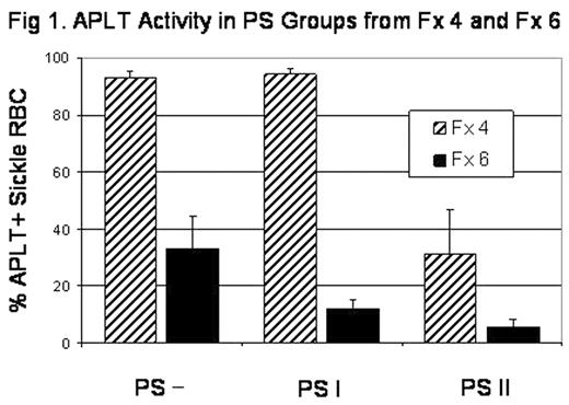
Red blood cell (RBC) membrane phosphatidylserine (PS) is normally confined to the inner leaflet. In sickle RBC, however, PS externalization has been observed and may contribute to thrombogenesis, endothelial adhesion, and shortened RBC lifespan. Increased calcium leads to PS externalization through inhibition of aminophospholipid translocase (APLT), which normally returns external PS to the inner leaflet, and activation of phospholipid scramblase, which allows nonspecific phospholipid equilibration between the leaflets. Sickle RBC in the light and dense (dehydrated) fractions exhibit increased PS externalization compared to sickle RBC in the normal density fractions. Sickle RBC with modest PS exposure (Type I PS+) occur in both light and dense fractions and, especially in the light fractions, include many reticulocytes. Sickle Cells with high levels of PS exposure (Type II PS+) occur predominantly in the dense fraction. Type II PS+ sickle cells have low levels of HbF, are more adherent than Type I PS+ cells, and tend to increase in number as sickle cells age in the circulation. Dense sickle cells have decreased APLT activity and this has been considered necessary (but probably not sufficient) for PS externalization. In the current studies we have examined the relationship between PS externalization and translocase activity in sickle cell density fractions. APLT activity and PS externalization were determined flow cytometrically using NBD-PS internalization and annexin V-phycoerythrin binding respectively. Sickle RBC density fractions were isolated using a discontinuous Optiprep® gradient, and two fractions were selected for further study, Fx 4 with essentially normal hydration (1.093< g/cc <1.100) and the dehydrated Fx 6 (> 1.120 g/cc). As expected, Fx 6 sickle RBC had a decreased percentage of APLT positive cells (29.9±10.8, N=4) compared to normal RBC (94.5±3.6, N=3) and Fx 4 sickle RBC (92.9±2.3, N=4). Fx 4 contained 5.7±2.8% Type I and 0.3±0.2% Type II PS+ cells. Fx 6 had 9.1±2.5% Type I and 3.2±2.3% Type II PS+ cells. Figure 1 shows the percentage of APLT positive cells in the PS negative (PS-), Type I PS+, and Type II PS+ groups for Fx 4 and Fx 6. Type I PS+ cells in Fx 4 are positive for APLT, whereas those in Fx 6 are mostly negative. In Fx 4, the Type I PS+ group most likely contains many reticulocytes, and in a separate experiment TfR+ reticulocytes had a uniform, slightly elevated APLT activity. The Type II PS+ cells in both Fx 4 and Fx 6 had decreased percentages of APLT positive cells. It thus appears that high levels of PS externalization, regardless of hydration state, are associated with low APLT activity. We therefore conclude:
In normally hydrated sickle cells, Type II PS+ cells, but not Type I PS+ cells, are associated with decreased APLT;
In dense sickle RBC, both Type I and Type II PS+ cells are associated with decreased APLT. These results further emphasize the pathologic nature of the Type II PS+ sickle cells.
APLT Activity is PS Groups form F × 4 and F × 6
Disclosure: No relevant conflicts of interest to declare.
Author notes
Corresponding author


