Abstract
Natural killer (NK) cells from patients with familial hemophagocytic lymphohistiocytosis because of PRF1 (FHL2, n = 5) or MUNC13-4 (FHL3, n = 8) mutations were cultured in IL-2 prior to their use in various functional assays. Here, we report on the surface CD107a expression as a novel rapid tool for identification of patients with Munc13-4 defect. On target interaction and degranulation, FHL3 NK cells displayed low levels of surface CD107a staining, in contrast to healthy control subjects or perforin-deficient NK cells. B-EBV cell lines and dendritic cell targets reveal the FHL3 NK-cell defect, whereas highly susceptible tumor targets were partially lysed by FHL3 NK cells expressing only trace amounts of Munc13-4 protein. Perforin-deficient NK cells were completely devoid of any ability to lyse target cells. Cytokine production induced by mAb-crosslinking of triggering receptors was comparable in patients and healthy control subjects. However, when cytokine production was induced by coculture with 721.221 B-EBV cells, FHL NK cells resulted in high producers, whereas control cells were almost ineffective. This could reflect survival versus elimination of B-EBV cells (ie, the source of NK-cell stimulation) in patients versus healthy control subjects, thus mimicking the pathophysiologic scenario of FHL.
Introduction
Hemophagocytic lymphohistiocytosis (HLH) is a rare, heterogeneous fatal disease of early infancy characterized by a hyperinflammatory syndrome with fever, hepatosplenomegaly, cytopenia, hypertriglyceridemia, hypofibrinogenemia, and, in some cases, central nervous system alteration. Histologic examination of involved organs (more commonly bone marrow aspiration) typically shows infiltration of lymphocytes and histiocytes with hemophagocytosis.1-3 Characteristic findings are also the high levels of various cytokines, such as interleukin 6 (IL-6), IL-8, IL-10, IL-18, interferon γ (IFN-γ), and tumor necrosis factor α (TNF-α) and also high plasma concentrations of sCD25 and sCD95-ligand.4-6 In some cases HLH may occur in patients of any age undergoing therapeutic immune suppression. In such cases, also defined as “secondary,” immune suppressive treatment withdrawal may result in control of HLH.7 The remaining (also called “primary”) forms are of genetic origin and often defined as familial hemophagocytic lymphohistiocytosis (FHL). A genetic heterogeneity underlies FHL. The most common genetic defects in patients with FHL involve the perforin gene (PRF1) on chromosome 10q21 and the MUNC13-4 gene on chromosome 17q25.8,9 Mutations of PRF1 account for approximately 30% to 40% of patients, defined as FHL2 subtype, and MUNC13-4 mutations are identified in an additional 25% to 30% of cases (FHL3). The proteins encoded by both genes are implicated in the killing machinery.10 Very recently, mutations in the syntaxin11 gene (6q24) have been reported in a small group of patients (FHL4) with common Kurdish origin.11 This defect is thought to alter intracellular vesicle trafficking of the phagocytic system.
PRF1 gene encodes perforin as an inactive precursor form, which, after processing in a post-Golgi apparatus by proteolysis and glycosylation, becomes an active protein.12 This mature form of perforin is stored with granzymes in specialized secretory lysosomes, known as lytic granules, which are present in natural killer (NK) and cytotoxic T lymphocytes (CTLs).13 On target-cell interaction, lytic granules polarize and release their content at the immunologic synapse.14 The secreted perforin then inserts into the lipid bilayer and following polymerization generates poly-perforin pores in the plasma membrane of target cells.15 This pore formation leads to the destruction of cells by osmotic lysis and by allowing entry of apoptosis-inducing granzymes.16 Munc13-4, a member of Munc13 family of proteins involved in vesicle priming function, has been described as a positive regulator of secretory lysosome exocytosis.9 Although Munc13-1 functions as a priming factor in neural cells, Munc13-4 is highly expressed in several hematopoietic cells. In patients with FHL3, Munc13-4 deficiency results in defective cytolytic granule exocytosis, despite polarization of the lytic granules and docking with the plasma membrane.9 Recent evidence shows that Munc13-4 binds to Rab27A; the 2 proteins colocalize on the membranes of secretory lysosomes in CTL and mast cells and promote the dense core granule secretion in platelets.17,18 Rab27A is highly expressed in melanocytes and hematopoietic and other secretory cells. Absence of functional Rab27A causes the Griscelli syndrome type 2, a genetic disorder characterized by defects of pigmentation and of granule exocytosis in CTLs, in which lytic granules fail to dock on the plasma membrane and therefore do not release their content.19 Thus, these fatal genetic disorders which display similar pathologic and clinical features all disrupt the release or function of cytotoxic proteins.
Defects in cellular cytotoxicity, excessive production of inflammatory cytokines, and abnormal macrophage activation characterize HLH.20 In these patients an impairment of cytolytic activity of NK cells, that provide the first line of host defense, and subsequently of CTL, results in a markedly reduced ability to control viral infection. Uncontrolled viral dissemination together with a parallel excessive inflammatory reaction results in extensive tissue damage. Two major mechanisms of cytotoxicity are perforin/granzyme- and death receptor (eg, FasL and TRAIL)–mediated pathways.21,22 Many studies, including experiments with perforin-deficient mice, led to the conclusion that NK cells primarily use the perforin/granzyme pathway to eliminate virus-infected or transformed cells.22 Function of NK cells is regulated by an array of different receptors.23-25 Human NK cells are equipped with activating receptors (ie, the NCRs NKp46, NKp30 and NKp44, NKG2D, DNAM-1) and coreceptors (ie, 2B4, NKp80, NTBA, and CD59) that once engaged by the specific ligands on target cells induce their lysis.26-30 The function of activating receptors is under the control of inhibitory NK receptors, namely KIRs (CD158), which recognize shared allelic determinants of classic HLA-A, -B, or -C, and the CD94/NKG2A heterodimeric receptor, which interacts with HLA-E.31-34 The general concept is that MHC class I–deficient aberrant cells are susceptible to NK-mediated lysis, whereas normal cells are protected from NK cells by the expression of HLA class I molecules.35 However, among normal cells an exception is represented by immature dendritic cells (iDCs), which are characterized by low amounts of surface HLA class I molecules, particularly HLA-E, which render them highly susceptible to lysis by autologous NK cells.36 In contrast, mDCs, expressing higher levels of HLA class I molecules, are protected from NK cytotoxicity in an autologous setting. Importantly, NK cells are capable of negatively selecting those DCs that did not acquire the capability of optimal Ag presentation and T-cell priming.37-40 It is of note that, in response to virus-infected or tumor-transformed cells, NK cells also release cytokines and chemokines which can activate or recruit multiple cell types, thus contributing to the inflammatory response.41,42
Differential diagnosis of HLH may be difficult.43 The presence of a family history of HLH-like episodes, or consanguinity, may induce the suspicion of a genetic defect underlying HLH. Yet analysis of genetic mutations is not widely accessible, because of being time consuming and expensive. Recently, cytofluorimetric analysis of perforin expression became available,44 whereas rapid screening of FHL3 has not been reported so far.
In the present study, we characterized the functional patterns of NK cells derived from patients with the 2 most frequent subtypes of FHL (FHL2 and FHL3). Our data also provide a novel tool for rapid identification of patients with FHL3 and discrimination between genetic defects.
Patients, materials, and methods
Patients
This study was approved by the institutional review board at the Istituto G. Gaslini. Peripheral blood samples were obtained from patients with HLH, diagnosed following current diagnostic criteria,2,3 after informed consent according to the Declaration of Helsinki. Five patients with FHL2 were included in this study, and the mutations in the PRF1 gene are listed in Table 1. Eight patients with FHL3 with MUNC13-4 gene mutations are described in Table 2. Main clinical features of these patients are summarized in Table 3. All patients were treated according to the HLH-94 protocol.45
PRF1 and MUNC13-4 gene sequencing
Sequences of PFR1 and MUNC13-4 genes were retrieved from the National Center for Biotechnology Information (NCBI). To analyze PFR1 and MUNC13-4 genes, exons and adjacent intronic regions were amplified, from genomic DNA, and directly sequenced in both directions (BigDye Terminator Cycle Sequencing Ready Reaction Kit; Applied Biosystems, Foster City, CA). Sequence primers used for amplification are available on request. Sequences obtained by ABI PRISM 3130 Sequence Detection System (Applied Biosystem) were analyzed and compared with the reported gene structure using the dedicated software SeqScape (Applied Biosystem).
Monoclonal antibodies and cytofluorimetric analysis
The following mAbs, produced in our laboratory, were used in this study: JT3A (IgG2a, anti-CD3), c127 (IgG1, anti-CD16), c218 (IgG1, anti-CD56), BAB281 and KL247 (IgG1 and IgM, respectively, anti-NKp46), Z231 and KS38 (IgG1 and IgM, respectively, anti-NKp44), A76 and F252 (IgG1 and IgM, respectively, anti-NKp30), BAT221 (IgG1, anti-NKG2D), MAR206 (IgG1, anti-CD2), PP35 (IgG1, anti-2B4), and A6-136 (IgM, anti–HLA class I).27,29,46 Anti–CD56-PC5 (N901, IgG1 mAb; Beckman Coulter, Marseille, France), anti–CD3-FITC (HIT3, IgG2a; BD Pharmingen, San Diego, CA), and anti–CD107a-PE (H4A3, IgG1; BD Pharmingen) were also used. Antibody concentrations were adjusted according to the protocol of the manufacturer. Surface phenotype of NK cells was assessed by indirect immunofluorescence using the appropriate mAb followed by PE-conjugated isotype-specific goat anti–mouse second reagent (Southern Biotechnology, Birmingham, AL). For perforin detection intracytoplasmic staining of NK cells was performed using cytofix/cytoperm (BD Pharmingen) and labeling with either anti–perforin-PE (δG9, IgG2b; Ancell, Bayport, MN) or a PE-conjugated isotype matched control as previously described.47
Flow cytometric analysis was performed by FACS (FACSCalibur) cytometer (BD Pharmingen).
Isolation and culture of NK-cell populations
NK cells from healthy donors and patients with FHL were purified using the RosetteSep method (StemCell Technologies, Vancouver, BC, Canada). Briefly, 5 to 10 × 106 peripheral blood mononuclear cells (PBMCs) mixed with autologous red blood cells (RBCs) (RBC/PBMC ratio of 30:1) were resuspended in 1 mL 10% FCS-RPMI and were incubated with 50 μL RosetteSep cocktail for 20 minutes at room temperature. The sample, diluted 2 times with medium, was layered on Ficoll-Hypaque gradients and centrifuged. Highly purified NK cells were recovered at the interface with optimal efficiency. NK cells were cultured on irradiated feeder cells in the presence of 2 μg/mL phytohemagglutinin (Sigma-Aldrich, Irvine, United Kingdom) and 100 U/mL rIL-2 (Proleukin; Chiron, Emeryville, CA) to obtain proliferation and great expansions of polyclonal NK-cell populations.
Cytolytic assay
Polyclonal NK-cell populations were tested in a 4-hour 51Cr-release assay for cytolytic activity against the erythroleukemia K562, the HLA-class I– melanoma FO-1, the HLA-class I– B-EBV cell line 721.221 (thereafter termed 221), and the HLA-class I+ B-EBV cell line AMALA. We also used iDCs, derived from healthy individuals as previously described,36 as target cells. Masking of HLA-class I molecules was accomplished with the addition of saturating amounts of A6-136 mAb. For redirected killing assays, the murine mastocytoma FcγRc+ P815 cell line was used as a target cell in the presence of mAbs specific to triggering receptors of IgG isotype at a concentration of 0.5 μg/mL. The E/T ratios are indicated in the text.
CD107a assay
We performed the degranulation assay quantifying cell surface CD107a expression, as previously described with minor modifications.48 Briefly, 2 × 10⁵ polyclonal NK-cell populations or 24 hour IL-2–activated PBMCs were cocultured with 2 × 10⁵ target cells (K562, FO-1, 221, or P815 cells) in 96 V-bottom well plates. In each well, containing 200 μL E/T cell suspension, 5 μL PE-conjugated anti-CD107a mAb (BD Pharmingen) were added prior to incubation. Cells were mixed by gentle pipetting and incubated for 2 hours at 37°C in 5% CO2. To induce r-ADCC against P815, NK cells were incubated with 50 μL IgG1 mAbs as indicated in the text. Thereafter, the cells were collected, washed in PBS, and stained with anti–CD3-FITC and anti–CD56-PC5 mAbs for flow cytometric analysis (FACSCalibur; Becton Dickinson). Surface expression of CD107a was assessed in the CD56+ cell fraction of either CD3– PBMC or polyclonal NK-cell populations.
NK-cell stimulation and cytokine analysis
Polyclonal NK-cell populations from patients and healthy donors were stimulated as follows. NK cells (1.5 × 10⁵/well) were cultured overnight in 96-well flat-bottom plastic plates (200 μL/well) precoated or not with the anti-NKp30 mAb (F252, 10 μg/mL), the anti-NKp46 mAb (KL247, 10 μg/mL), the anti-NKp44 (KS38, 10 μg/mL), anti-CD16 (c127, 1 μg/mL), or anti-CD56 as control mAb (A6/220, 10 μg/mL). NK cells (1.5 × 10⁵/well) were also stimulated by overnight coculture with 221 B-EBV cell line (5 × 10⁴/well) in U-bottom plastic plates (200 μL/well). The culture supernatants were then collected and analyzed for the presence of TNF-α and IFN-γ. Cytokine analysis was carried out using enzyme-linked immunosorbent assay (ELISA) kits from Bio-Source International (Camarillo, CA) according to the manufacturer's instructions.
Immunoblotting
Polyclonal activated NK cells were washed in PBS and lysed at 2 × 10⁷ cells/mL in 50 mM Tris [tris(hydroxymethyl)aminomethane] HCl [pH 8], 150 mM NaCl, 1 mM MgCl2, 1% Triton X-100 with complete protease inhibitor (Roche Diagnostics, Lewes, United Kingdom) for 15 minutes on ice, vortexing every 5 minutes. Nuclei and membranes were spun at 16 000g for 15 minutes at 4°C. Lysates were resolved by SDS gel electrophoresis (SDS-PAGE) on NuPAGE 4% to 12% Bis-Tris gels (Invitrogen, Paisley, United Kingdom) under reducing conditions. Proteins were transferred to nitrocellulose membranes (Invitrogen) using an XCellII blot module (Invitrogen) in 25 mM Tris (pH 8.3), 192 mM glycine, and 20% methanol. Membranes were blocked in PBS, 5% milk powder, and 0.1% Tween20 for 1 hour at room temperature, incubated overnight at 4°C with rabbit anti–Munc13-4 antibody raised against amino acids 1 to 262 (a gift from Hisanori Horiuchi).18 Membranes were washed 3 times in PBS/0.1% Tween20 for 10 minutes each and incubated for 1 hour with HRP-labeled anti–rabbit Ig secondary antibody (Jackson ImmunoResearch Laboratories, West Grove, PA) diluted in blocking buffer. Excess HRP was removed by washing 3 times in PBS/0.1% Tween20 for 10 minutes each and developed for 5 minutes in Supersignal (Perbio Science UK, Cramlington, United Kingdom), exposed for 1 minute to 1 hour using Biomax Film (Kodak, Sigma-Aldrich). SeeBlue Plus2 (Invitrogen) was loaded on each gel as molecular weight standards. The membranes were normalized using a rabbit antiactin antibody (Sigma-Aldrich).
Immunofluorescence microscopy
NK cells were conjugated with 221 B-EBV target cells and attached to slides in serum-free RPMI-1640 at 37°C for 15 minutes and stained as previously described.47 Primary mouse monoclonal antibodies antiperforin (δG9; BD Biosciences, Oxford, United Kingdom) and antitubulin (TAT-1; a gift from Keith Gull, Sir William Dunn School of Pathology, Oxford University, United Kingdom), or rabbit antiserum against cathepsin D (Upstate, Lake Placid, NY) and either FITC- or Cy3-labeled secondary antibodies (Jackson ImmunoResearch) were used. Samples were analyzed using a Zeiss Axioplan 2 microscope (Carl Zeiss, Hertfordshire, United Kingdom) equipped with a Zeiss Plan-NEOFLUAR 100×/1.30 objective lens and mounted with a CoolSnap HQ Camera (Roper Scientific, Tucson, AZ). Images were processed using Metamorph software (Molecular Devices, Downington, PA) and AutoDeblur + AutoVisualize software (AutoQuant Imaging, Watervliet, NY).
Results
Perforin and Munc13-4 expression in FHL NK cells
Five patients, identified by FACS analysis as perforin-deficient and confirmed by genetic analysis to carry nonsense perforin mutations (FHL2 subtype), were included in this study (Table 1). Figure 1 shows the FACS profiles of perforin from a representative patient with FHL2 (UPN 314; Figure 1C) in comparison to a healthy control subject (Figure 1A) and a patient with FHL3 (UPN 336; Figure 1B).
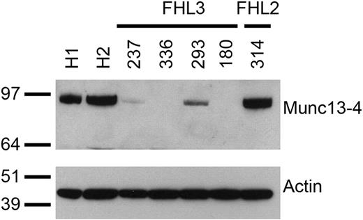
Genetic analysis of MUNC13-4 identified 8 patients with FHL3 with mutations illustrated in Table 2. Figure 2 shows the analysis of Munc13-4 expression in activated polyclonal NK-cell populations derived from 2 healthy donors (H1 and H2), 4 patients with FHL3 (UPNs 237, 336, 293, and 180) and a representative patient with FHL2 (UPN 314). Although equal levels of Munc13-4 are expressed in healthy donors and the patient with FHL2, protein expression is not detected in UPNs 336 and 180, reduced protein levels are detectable in UPN 293, in which the predicted protein possesses a 4 amino acid deletion, and only a trace amount of protein is detected in UPN 237. These results show that the mutations summarized in Table 2 result in greatly reduced or complete loss of Munc13-4 protein expression in NK cells.
Confocal microscopy analysis of cytotoxic granules in perforin and Munc13-4–defective NK cells
To analyze the perforin localization as well as the polarization of lytic granules in patient or donor NK cells, granules were labeled using antibodies against perforin, cathepsin D, and microtubules using an antibody against tubulin. NK cells were visualized either alone or conjugated to the 221 B-EBV cell line. Perforin and cathepsin D colocalize in the same granules in control subject (Figure 1D) and the patient with FHL3 patient (UPN 336) (Figure 1E), whereas only cathepsin D is detectable in the patient with FHL2 (UPN 314) (Figure 1F), thus confirming the loss of perforin expression revealed by FACS analysis (Figure 1C). Lytic granules are distributed along microtubules in healthy donor and patients with FHL2 and FHL3 (Figure 1G-I) and polarize tightly at the immunologic synapse (Figure 1J-L), consistent with previous results in CTLs.9
Perforin expression and granule polarization of patients with FHL compared with control subjects. Polyclonal activated NK cells derived from healthy donor (A,D,G,J) and patients with FHL3 (UPN 336; B,E,H,K) and with FHL2 (UPN 314; C,F,I,L) were analyzed by flow cytometry (A-C) and confocal microscopy (D-L). Filled curves indicate perforin expression, whereas open curves indicate isotypic control (A-C). Confocal staining of perforin (green) and cathepsin D (red) (D-F) or cathepsin D (red) and tubulin (green) (G-L) is analyzed in isolated NK cells (D-I) or conjugates between NK and target 221 (J-L). Bar indicates 10 μm.
Perforin expression and granule polarization of patients with FHL compared with control subjects. Polyclonal activated NK cells derived from healthy donor (A,D,G,J) and patients with FHL3 (UPN 336; B,E,H,K) and with FHL2 (UPN 314; C,F,I,L) were analyzed by flow cytometry (A-C) and confocal microscopy (D-L). Filled curves indicate perforin expression, whereas open curves indicate isotypic control (A-C). Confocal staining of perforin (green) and cathepsin D (red) (D-F) or cathepsin D (red) and tubulin (green) (G-L) is analyzed in isolated NK cells (D-I) or conjugates between NK and target 221 (J-L). Bar indicates 10 μm.
Cytotoxic activity of NK lymphocytes in patients with FHL
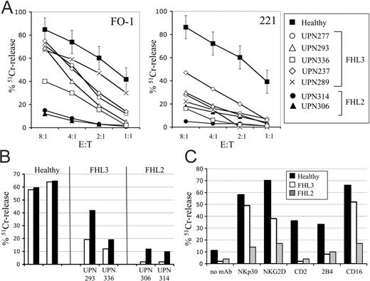
Cytolytic activity of freshly derived peripheral blood lymphocytes (PBLs) against K562 target cells (usually referred to as NK activity) was preliminarily tested in patients with FHL3 and found to be markedly reduced.49 To further characterize this functional impairment, purified NK cells were expanded in IL-2. These activated polyclonal NK cells were tested against a variety of target cells by using a standard 51Cr-release assay (Figure 3). These included the melanoma FO-1 (Figure 3A, left quadrant) and K562 (not shown) tumor cell lines, the 221 (HLA-class I–; Figure 3A, right quadrant) and AMALA (HLA-class I+, not shown) B-EBV cell lines, and iDCs (Figure 3B). In addition, the FcγRc+ P815 was tested to assess the function of individual activating receptors in redirected killing assay (Figure 3C). NK cells from patients with FHL2 and healthy donors were tested for comparison. NK cells from patients with FHL3 displayed intermediate levels of cytotoxicity with respect to the high efficiency of killing by NK cells from healthy donors and the inability of killing by perforin-deficient NK cells from patients with FHL2. The defect of FHL3 NK cells was more evident against the B-EBV cell lines 221 and AMALA (on mAb-mediated masking of HLA class I) than using the tumor cell lines as target cells. Indeed, killing of K562 was clearly impaired only in patient UPN 336. FO-1 target cells elicited a better discrimination of killing capability and also in this assay UPN 336 appeared the most defective patient. Notably, Munc13-4 expression was completely absent in this patient (Table 2).
Munc13-4 expression in control and FHL NK cells. Cell extracts from polyclonal NK cells of healthy donors (H1, H2), patients with FHL3 (UPNs 237, 336, 293, 180), and patient with FHL2 (UPN 314) were analyzed by Western blot with anti–Munc13-4 antibody. Blots were reprobed with antiactin antibody. Molecular weight standards are shown on the left (kDa).
Munc13-4 expression in control and FHL NK cells. Cell extracts from polyclonal NK cells of healthy donors (H1, H2), patients with FHL3 (UPNs 237, 336, 293, 180), and patient with FHL2 (UPN 314) were analyzed by Western blot with anti–Munc13-4 antibody. Blots were reprobed with antiactin antibody. Molecular weight standards are shown on the left (kDa).
Immature DCs, characterized by low surface expression of HLA-class I molecules, are usually highly susceptible to lysis by normal NK cells, in both allogeneic and autologous combination. Indeed, as shown in Figure 3B, NK cells from healthy donors efficiently killed allogeneic iDCs, and lysis was not increased by the addition of anti–HLA class I mAb. Notably, the same iDCs were poorly lysed by the NK cells derived from patients with FHL3, and restoration of lysis was observed on mAb-mediated masking of HLA-class I molecules. Although the values did not reach those of normal NK cells, these data indicate that, because of an inefficient mechanism of lysis induction, the inhibitory receptors can predominate and elicit a strong inhibition of target-cell lysis. As expected, the FHL2 NK cells did not kill even following addition of anti–HLA class I mAb.50
We then assessed the ability of different triggering receptors, including NCR, NKG2D, CD16, CD2, and 2B4, to induce killing of P815. In Figure 3C, in UPN 293 strong NK-cell cytotoxicity could be induced by mAbs to NCR (the representative NKp30 is shown), CD16, and NKG2D, although to a lesser extent than NK cells from the healthy control subject. Remarkably, triggering via CD2 and 2B4, that in normal NK deliver a weaker activation signal, were virtually inactive in UPN 293 NK cells. However, in the perforin-deficient NK cells, none of the activation pathways initiated by different receptors resulted in efficient target-cell lysis.
Taken together, these data support the notion that activated NK cells from patients with FHL3 display an impaired cytolytic activity although less marked than in patients with FHL2.
CD107a expression in patients with FHL after coculture with target cells
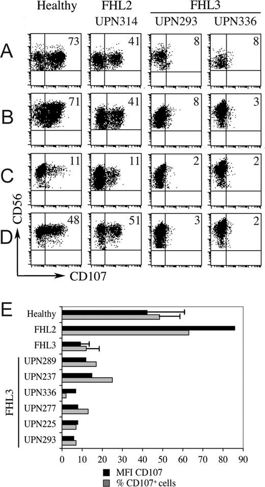
Patients with FHL3 are characterized by critical mutations in the gene encoding Munc13-4, a protein essential for cytolytic granule fusion to the cell surface membrane.9 Therefore, we further analyzed whether the cell surface expression of CD107a molecule, which marks degranulation in NK cells, was altered.48,51 Consistent with previous data, resting normal NK cells (CD3–CD56+ gated in PBMCs) were stained with anti-CD107a mAb (range, 5%-25% CD107a+ cells) when cocultured with K562 (not shown). The percentage of CD107a+ cells increased when they were incubated overnight with IL-2 before addition of target cells (range, 27%-75% CD107a+ cells). To allow an optimal discrimination between normal and pathologic samples, we standardized the assay using a short-term IL-2 cell culture. As shown in Figure 4A, NK cells from a representative healthy individual were compared with those from patients with FHL2 (UPN 314) and 2 different patients with FHL3 (UPNs 293 and 336). Although normal NK cells displayed 73% CD107a+ cells, NK cells with Munc13-4 defect had only 8% CD107a+ cells with a dim staining. In contrast, perforin-deficient NK cells, although unable to lyse K562 target cells, showed a very high proportion (41%) of CD107a+ cells. In Figure 4 (panels B-D), CD107a expression in NK cells from the same donors was also tested using purified polyclonal NK-cell populations. These were incubated with different target cells, such as FO-1 (Figure 4B), K562 (not shown), 221 (Figure 4C), and P815 (redirected killing assay using anti-NKp30 mAb; Figure 4D). The highly susceptible FO-1 and K562 tumor cell lines induced a strong CD107a expression in both healthy and perforin-deficient NK cells (consistently greater than 40%), whereas in Munc13-4–deficient NK cells CD107a expression was lower in terms of both percentage of positive cells and mean fluorescence intensity (MFI; 7 compared with 75 of the healthy donor and 52 of the patient with FHL2). Although the B-EBV cell line 221 was less efficient in inducing CD107a expression by NK cells, differences between healthy and perforin-deficient NK cells as compared with Munc13-4–deficient NK cells were still clearly detectable (11% versus 2%). Finally, when NK cells from healthy individuals were tested against P815 on addition of mAb specific for various activating receptors, similar data (∼ 50% CD107a+ cells) were obtained with NKp30, NKp46, NKp44, and CD16 (results of NKp30 triggering are shown in Figure 4D); in controls, in which no mAb was added, CD107a+ cells were less than 4%. Thus, in normal NK cells the cytofluorimetric analysis using CD107a correlates well with the results of the 51Cr-release assay (see also Figure 3C). Also in this case, perforin-deficient NK cells displayed normal levels of CD107a expression, whereas FHL3 NK cells were clearly defective. Figure 4E further documents that the defect of CD107a expression (on exposure to various target cells) is a common feature to all patients with FHL3, analyzed as a group as well as individually. In particular CD107a expression was observed in 11.8% ± 7.5% of cells in 6 patients with FHL3, whereas it was 48.5% ± 11.5% in 9 healthy individuals (P < .001). The MFI was also significantly lower in the patients with FHL3 compared with the controls (9.3 ± 3.1 versus 42.6 ± 21.9; P < .001). In contrast to FHL3, the mean values of CD107a expression obtained from 3 patients with FHL2 were even higher than healthy control subjects in terms of both percentage (63.3%) and MFI (86.6).
Cytotoxic activity in patients with FHL. Using the standard 51Cr-release assay, polyclonal-activated NK cells derived from patients with FHL3 and FHL2 were compared with those from age-matched healthy individuals for cytolytic activity against various target cells. (A) NK cells from a large group of healthy donors (mean of values, ▪, error bars showing ± SD), from 5 different patients with FHL3 (open symbols and crosses) and 2 different patients with FHL2 (• and ▾) were tested against the melanoma FO-1 and the B-EBV cell line 221 at different E/T ratios, as indicated. (B) NK cells from 2 representative healthy individuals and patients with FHL3 and FHL2 were tested against iDCs derived from a single allogeneic healthy individual either in the absence (□) or in the presence (▪) of anti–HLA class I mAb. The E/T ratio used was 10:1. (C) NK cells from 1 representative healthy individual and 1 patient with FHL3 (UPN 293), and 1 patient with FHL2 (UPN 314) were tested against the Fcγ-Rc+ P815 in the absence or in the presence of mAb to different triggering receptors, as indicated. The E/T ratio used was 4:1. Error bars indicate standard error of the mean.
Cytotoxic activity in patients with FHL. Using the standard 51Cr-release assay, polyclonal-activated NK cells derived from patients with FHL3 and FHL2 were compared with those from age-matched healthy individuals for cytolytic activity against various target cells. (A) NK cells from a large group of healthy donors (mean of values, ▪, error bars showing ± SD), from 5 different patients with FHL3 (open symbols and crosses) and 2 different patients with FHL2 (• and ▾) were tested against the melanoma FO-1 and the B-EBV cell line 221 at different E/T ratios, as indicated. (B) NK cells from 2 representative healthy individuals and patients with FHL3 and FHL2 were tested against iDCs derived from a single allogeneic healthy individual either in the absence (□) or in the presence (▪) of anti–HLA class I mAb. The E/T ratio used was 10:1. (C) NK cells from 1 representative healthy individual and 1 patient with FHL3 (UPN 293), and 1 patient with FHL2 (UPN 314) were tested against the Fcγ-Rc+ P815 in the absence or in the presence of mAb to different triggering receptors, as indicated. The E/T ratio used was 4:1. Error bars indicate standard error of the mean.
CD107a surface expression on NK cells, on target interaction, identifies the Munc13-4 defect. (A) PBMCs from a representative healthy donor, a patient with FHL2 (UPN 314), and 2 patients with FHL3 (UPNs 293 and 336) were cultured overnight in the presence of IL-2 and then cocultured with K562. (B-D) Polyclonal-activated NK-cell populations derived from the same donors were cocultured with FO-1 (B), 221 (C), and P815 with anti-NKp30 (D) mAbs. Cells were stained with anti–CD56-PC5 mAb and anti–CD107a-PE mAb and then analyzed by double fluorescence gating on CD56+ cells. Numbers indicate the percentage of CD107a+ cells. In panel E, histograms refer to the percentage of CD107a+ cells (▦) and to the mean fluorescence intensity (MFI) of CD107a surface expression (▪) considering CD56+ cells in a group of patients with FHL3 (mean of 6 ± SD) compared with healthy individuals (mean of 9 ± SD) and patients with FHL2 (mean of 3) after coculture with susceptible target cells. In patients with FHL3, both percentage and MFI were significantly lower than in healthy controls (P < .001; Student t test). Patients with FHL3 are also shown individually.
CD107a surface expression on NK cells, on target interaction, identifies the Munc13-4 defect. (A) PBMCs from a representative healthy donor, a patient with FHL2 (UPN 314), and 2 patients with FHL3 (UPNs 293 and 336) were cultured overnight in the presence of IL-2 and then cocultured with K562. (B-D) Polyclonal-activated NK-cell populations derived from the same donors were cocultured with FO-1 (B), 221 (C), and P815 with anti-NKp30 (D) mAbs. Cells were stained with anti–CD56-PC5 mAb and anti–CD107a-PE mAb and then analyzed by double fluorescence gating on CD56+ cells. Numbers indicate the percentage of CD107a+ cells. In panel E, histograms refer to the percentage of CD107a+ cells (▦) and to the mean fluorescence intensity (MFI) of CD107a surface expression (▪) considering CD56+ cells in a group of patients with FHL3 (mean of 6 ± SD) compared with healthy individuals (mean of 9 ± SD) and patients with FHL2 (mean of 3) after coculture with susceptible target cells. In patients with FHL3, both percentage and MFI were significantly lower than in healthy controls (P < .001; Student t test). Patients with FHL3 are also shown individually.
Altogether, these data obtained by flow cytometry show that the pattern of CD107a expression represents a novel tool to identify, among patients with FHL, the defect of degranulation which is characteristic of Munc13-4 deficiency. Importantly, in FHL2 NK cells, whose granules lack perforin, the degranulation pattern is normal.
Cytokine production from NK cells in patients with FHL
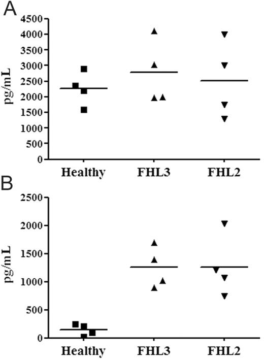
Polyclonal NK-cell populations derived from 4 patients with FHL3 and 4 patients with FHL2 were examined for cytokine production and compared with healthy donors. TNF-α (Figure 5) and IFN-γ (not shown) production was induced by NK-cell triggering via anti-NKp30 mAb (Figure 5A) or by overnight coculture with 221 B-EBV cell line (Figure 5B). NKp30 stimulation induced the production of high amounts of both cytokines with no substantial differences between patients and healthy donors. Similar data were obtained on cell stimulation via NKp46, NKp44, and CD16 (not shown). The basal cytokine production (ie, NK cells incubated with no mAb or with anti-CD56 mAb) was similarly low in patients and healthy individuals. However, when NK cells were cultured with 221 cells, a clear difference existed between patients, characterized by high production, and healthy controls, characterized by low production. This difference could be explained by the fact that, in contrast to normal NK cells, those from patients with FHL are inefficient in killing the B-EBV cells (see Figure 3A), which therefore remain alive and continue to provide a stimulatory signal to NK cells. Normal NK cells, efficiently killing 221, rapidly eliminate the source of stimulating interactions. That this might be a likely explanation is further supported by the finding that target cells which are partially lysed by FHL3, such as FO-1 and P815 in the presence of anti-NKp30 mAb (not shown), induced in FHL3 NK-cell cytokine production only slightly above normal cells. In addition NK cells from patients with FHL2 that do not lyse any of the target cells analyzed, consistently produced high amounts of TNF-α when cocultured with these targets.
Discussion
The present study focuses on the NK-cell function in patients with different FHL subgroups. We show that, by using appropriate target cells, it is possible to detect both perforin and Munc13-4 defects. Remarkably, we first report that the analysis of CD107a, a marker of granule exocytosis (expressed at the surface of NK cells after their interaction with suitable target cells), in combination with the detection of intracytoplasmic perforin, allows a rapid discrimination between FHL2 and FHL3.
TNF-α production by patients with FHL compared with healthy donors. Polyclonal NK-cell populations from 4 different healthy donors, patients with FHL3 (UPNs 249, 237, 225, and 289), and patients with FHL2 (UPNs 210, 235, 256, and 314) were stimulated overnight by plastic-bound anti-NKp30 mAb (A) or by coculture with 221 cell line (B). Supernatants were harvested and analyzed by specific ELISA for their TNFα content. Bars represent the mean of values within a group. Differences among groups were not significant in panel A (P > .05), whereas both FHL3 and FHL2 were different (P < .05) from the healthy group in panel B (Kruskal-Wallis test).
TNF-α production by patients with FHL compared with healthy donors. Polyclonal NK-cell populations from 4 different healthy donors, patients with FHL3 (UPNs 249, 237, 225, and 289), and patients with FHL2 (UPNs 210, 235, 256, and 314) were stimulated overnight by plastic-bound anti-NKp30 mAb (A) or by coculture with 221 cell line (B). Supernatants were harvested and analyzed by specific ELISA for their TNFα content. Bars represent the mean of values within a group. Differences among groups were not significant in panel A (P > .05), whereas both FHL3 and FHL2 were different (P < .05) from the healthy group in panel B (Kruskal-Wallis test).
For most patients with FHL, viral infection may represent a serious challenge sometimes difficult to overcome. During viral infection, the balance between the virus and the host may vary and result in different scenarios. When the cytotoxic response is rapid and efficient, infected cells are rapidly killed, and the infection is terminated as a result of the clearance of the virus. If the killing mechanism is inefficient, as in the case of patients with FHL, both T and NK cells become activated and undergo proliferation; however, they fail to kill infected cells and do not arrest virus dissemination. However, because the source of antigen stimulation is not removed, a persistent T-cell and NK-cell activation occurs and results in the production of large amounts of cytokines, including IFN-γ, granulocyte-macrophage colony-stimulating factor (GM-CSF) (ie, major macrophage activators), and TNFα. In turn, the macrophage homing to the sites of T-cell and NK-cell triggering and activation results in tissue infiltration and in the production of high levels of primary inflammatory cytokines, including TNFα, IL-1, and IL-6, which play a major role in tissue damage and in the various clinical symptoms. It is of note that HLH represents a clinical syndrome, resulting from inefficient cytolytic function and macrophage hyperactivation, which may be common not only to FHL subtypes but also to different congenital immune defects (including Chédiak-Higashi and Griscelli syndromes). However, a different pathogenic mechanism occurs in male children with X-linked lymphoproliferative disease (XLP), a condition which, in some situations, may be barely distinguishable from HLH.20,52 Thus, patients with XLP are unable to control Epstein-Barr virus (EBV) infection as a consequence of a major dysfunction of 2B4 receptor which exerts inhibitory instead of activating function.53 Remarkably, our present data provide a simple in vitro model which mimics the effect of the inability of effector cells to clear the source of (antigen) stimulation. Thus, NK cells from healthy individuals, when cocultured with the B-EBV cell line 221, released small amounts of cytokines, whereas those isolated from patients with FHL2 and FHL3 were consistently high producers. The explanation for this difference is that normal NK cells, but not FHL NK cells, rapidly killed target cells, thus removing the source of NK-cell stimulation. Notably, 221 target cells were largely viable after 24 hours of coculture with FHL NK cells. Remarkably, in patients with FHL the overwhelming activation of the immune system may also result from the marked impairment of NK-mediated killing of iDCs (see Figure 3B and Vitale et al50 ). Indeed, the lack of an efficient DC editing may further lead to an excessive T-cell activation and cytokine release. It should be stressed that potential target cells for NK-mediated lysis in vivo are likely to be mostly represented by virus-infected cells and iDCs. Therefore, the insight into NK-cell function provided by our present study may be more representative of the pathophysiology of HLH.
Activated NK cells derived from various patients with FHL3 showed a variable degree of cytotoxic activity against highly susceptible tumor targets. This correlated with the amounts of Munc13-4 protein detected by Western blot analysis. Our results show that even trace amounts of Munc13-4 protein, associated with certain mutations, were sufficient to allow some killing, whereas the complete absence (eg, UPN 336) led to a marked cytolytic defect. FHL2 NK cells were unable to lyse any target, including the highly susceptible ones.
In addition to providing information useful for the pathophysiology of FHL and for a functional correlation with the different mutations, our data may impact on the diagnosis of this disease. Indeed, we report the first use of surface CD107a expression as a novel screening tool for identification, among patients with FHL, those with Munc13-4 defect (FHL3). It is conceivable that other diseases characterized by defects in granule exocytosis may display the same CD107a defective pattern. However, these diseases (eg, Griscelli or Chédiak-Higashi syndromes) can be distinguished from FHL on a clinical ground. On coculture with target cells, NK lymphocytes from patients with FHL3 showed a sharply lower frequency and MFI of CD107a staining compared with healthy control subjects. Thus, the defect of granule exocytosis could be clearly detected. Differently, FHL2 NK cells, lacking perforin in their granules, showed a normal pattern of CD107a staining. Although the use of purified activated NK-cell populations may increase the discrimination power of this test, it is remarkable that even PBMCs (IL-2 activated for 24 hours) could be used to reveal a defect of granule exocytosis. Thus, the combined use of surface expression of CD107a, together with intracytoplasmic staining with anti-perforin mAb,44 allows to promptly dissect FHL3 and FHL2 defects by the simple analysis of PBMCs, so directing further genetic analysis.
Prepublished online as Blood First Edition Paper, June 15, 2006; DOI 10.1182/blood-2006-04-015693.
Supported by grants from the Associazione Italiana per la Ricerca sul Cancro (AIRC; L.M., D.P., A.S., and M.A.); the Istituto Superiore di Sanità (ISS; L.M. and D.P.); the Ministero della Salute (L.M., D.P., M.A., and A.S.); the Associazione per la Ricerca sulle Sindromi Emofagocitiche-Istiocitosi (ARSE) (M.A.); the Ministero dell'Istruzione dell'Università e della Ricerca (MIUR), the Fondazione Compagnia di San Paolo, the European Union FP6, and grant LSHB-CT-2004-503 319-Allostem (L.M. and D.P.); the Wellcome Trust, United Kingdom (G.M.G. and F.G.); and by a fellowship from the Fondazione Italiana per la Ricerca sul Cancro (FIRC; S.M.).
The publication costs of this article were defrayed in part by page charge payment. Therefore, and solely to indicate this fact, this article is hereby marked “advertisement” in accordance with 18 U.S.C. section 1734.
The European Commission is not liable for any use that may be made of the information contained.


