Abstract
ADP-induced TXA2 generation requires the costimulation of P2Y1, P2Y12, and the GPIIb/IIIa receptors. Signaling events downstream of the P2Y receptors that contribute to ADP-induced TXA2 generation have not been clearly delineated. In this study, we have investigated the role of G-protein–gated inwardly rectifying potassium channels (GIRKs), a recently identified functional effector for the P2Y12 receptor, in the regulation of ADP-induced TXA2 generation. At 10-μM concentrations, the 2 structurally distinct GIRK channel blockers, SCH23390 and U50488H, caused complete inhibition of ADP-induced cPLA2 phosphorylation and TXA2 generation, without affecting the conversion of AA to TXA2 or ADP-induced primary platelet aggregation in aspirin-treated platelets. In addition, Src family kinase selective inhibitors abolished 2MeSADP-mediated cPLA2 phosphorylation and TXA2 generation. Furthermore, these GIRK channel blockers completely blocked Gi-mediated Src kinase activation, suggesting that GIRK channels are upstream of Src family tyrosine kinase activation. In weaver mouse platelets, which have dysfunctional GIRK2 subunits, ADP-induced TXA2 generation was impaired. However, we did not observe any defect in 2MeSADP-induced platelet functional responses in GIRK2-null mouse platelets, suggesting that functional channels composed of other GIRK subunits contribute to ADP-induced TXA2 generation, via the regulation of the Src and cPLA2 activity.
Introduction
Platelets are involved in the maintenance of hemostasis, and abnormal platelet activation could initiate acute thrombotic events. Platelet activation is associated with platelet shape change, granule secretion, aggregation, and the generation of positive feedback mediators such as adenosine diphosphate (ADP) (secreted), thrombin, and thromboxane A2 (TXA2) (generated). ADP, a very important platelet agonist and a key mediator of hemostasis and thrombosis,1,2 is secreted from the platelet-dense granules following their stimulation by physiological platelet agonists such as TXA2, collagen, and thrombin. Released ADP consolidates and stabilizes the growing thrombus by acting via the G-protein–coupled P2Y1 and P2Y12 receptors.3-5 Gq-coupled P2Y1 receptor stimulation results in phospholipase C β (PLC β)–mediated platelet shape change, whereas the P2Y12 receptor stimulation results in Gαi-mediated inhibition of adenylyl cyclase and Gβγ-mediated activation of PI3-Kγ,6,7 Rap1B,8,9 and GIRKs.10 Marked inhibition of platelet aggregation and prolonged bleeding times in patients with mutations in the P2Y12 receptor,11 in P2Y12- or Gαi2-null mice,12,13 or in the presence of P2Y12 receptor antagonists such as clopidogrel14 confirm their critical role in the maintenance of hemostasis.
TXA2 is a potent platelet agonist and an arachidonic acid (AA) metabolite, produced via the cyclooxygenase pathway.15 Activation of cytosolic phospholipase A2 (cPLA2) has been shown to be important for the release of AA from the sn-2 position of membrane-bound phospholipids.16 Phosphorylation of cPLA2 at the Ser505 residue,16-20 and its translocation to the membranes in a calcium-dependent manner,21 have been demonstrated to be important for cPLA2 activity. In platelets, cPLA2 phosphorylation has been noted to occur following stimulation with thrombin, thrombin receptor–activating peptides, or collagen.17,22,23 Studies from our laboratory have shown that ADP-induced TXA2 generation requires concomitant signaling from P2Y1, P2Y12, and GPIIb/IIIa receptors, as antagonism of any of these receptors blocks the generation of TXA2.24 The generated TXA2 triggers the second wave of robust platelet aggregation and concomitant dense granule secretion by acting via the Gq-coupled thromboxane-prostanoid (TP) receptor. Therefore, in the absence of generated TXA2 (as seen in aspirin-treated platelets), ADP fails to induce platelet-dense granule secretion.
GIRKs are G-protein–gated inwardly rectifying potassium channels and are activated by the βγ subunits of pertussis toxin–sensitive G-protein–coupled receptors.25 They play an important role in controlling the membrane excitability in the brain and the heart by modulating the potassium ion conductance. Mammalian cells express 4 different GIRK subunits, GIRK1-4. Functional GIRK channels exist as homo- or heterotetrameric channels, composed of 4 similar or dissimilar subunits,26-29 for example, GIRK1/4 and GIRK4/4 are present in the heart, whereas GIRK2/3 and GIRK1/2 complexes are prevalent in the brain. GIRK2, -3, and -4 can form functional homomultimers, while GIRK1 requires the coassociation with GIRK2, -3, or -4 to achieve membrane surface expression and for the formation of functional channels. In our previous investigation, we demonstrated that GIRK1, 2, and 4 subunits are expressed on human platelets and are important for P2Y12 receptor–mediated platelet functional responses such as fibrinogen receptor activation, platelet aggregation, potentiation of dense granule release, and Akt phosphorylation but not for Gαi-mediated adenylyl cyclase inhibition or Gαq-mediated platelet shape change or intracellular calcium mobilization.10 The role of GIRK2-subunit–containing channels in neurologic responses has been extensively studied by using weaver (WV) and GIRK2-deficient (G2KO) mice. The WV mouse is a neurologic mutant and possesses a missense mutation (G156S) in the pore region of the GIRK2 subunit, leading to the loss of potassium ion selectivity.30 The WV mouse, similar to the G2KO mouse, exhibits the loss of GTPγs-mediated inwardly rectifying currents,31 reduction in the GIRK1 and GIRK2 expression levels in the brain,32 and spontaneous seizure activity.32,33
In the current investigation, we have evaluated the role GIRK channels in ADP-induced TXA2 generation by using pharmacological GIRK channel blockers and 2 different mouse models, namely the WV and G2KO mice. Furthermore, we have attempted to characterize the signaling effectors downstream of GIRK activation that contribute to ADP-induced TXA2 generation response.
Materials and methods
Materials
GIRK channel blockers, SCH23390 (R (+)-7-chloro-8-hydroxy-3-methyl-1-phenyl-2,3,4,5-tetrahydro-1H-3-benzazepine hydrochloride), U50488H (trans-(±)-3,4-dichloro-N-methyl-N-[2-(pyrrolidinyl)cyclohexyl] benzeneacetamide methanesulfonate salt), SKF38393 ((±)-1-phenyl-2,3,4,5-tetrahydro-(1H)-3-benzazepine-7,8-diol hydrochloride), 2MeSADP, apyrase grade VII, human fibrinogen, thrombin, and acetylsalicylic acid were obtained from Sigma (St Louis, MO). ADP was purchased from Chrono-Log (Havertown, PA). AR-C69931MX was a generous gift from Astra-Zeneca Research Laboratories (Charnwood, Loughborough, United Kingdom). Luciferin-Luciferase reagent was purchased from Chrono-Log. Polyclonal antibodies, phospho-cPLA2 (Ser505), and phospho-Src (Y416) were obtained from Cell Signaling Technologies (Beverly, MA). PKCδ antibody was obtained from Santa Cruz Biotechnologies (Santa Cruz, CA). The WV mice and the corresponding age- and sex-matched wild-type (WT) controls were purchased from Jackson Laboratories (Bar Harbor, ME). G2KO mice and WT littermates were kindly provided by Dr Kevin Wickman, University of Minnesota, Minneapolis, MN. All the other reagents were of reagent grade, and deionized water was used throughout.
Preparation of washed human platelets
Whole blood was drawn from healthy, consenting human volunteers selected from students, staff, or workers at Temple University (Philadelphia, PA). This study was approved by the institutional review board of Temple University. Donated blood was collected in tubes containing one-sixth volume of acid citrate dextrose (ACD; 2.5 g sodium citrate, 1.5 g citric acid, and 2 g glucose in 100 mL deionized water). Citrated blood was centrifuged (Eppendorf 5810R centrifuge; Eppendorf, Hamburg, Germany) at 230 RCF (relative centrifugal force) for 20 minutes at room temperature (RT) to obtain platelet-rich plasma (PRP). If required, the PRP was incubated with 1 mM acetylsalicylic acid (aspirin) for 30 minutes at 37°C, and then allowed to remain at RT for 15 minutes. The PRP was then centrifuged for 10 minutes at 980 RCF at RT. Platelet pellet was resuspended in calcium-free Tyrode buffer (138 mM NaCl, 2.7 mM KCl, 1 mM MgCl2,3mMNaH2PO4, 5 mM glucose, 10 mM Hepes adjusted to pH 7.4) containing 0.01 units/mL apyrase. Cells were counted using the Z1 Coulter Particle Counter (Beckman Coulter, Miami, FL) and adjusted to the desired concentration.
Preparation of washed mouse platelets
Blood was collected from anesthetized mice by cardiac puncture into syringes containing 3.8% sodium citrate as anticoagulant. The whole blood was centrifuged (IEC Micromax centrifuge; International Equipment, Needham Heights, MA) at 100 RCF for 10 minutes to isolate the PRP. Prostaglandin E1 (PGE1, 1 μM) was added to PRP. The platelets were centrifuged at 400 RCF for 10 minutes and the pellet was resuspended in Tyrode buffer containing 0.01 unit/mL apyrase.
Measurement of TXA2 generation in human and mouse platelets
Washed human platelets (500 μL; 2 × 10⁸ platelets/mL) or mouse platelets (250 μL; 4-5.5 × 10⁷ platelets/mL) were stimulated with various agonists as mentioned in a lumiaggregometer at 37°C with stirring at 900 rpm, in the presence or absence of SCH23390 (10 μM), U50488H (10 μM), SKF38393 (10 μM), PP3 (10 μM), PP2 (10 μM), and PD173952 (25 μM). Ten-minute preincubations at 37°C were performed for PP3 (10 μM), PP2 (10 μM), PP1 (10 μM), and PD173952 (25 μM). Fibrinogen (1 mg/mL) was added to ADP- and 2MeSADP-stimulated human platelets only. GIRK channel blockers, SCH23390 (10 μM), U50488H (10 μM), and the negative control, SKF38393 (10 μM), were added just prior to the addition of agonist. The baseline was set using Tyrode buffer as blank. The chart recorder (Kipp and Zonen, Bohemia, NY) was set for 0.2 mm/s. After 3.5 minutes of stimulation, the reaction was stopped by quickly freezing the sample in a dry ice–methanol bath. Samples were stored at –80°C and were brought to RT prior to analysis. The samples were centrifuged for 10 minutes at 4°C at 15 000g to remove lysed platelets. The supernatants of human platelets were diluted (1:10 for ADP- and 2MeSADP-treated platelets; 1:400 for AA-treated platelets) with standard diluent (assay buffer), whereas in the case of 2MeSADP (100 nM)–stimulated mouse platelets, 1:5 dilution was performed. The level of TXB2, the stable metabolite of TXA2, was measured in duplicate using the Thromboxane B2 Enzyme Immunoassay Kit according to the manufacturer's instructions (Assay Designs, Ann Arbor, MI). Data represent average of at least 3 days ± standard error (SE).
Measurement of platelet-dense granule secretion in human and mouse platelets
ATP secretion from platelet-dense granules was determined in non–aspirin-treated human or mouse platelets by using the Luciferin-Luciferase assay. The platelets were stimulated in a lumiaggregometer at 37°C with stirring at 900 rpm, and the corresponding luminescence was measured. The data are represented as either percent maximum ATP released (normalized to ADP-mediated platelet-dense granule secretion as 100%) or in the form of actual secretion tracings.
Western blotting
Aliquots of aspirin-treated and washed human platelets were lysed using half the volume of 3X sample loading buffer (with dithiothreitol [DTT], 300 mM) and boiled for 10 minutes. The platelet lysates were loaded onto a 10% or 8% Tris-glycine gel for the measurement of Src phosphorylation and cPLA2 phosphorylation, respectively, subjected to sodium dodecyl sulfate–protein agarose gel electrophoresis (PAGE), and transferred to PVDF membrane. Nonspecific binding sites were blocked by incubating the membrane in Tris-buffered saline–Tween (TBST; 20 mM Tris, 140 mM NaCl, 0.1% [vol/vol] Tween 20) containing 5% (wt/vol) bovine serum albumin (BSA) for 30 minutes at RT, followed by incubating it overnight at 4°C with gentle agitation in the primary antibody (1:1000 dilution for anti–phospho-Src and anti-PKCδ and 1:500 dilution for anti–phospho-cPLA2, in TBST with 2% BSA). After washing with TBST, the membranes were probed with an alkaline phosphatase–labeled secondary antibody (1:5000 dilution in TBST with 2% BSA) for 1 hour at RT. After additional washing steps, membranes were then incubated with chlordiazepoxide (CDP)–Star and chemiluminescent substrate (Tropix, Bedford, MA) for 10 minutes at RT, and immunoreactivity was detected using a Fuji Film Luminescent Image Analyzer (LAS-1000 CH; Tokyo, Japan).
Results
Effect of low concentrations of GIRK channel blockers on ADP-induced platelet aggregation in non–aspirin-treated human platelets
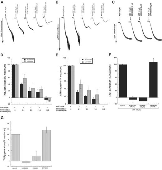
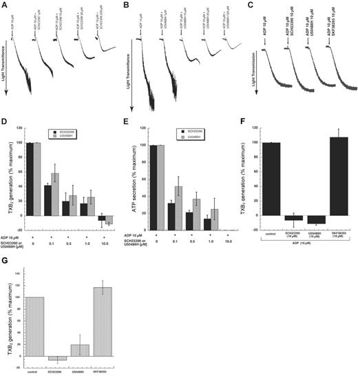
The effect of different concentrations of GIRK channel blockers on ADP-induced platelet aggregation, in the presence and absence of aspirin treatment, was tested. Low concentrations of GIRK channel blockers inhibited ADP-induced platelet aggregation in non–aspirin-treated human platelets (Figure 1A-B), and did not have any effect in aspirin-treated human platelets (Figure 1C). However, 30-μM and higher concentrations of GIRK channel blockers inhibited ADP-induced platelet aggregation in aspirin-treated platelets as well.10 These results suggest that the observed inhibition of platelet aggregation by lower concentrations of GIRK channel blockers in the absence of aspirin treatment might be due to the inhibition of TXA2 generation.
Inhibition of ADP-induced TXA2 generation by low concentrations of GIRK channel blockers
To confirm our hypothesis, we measured the effect of low concentrations of these channel blockers on ADP-induced TXA2 formation (correlated with the levels of TXB2, its stable metabolite) in non–aspirin-treated human platelets. Simultaneous estimation of ADP-induced platelet-dense granule release, a TXA2-dependent response, was also performed. Both of these responses were inhibited in a concentration-dependent manner with maximal inhibition noted at 10-μM concentrations of SCH23390 and U50488H (Figure 1C-D). However, 10 μM SKF38393, the inactive analog of SCH23390, failed to inhibit TXA2 generation under similar conditions, confirming the specificity of these reagents (Figure 1E). Similar experiments were performed with another platelet P2Y receptor agonist 2MeSADP (100 nM) and comparable observations were made (Figure 1F). We have also confirmed that these channel blockers (10 μM) fail to inhibit U46619-induced platelet aggregation, indicating that they do not directly interfere with TXA2-induced signaling (data not shown). Based on these findings, we conclude that GIRK channels are essential for ADP-induced TXA2 generation.
Evaluation of the role of GIRK2 subunit–containing GIRK channels in ADP-induced TXA2 generation by molecular genetic approaches
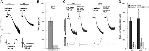
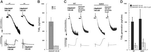
The WV mouse has dysfunctional GIRK2 homo- and heteromultimers and is commercially available. Therefore, we used it as a model for evaluating the role of GIRK2 channels in ADP-induced TXA2 generation. We noticed that in non–aspirin-treated WV mouse platelets, 2MeSADP (100 nM)–induced platelet aggregation, ATP secretion from the platelet-dense granules (Figure 2A), and TXA2 generation were dramatically impaired in comparison with the WT platelets (Figure 2B).
To validate these findings obtained with the WV mouse platelets, we measured TXA2 generation in platelets obtained from G2KO mice. In contrast to the results obtained with the WV mouse platelets, G2KO mouse platelets did not show significant impairment of 2MeSADP (100 nM)–induced platelet aggregation, dense granule release (Figure 2C), or TXA2 generation (Figure 2D). In addition, pretreatment of the G2KO mouse platelets with SCH23390 (10 μM) caused significant inhibition of these aforementioned responses (Figure 2C-D) that was comparable with the results obtained with WT controls.
GIRK channel blockers do not impair the conversion of AA to TXA2
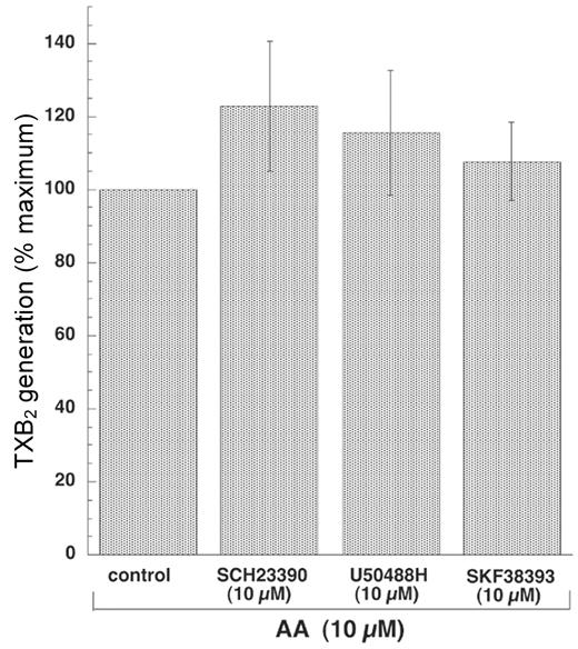
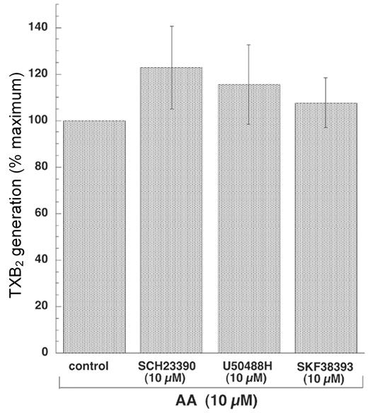
Since some pharmacological compounds such as SB203580, the p38MAPK inhibitor, and PD98059, the MEK inhibitor, have been shown to inhibit agonist-induced TXA2 generation by directly inhibiting the enzymes of the COX pathway,34 we wanted to eliminate the possibility that the GIRK channel blockers nonspecifically exert any such effects. Therefore, we measured AA (100 μM)–stimulated TXB2 levels in the presence and absence of GIRK channel blockers. SQ29548, the TP receptor antagonist, was added to all samples to prevent the positive feedback amplification of TXA2 formation via the TP receptor stimulation. As shown in Figure 3, neither SCH23390 (10 μM) nor U50488H (10 μM) inhibited AA-induced TXA2 formation, implying that they do not impair the activity of the COX enzymes. These results confirm that the noted inhibition on ADP-induced TXA2 generation is not due to interference with the conversion of AA to TXA2.
GIRK channel blockers abolish the phosphorylation of cPLA2
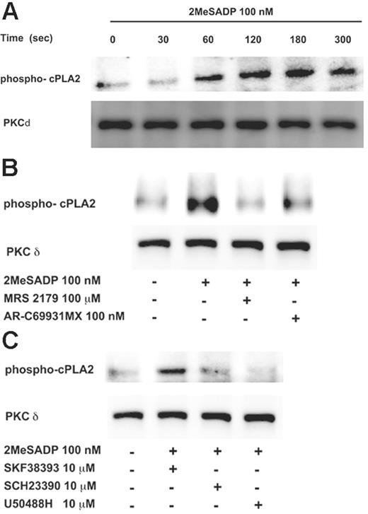
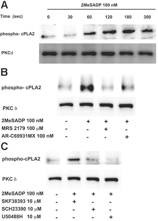
Next, we sought to investigate whether the GIRK channels mediate ADP-induced TXA2 generation by regulating the phosphorylation of cPLA2 at Ser505 residue, a measure of its activity. 2MeSADP (100 nM) caused a time-dependent increase in phosphorylation of cPLA2 (Ser505) in aspirin-treated platelets (Figure 4A). In addition, 2MeSADP-induced cPLA2 phosphorylation was abolished by antagonism of either P2Y1 or P2Y12 receptors (Figure 4B). Furthermore, 2MeSADP-induced cPLA2 phosphorylation was also abolished by SCH23390 (10 μM) or U50488H (10 μM) (Figure 4C). These observations suggest that costimulation of P2Y1 or P2Y12 receptors is essential for MeSADP-induced cPLA2 phosphorylation and that GIRK channel activation, downstream of P2Y12 receptor stimulation, is an important mediator of this phosphorylation response.
Role of Src family tyrosine kinases in GIRK channel–mediated cPLA2 phosphorylation and TXA2 generation
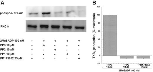
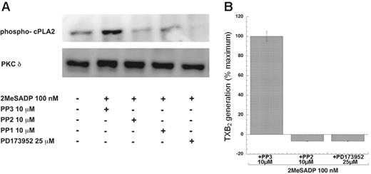
The Src family of tyrosine kinases have been implicated in TXA2 generation downstream of different receptors.35-37 As shown in Figure 5, 2MeSADP-induced cPLA2 phosphorylation at Ser505 residue (Figure 5A) and TXA2 generation (Figure 5B) were completely abolished by the pretreatment of platelets with Src kinase–specific inhibitors such as PD173952 (25 μM) and PP2 (10 μM), but not by PP3 (10 μM), the inactive analog of PP2. These results confirm that the Src family of tyrosine kinases is upstream of cPLA2 phosphorylation and is essential for ADP-induced TXA2 generation.
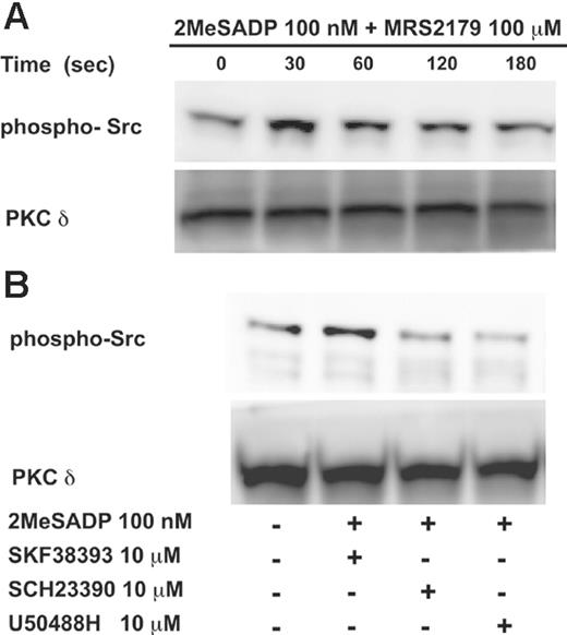
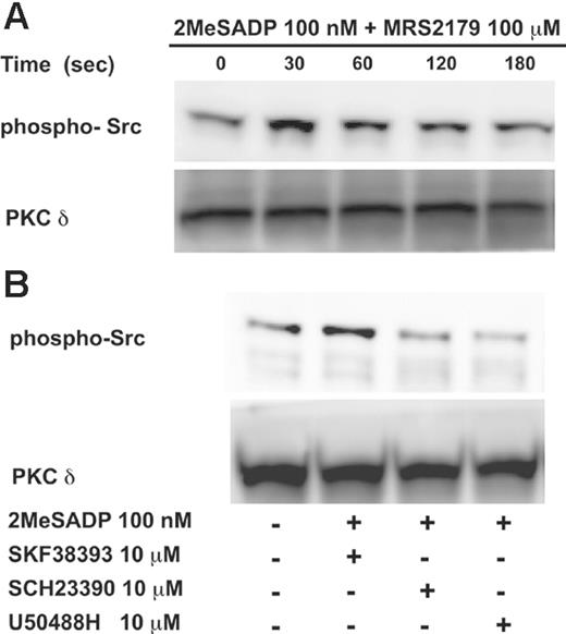
Furthermore, reports from our laboratory have shown that selective stimulation of the Gi-coupled P2Y12 receptor can induce Src kinase activation.35 Therefore, we investigated whether the GIRK channels contribute to Gi-mediated Src kinase activation. Selective Gi activation was achieved by stimulating platelets with 2MeSADP (100 nM) in the presence of MRS2179 (100 μM), a P2Y1 receptor selective antagonist. As shown in Figure 6, a time-dependent increase in Src kinase Y416 phosphorylation was observed (Figure 6A). Both SCH23390 (10 μM) and U50488H (10 μM) inhibited Gi-mediated Src kinase activation (Figure 6B), suggesting that GIRK channels contribute to ADP-induced cPLA2 phosphorylation and TXA2 generation by regulating the activation of the Src family of tyrosine kinases. Similar inhibition of Src activity was observed in the WV mouse platelets compared with WT mouse platelets (data not shown).
Effect of GIRK channel blockers on ADP-induced platelet aggregation, TXA2 generation, and dense granule secretion in non–aspirin-treated human platelets. Washed non–aspirin-treated human platelets (A-B,D-G) or aspirin-treated human platelets (C) were stimulated with ADP (10 μM) (A-F) or 2MeSADP (100 nM) (G) for 3.5 minutes under stirring conditions at 37°C in the presence and absence of different concentrations of SCH23390, or U50488H. Fibrinogen (1 mg/mL) was added to all the samples prior to the addition of the agonist. In panels A, B, and C, platelet aggregation was measured by measuring light transmittance. In panels D and F, and G, TXB2 levels were analyzed as described in “Materials and methods.” In panel E, ATP secretion was measured as described in “Materials and methods.” In panels D and E, different concentrations of SCH23390 or U50488H were used as indicated. In panels F and G, 10-μM concentrations of SCH23390, U50488H, or SKF38393 were used. The data are represented as percent maximal TXB2 generated by ADP or 2MeSADP in the absence of antagonists. Each bar is the average of 3 experiments ± SEM from 3 different donors.
Effect of GIRK channel blockers on ADP-induced platelet aggregation, TXA2 generation, and dense granule secretion in non–aspirin-treated human platelets. Washed non–aspirin-treated human platelets (A-B,D-G) or aspirin-treated human platelets (C) were stimulated with ADP (10 μM) (A-F) or 2MeSADP (100 nM) (G) for 3.5 minutes under stirring conditions at 37°C in the presence and absence of different concentrations of SCH23390, or U50488H. Fibrinogen (1 mg/mL) was added to all the samples prior to the addition of the agonist. In panels A, B, and C, platelet aggregation was measured by measuring light transmittance. In panels D and F, and G, TXB2 levels were analyzed as described in “Materials and methods.” In panel E, ATP secretion was measured as described in “Materials and methods.” In panels D and E, different concentrations of SCH23390 or U50488H were used as indicated. In panels F and G, 10-μM concentrations of SCH23390, U50488H, or SKF38393 were used. The data are represented as percent maximal TXB2 generated by ADP or 2MeSADP in the absence of antagonists. Each bar is the average of 3 experiments ± SEM from 3 different donors.
Discussion
We recently showed that platelets express GIRK1, GIRK2, and GIRK4 subunits.10 Using the 2 structurally distinct GIRK channel blockers, SCH23390 and U50488H, we demonstrated that GIRK channels are one of the functional effectors for the P2Y12 receptor–mediated platelet aggregation.10 When the effect of different concentrations of GIRK channel blockers on ADP-induced platelet aggregation was evaluated, it was observed that varying concentrations of GIRK channel blockers produced a differential pattern of inhibitory effect in the presence and absence of aspirin treatment. At concentrations 30 μM and higher, these channel blockers inhibited aggregation of platelets treated with aspirin,10 whereas the lower concentrations inhibited platelet aggregation only in non–aspirin-treated human platelets. Based on these results, we speculated that impairment of the platelet aggregation response by lower concentrations of GIRK channel blockers could be due to the inhibition of TXA2 generation. Therefore, in the current study, we have focused our attempts on understanding the role of GIRK channels in ADP-induced TXA2 generation.
2MeSADP-induced platelet aggregation, dense granule secretion, and TXA2 generation in WV and G2KO mouse platelets. Washed and non–aspirin-treated WT and WV mouse platelets (A-B) or WT and G2KO mouse platelets (C-D) were stimulated with 2MeSADP (100 nM), in the absence of exogenously added fibrinogen, for 3.5 minutes at 37°C under stirring conditions, and platelet aggregation (A,C) or TXB2 levels (B,D) were measured as described in “Materials and methods.” The data are represented as the percent maximal TXB2 generated by 2MeSADP in WT platelets. In panels A and C, corresponding luminescence in each sample was recorded. Each bar is the average of 3 experiments ± SEM from 3 different donors.
2MeSADP-induced platelet aggregation, dense granule secretion, and TXA2 generation in WV and G2KO mouse platelets. Washed and non–aspirin-treated WT and WV mouse platelets (A-B) or WT and G2KO mouse platelets (C-D) were stimulated with 2MeSADP (100 nM), in the absence of exogenously added fibrinogen, for 3.5 minutes at 37°C under stirring conditions, and platelet aggregation (A,C) or TXB2 levels (B,D) were measured as described in “Materials and methods.” The data are represented as the percent maximal TXB2 generated by 2MeSADP in WT platelets. In panels A and C, corresponding luminescence in each sample was recorded. Each bar is the average of 3 experiments ± SEM from 3 different donors.
ADP-induced TXA2 generation was completely abolished by 10-μM concentrations of SCH23390 and U50488H. Consistent with this observation, TXA2-dependent platelet aggregation and dense granule release were also completely inhibited at these concentrations. However, as discussed earlier, 10-μM concentrations of SCH23390 and U50488H did not inhibit ADP-induced platelet aggregation in aspirin-treated platelets, confirming that the noted inhibition of TXA2 generation is not due to the direct inhibition of GPIIb/IIIa activation, which along with the P2Y1 and P2Y12 receptor signaling pathway is required for ADP-induced TXA2 generation.24
Effect of GIRK channel blockers on AA-induced TXA2 generation. Washed non–aspirin-treated human platelets were stimulated with AA (100 μM) for 3.5 minutes at 37°C under stirring conditions in the presence of SQ29548 (10 μM), in the presence and absence of SCH23390 (10 μM), U50488H (10 μM), or SKF38393 (10 μM). Reactions were stopped after 3.5 minutes and TXB2 levels were analyzed as described in “Materials and methods.” The data are represented as the percent maximal TXB2 generated by AA obtained in the absence of antagonists. Each bar is the average of 3 experiments ± SEM from 3 different donors.
Effect of GIRK channel blockers on AA-induced TXA2 generation. Washed non–aspirin-treated human platelets were stimulated with AA (100 μM) for 3.5 minutes at 37°C under stirring conditions in the presence of SQ29548 (10 μM), in the presence and absence of SCH23390 (10 μM), U50488H (10 μM), or SKF38393 (10 μM). Reactions were stopped after 3.5 minutes and TXB2 levels were analyzed as described in “Materials and methods.” The data are represented as the percent maximal TXB2 generated by AA obtained in the absence of antagonists. Each bar is the average of 3 experiments ± SEM from 3 different donors.
Effect of P2Y1 and P2Y12 receptor antagonists or GIRK channel blockers on 2MeSADP-induced cPLA2 phosphorylation. Washed and aspirin-treated human platelets were stimulated with 2MeSADP (100 nM) in the presence of fibrinogen (1 mg/mL) for various time points at 37°C (A), or for 60 seconds at 37°C, in the presence of MRS2179 (100 μM) or AR-C69931MX (100 nM) (B), and SCH23390 (10 μM), U50488H (10 μM), or SKF38393 (10 μM) (C). The reaction was stopped using 3X sample loading buffer (with DTT). The lysates were subjected to Western blotting analysis and probed with phospho-cPLA2 (Ser505) and PKCδ (lane loading control).
Effect of P2Y1 and P2Y12 receptor antagonists or GIRK channel blockers on 2MeSADP-induced cPLA2 phosphorylation. Washed and aspirin-treated human platelets were stimulated with 2MeSADP (100 nM) in the presence of fibrinogen (1 mg/mL) for various time points at 37°C (A), or for 60 seconds at 37°C, in the presence of MRS2179 (100 μM) or AR-C69931MX (100 nM) (B), and SCH23390 (10 μM), U50488H (10 μM), or SKF38393 (10 μM) (C). The reaction was stopped using 3X sample loading buffer (with DTT). The lysates were subjected to Western blotting analysis and probed with phospho-cPLA2 (Ser505) and PKCδ (lane loading control).
Effect of Src kinase inhibitors on 2MeSADP-induced cPLA2 phosphorylation and TXA2 generation. Washed and aspirin-treated human platelets were stimulated with 2MeSADP (100 nM), in the presence of fibrinogen (1 mg/mL) under stirring conditions for 60 seconds at 37°C (A). As per the requirement, platelets were preincubated with PP3 (10 μM), PP2 (10 μM), or PD173952 (25 μM) for 10 minutes at 37°C. The reaction was stopped using 3X sample loading buffer (with DTT). The lysates were subjected to Western blotting analysis and probed with phospho-cPLA2 (Ser505) and PKCδ (lane loading control) (A). In the case of TXB2 measurement (B), platelets were stimulated in the presence of Src kinase inhibitors or the inactive analog for 3.5 minutes at 37°C, following which the reactions were stopped and TXB2 levels were analyzed as described in “Materials and methods.” The data are represented as the percent maximal TXB2 generated by 2MeSADP in the absence of inhibitors. Each bar is the average of 3 experiments ± SEM from 3 different donors.
Effect of Src kinase inhibitors on 2MeSADP-induced cPLA2 phosphorylation and TXA2 generation. Washed and aspirin-treated human platelets were stimulated with 2MeSADP (100 nM), in the presence of fibrinogen (1 mg/mL) under stirring conditions for 60 seconds at 37°C (A). As per the requirement, platelets were preincubated with PP3 (10 μM), PP2 (10 μM), or PD173952 (25 μM) for 10 minutes at 37°C. The reaction was stopped using 3X sample loading buffer (with DTT). The lysates were subjected to Western blotting analysis and probed with phospho-cPLA2 (Ser505) and PKCδ (lane loading control) (A). In the case of TXB2 measurement (B), platelets were stimulated in the presence of Src kinase inhibitors or the inactive analog for 3.5 minutes at 37°C, following which the reactions were stopped and TXB2 levels were analyzed as described in “Materials and methods.” The data are represented as the percent maximal TXB2 generated by 2MeSADP in the absence of inhibitors. Each bar is the average of 3 experiments ± SEM from 3 different donors.
Functional GIRK channels exist as homo- or heterotetramers, which are composed of 4 similar or dissimilar subunits, respectively. Therefore, several functional GIRK channel complexes based on different subunit combinations could possibly exist in human platelets. It is also known that SCH23390 and U50488H, when used at differing concentrations, have the ability to inhibit distinct GIRK channel complexes.38 Hence, based on the previous findings and the results presented here, we contend that P2Y12 receptor stimulation results in activation of different functional GIRK channel complexes that mediate diverse platelet functional responses. One subpopulation of the GIRK channels susceptible to inhibition by lower concentrations of SCH23390 and U50488H is important for ADP-induced TXA2 generation, whereas higher concentrations of these blockers inhibit all combinations of functional GIRK channels and hence inhibit P2Y12 receptor–mediated platelet responses such as platelet aggregation, potentiation of dense granule release, and Akt phosphorylation.10
Since the WV mouse has a G156S mutation in the pore region the GIRK2 subunit, it has been used extensively to evaluate the of GIRK2-subunit–containing channels in a number of neurologic responses. Hence, we first used the WV mouse platelets as a model system to confirm the role of GIRK2 subunit–containing GIRK channels in ADP-induced TXA2 generation response. Compared with WT controls, the WV mouse platelets demonstrated significant impairment of ADP-induced platelet aggregation, dense granule secretion, and TXA2 generation. Unexpectedly, the G2KO mouse platelets failed to elicit any inhibition of platelet aggregation, dense granule secretion, or TXA2 generation compared with WT controls. However, this could be a reflection of compensatory mechanisms provided by the overexpression of other GIRK channel subunits.
Effect of GIRK channel blockers on Gi-mediated Src activation. Washed aspirin-treated human platelets were stimulated with 2MeSADP (100 nM) in the presence of MRS2179 (100 μM) for different time points at 37°C as indicated under stirring conditions (A), in the absence of exogenously added fibrinogen. In panel B, they were stimulated with 2MeSADP (100 nM) in the presence of MRS2179 (100 μM) and SCH23390 (10 μM), U50488H (10 μM), or SKF38393 (10 μM). The reaction was stopped using 3X sample loading buffer (with DTT). The lysates were subjected to Western blotting analysis and probed with phospho-Src (Y416) (A-B) or PKCδ (lane loading control).
Effect of GIRK channel blockers on Gi-mediated Src activation. Washed aspirin-treated human platelets were stimulated with 2MeSADP (100 nM) in the presence of MRS2179 (100 μM) for different time points at 37°C as indicated under stirring conditions (A), in the absence of exogenously added fibrinogen. In panel B, they were stimulated with 2MeSADP (100 nM) in the presence of MRS2179 (100 μM) and SCH23390 (10 μM), U50488H (10 μM), or SKF38393 (10 μM). The reaction was stopped using 3X sample loading buffer (with DTT). The lysates were subjected to Western blotting analysis and probed with phospho-Src (Y416) (A-B) or PKCδ (lane loading control).
Several studies have shown that even though WV and G2KO mice are similar in certain aspects, they differ markedly in a number of phenotypical and morphologic responses mainly due to the anomalous sodium ion flux observed in the WV mouse.32,39 The WV GIRK2 channel lacks potassium ion selectivity,39-41 and it possesses significant cerebellar granule cell degeneration and ataxia due to intracellular accumulation of sodium ions39,40 and calcium ions,42 in addition to male infertility.43 In contrast, G2KO mice reveal much less severe phenotype and are almost indistinguishable from the WT littermates. They have morphologically normal cerebellar granule cells as they lack the aberrant sodium ion flux and are fertile. It has been reported that the phenotypical and morphologic abnormalities in the WV mouse are due to gain of function rather than loss of function for the homomeric or heteromeric GIRK2 channels.31,32,44 Furthermore, reversal of some of these defects in the WV mouse with pharmacological blockade of sodium influx substantiates the gain-of-function characteristic in the WV mouse.39 In platelets, it has been shown that modulation of the sodium-proton exchanger activity by regulating the extracellular sodium concentration, or extracellular pH, significantly affects AA mobilization and dense granule release induced by weaker agonists such as ADP or low-dose thrombin.45 Therefore, based on these published findings, we hypothesize that the noticeable impairment of ADP-induced platelet aggregation, dense granule secretion, and TXA2 generation in the WV mouse is due to altered sodium ion fluxes and not due to loss of function for the GIRK2-subunit–containing channels.
It is well documented that phosphorylation of cPLA2 is essential for its activity and AA release. In cells such as macrophages, fibroblasts, platelets, and CHO cells, treatment with diverse physiological agonists such as thrombin, ATP, colony-stimulating factor-1 (CSF-1), growth factors, vasopressin, or zymosan causes increases in phosphorylation of cPLA2 on serine residues that correlate well with increases in its activity.16,17,46-49 Of importance, Lin et al16,20 have shown that phosphorylation of cPLA2 at serine 505 residue is important for the hormonally regulated release of AA from the membrane-bound phospholipids, since S505A mutation prevents normal agonist-mediated AA release. Using phospho-specific antibody, we now show that ADP-induced cPLA2 phosphorylation (Ser505) requires both P2Y1 and P2Y12 receptors since antagonism of either receptor inhibits its phosphorylation. Therefore, we believe that costimulation of Gq- and Gi-mediated signaling events is necessary for Ser505 phosphorylation of cPLA2. Consistent with these findings, Jin et al24 have shown that ADP-induced AA release and TXA2 generation also require costimulation of P2Y1 and P2Y12 receptors. Similar to the TXA2 generation response, low concentrations of GIRK channel blockers also abrogated 2MeSADP-induced cPLA2 phosphorylation, confirming that they regulate TXA2 generation by regulating cPLA2 activity, downstream of P2Y12 receptor stimulation.
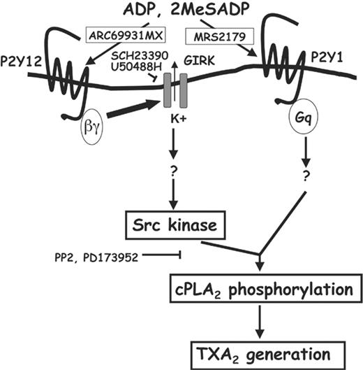
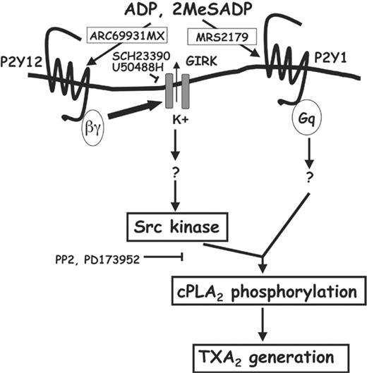
Recent reports from our laboratory have shown that the Src family of tyrosine kinases are activated following P2Y12 receptor stimulation50 and are an important player in TXA2 generation downstream of different receptors.35-37 Also, Cho et al51 have shown that in Lyn-deficient mouse platelets, TXA2 generation is impaired. Src kinase family selective inhibitors completely abrogated ADP receptor–mediated cPLA2 phosphorylation (Ser505) and TXA2 generation, indicating that Src kinase activation is important for both these events. Furthermore, Gi-mediated Src activation was abolished by low concentrations of GIRK channel blockers. Taking these data into consideration, we conclude that GIRK channel activation following P2Y12 receptor stimulation mediates the Src activation (depicted in Figure 7). It is important to note that selective stimulation of the Gq-coupled P2Y1 signaling can also activate the Src tyrosine kinases.52 Therefore, it is possible that Src kinases downstream of Gq could be contributing to ADP-induced cPLA2 phosphorylation as well. Since the Src family kinase selective inhibitors used in this study fail to distinguish between the different members of the Src family, it is difficult to ascertain the exact identity of the particular kinase (or kinases) that transduces this phosphorylation event. However, since 10-μM concentrations of GIRK channel blockers abolish Gi-mediated Src activity and ADP-induced cPLA2 activity, we believe that Src kinases downstream of the Gi pathway are important for this response.
There is evidence that GIRK1 and GIRK4 subunits can be phosphorylated on select tyrosine residues in their cytosolic domain.53 It is therefore possible that the tyrosine-phosphorylated GIRK subunits could activate the Src kinase(s) by direct interaction. However, more elaborate studies need to be performed to conclusively describe the mechanism of GIRK-mediated Src activation.
Schematic representation of the role of GIRK channels in ADP-induced TXA2 generation. ADP induces platelet aggregation by activating the P2Y1 and P2Y12 receptors. P2Y12 receptor stimulation causes the release of the βγ dimers from the Gαi subunit, which activates the GIRK channels by binding to their cytosolic regions. Costimulation of P2Y1 and P2Y12 receptors is important for ADP-induced cPLA2 phosphorylation on the serine 505 residue, AA release, and TXA2 generation. Selective stimulation of the P2Y12 receptor leads to Src tyrosine kinase activation, which is inhibited by GIRK channel blockade. Activation of both GIRK channels and Src family of tyrosine kinases, following P2Y12 receptor stimulation, is essential for ADP-induced cPLA2 phosphorylation and for TXA2 generation. SCH23390 and U50488H are the 2 structurally distinct GIRK channel blockers that have been used in our study.
Schematic representation of the role of GIRK channels in ADP-induced TXA2 generation. ADP induces platelet aggregation by activating the P2Y1 and P2Y12 receptors. P2Y12 receptor stimulation causes the release of the βγ dimers from the Gαi subunit, which activates the GIRK channels by binding to their cytosolic regions. Costimulation of P2Y1 and P2Y12 receptors is important for ADP-induced cPLA2 phosphorylation on the serine 505 residue, AA release, and TXA2 generation. Selective stimulation of the P2Y12 receptor leads to Src tyrosine kinase activation, which is inhibited by GIRK channel blockade. Activation of both GIRK channels and Src family of tyrosine kinases, following P2Y12 receptor stimulation, is essential for ADP-induced cPLA2 phosphorylation and for TXA2 generation. SCH23390 and U50488H are the 2 structurally distinct GIRK channel blockers that have been used in our study.
In conclusion, we have revealed a novel role for GIRK channels as a functional effector for ADP-mediated cPLA2 phosphorylation and TXA2 generation via the regulation of Src activity. We conclude that different GIRK channel complexes mediate the different platelet functional responses upon ADP receptor stimulation, namely platelet aggregation and TXA2 generation. Finally, we also believe that GIRK2-subunit–containing functional channels probably do not contribute significantly to ADP-induced TXA2 generation.
Prepublished online as Blood First Edition Paper, July 20, 2006; DOI 10.1182/blood-2006-03-010330.
Supported by research grants HL60683 and HL80444 from the National Institutes of Health (S.P.K.) and predoctoral fellowship from the American Heart Association, Pennsylvania-Delaware affiliate (H.S.).
H.S. designed research, performed research, collected data, analyzed data, and wrote the paper; B.N.K. designed experiments, performed experiments, collected data, and analyzed data; J.P. performed experiments, collected data, and analyzed data; P.L. designed experiments, performed experiments, collected data, and analyzed data; S.K. analyzed data; and S.P.K. designed research, analyzed data, and provided overall guidance to the manuscript.
The publication costs of this article were defrayed in part by page charge payment. Therefore, and solely to indicate this fact, this article is hereby marked “advertisement” in accordance with 18 U.S.C. section 1734.
We thank Dr Kevin Wickman, University of Minnesota, Minneapolis, MN, for the generous gift of GIRK2-null mice.

