Abstract
Murine CD4+CD25+ regulatory T cells (Treg cells) reduce acute graft-versus-host disease (aGvHD). However, surface molecules critical for suppression are unclear. Deficiency of CD30 (CD30−/−) leads to impaired thymic negative selection and augmented T-cell autoreactivity. Therefore, we investigated the role of CD30 signaling in Treg-cell function during aGvHD. Treg cells derived from CD30−/− animals were significantly less effective in preventing aGvHD lethality. Early blockade of the CD30/CD153 pathway with a neutralizing anti-CD153 mAb reduced Treg-mediated protection from proinflammatory cytokine accumulation and donor-type T-cell apoptosis. In vivo bioluminescence imaging demonstrated intact homing but reduced expansion of luciferase-expressing Treg cells when CD153 was blocked during the early phase after adoptive transfer. CD30 surface expression on Treg cells increased with alloantigen exposure, and CD153 expression on recipient-type dendritic cells increased in the presence of a proinflammatory environment. These data demonstrate that early CD30 signaling is critical for Treg-mediated aGvHD protection after major MHC-mismatch bone marrow transplantation.
Introduction
Acute graft-versus-host disease (aGvHD) is one of the major complications after allogeneic bone marrow transplantation (BMT), which limits the success of this otherwise life-saving strategy.1 Our group and others have demonstrated that naturally occurring CD4+CD25+ regulatory T cells (Treg cells) can reduce the incidence and severity of murine aGvHD.2–5 In spite of findings that cell-cell contact is critical for Treg-mediated suppression in vitro and that IL-10 production6 and TGF-β surface expression7 are relevant in vivo, the mechanisms by which Treg cells exert suppressor activity in the setting of aGvHD are poorly understood. Recent reports have described the role of apoptosis induction by activated Treg cells of conventional T cells (Tconv cells) and B cells as a mechanism of suppression.8–12
The TNF-R superfamily member CD30 has been shown to be expressed on Tr1 regulatory cells that down-modulate nickel-specific immune responses13 and to be relevant for Treg-mediated protection from allograft rejection.9 In human immune-mediated diseases, CD30 is expressed on T cells that serve a regulatory role in rheumatoid arthritis14 and on cells that accumulate at the inflammatory sites of patients with systemic sclerosis15,16 and chronic GvHD.15 Expression of CD30 is detected late after T-cell activation in vitro with immobilized CD3 mAb17 and activation-induced CD30 is found on T helper 1 (Th1), Th0, and Th2 cell clones.18,19 CD30 signaling up-regulates the lymph node homing molecule CCR7,20 provides costimulatory signals to T cells, and enhances their proliferative responses to suboptimal stimulation via T-cell receptor (TCR) engagement.17,21
The ligand for CD30 (CD30L, or CD153) is a membrane-associated glycoprotein related to TNF,21 which is known to be expressed on thymic epithelial cells (TECs), antigen presenting cells (APCs), activated T cells, neutrophils, eosinophils, and resting B cells.22,23 CD30-deficient C57B/6 mice display elevated numbers of thymocytes and a gross defect in negative selection,24 although this was not found in all mouse strains.25 Overexpression of CD30 on T cells results in augmented thymocyte depletion upon treatment with a superantigen,26 suggesting an important role for CD30/CD153 interactions in thymic deletion of autoreactive T cells. Recently, the thymic medulla has been demonstrated to be an anatomic site where Treg cells interact with activated dendritic cells (DCs) and Hassall corpuscles27 that express CD153.23
In this report, we investigated the role of CD30 signaling in Treg-cell function, demonstrating a critical role for CD30/CD153 interactions early after adoptive transfer.
Materials and methods
Mice
FVB/N (H-2kq, Thy-1.1), C57B/6 (H-2kb, Thy-1.1 or Thy-1.2), C57B/6eGFP+, and Balb/c (H-2kd, Thy-1.2) mice were purchased from Jackson Laboratory (Bar Harbor, ME) or Charles River Laboratory (Wilmington, MA). CD30-deficient C57B/6 mice were kindly provided by Dr T. Mak (University of Toronto, ON, Canada); Balb/c Rag2−/− common γ-chain−/− (Rag2−/− γc−/−) mice were kindly provided by Dr I. Weissman (Stanford University, Stanford, CA) and bred in the Stanford University animal facility. Mice were used between 6 and 12 weeks of age. Only sex-matched combinations were used for transplant experiments. The luciferase-expressing (luc+) transgenic FVB/N line (L2G85) was generated as previously described.28 To generate luc+ C57B/6 animals, FVB-L2G85 mice were backcrossed into the C57B/6 background and luc+ C57B/6 animals were used for the transplant experiments after generation 10. All animal protocols were approved by the University Committee on Use and Care of Laboratory Animals at Stanford University.
Antibody treatment
To block the CD30/CD153 interaction, mice were injected intraperitoneally with anti-CD153 blocking Ab (RM153; Biolegend, San Diego, CA) or isotype control Ab (rat IgG2b; Biolegend) at a dosage of 0.1 mg on days −2, 0, 2, and 4 (early blockade) or days 4, 6, 8, and 10 (late blockade) after transplantation as indicated for the respective experiments.
Flow cytometric cell purification and analysis
The following antibodies were used for flow cytometric analysis: unconjugated anti-CD16/32 (2.4G2), -CD4 (RM4-5), -CD8α (53-6.7), -CD25 (PC61), -CD11c (M1/70), -CD45R/B220 (RA3-6B2), –H-2Kq (KH114), –H-2Kd (34-2-12), –Thy-1.1 (H1S51), –Thy-1.2 (53-2.1), -Foxp3 (FJK-16s), -CD153 (RM153), and -CD30 (mCD30.1). All reagents were purchased from BD Pharmingen and eBiosciences (both San Diego, CA). Staining was performed in the presence of purified anti-CD16/32. All analytical flow cytometry was done on a dual laser LSRScan (BD Immunocytometry Systems, San Diego, CA).
Cell isolation and sorting
Single-cell suspensions from cervical lymph nodes (cLNs), axillary lymph nodes (aLNs), inguinal lymph nodes (iLNs), mesenteric lymph nodes (mLNs), and spleens were enriched for CD25+ cells after sequential staining with anti-CD25 PE (BD PharMingen) and anti-PE magnetic beads using the autoMACS system (Miltenyi Biotech, Auburn, CA). CD25+ cells were then stained with anti-CD4 APCs and sorted on a MoFlo cell sorter (Becton Dickinson, Mountain View, CA) for the CD4+CD25high population (15%-20% of the enriched CD25+ cells). The sorted cell population was 96% or more Foxp3+. T-cell–depleted bone marrow (TCD-BM) was obtained through negative depletion using CD4/CD8-conjugated magnetic beads (Miltenyi Biotech). CD4+CD25− T cells for in vitro studies were purified from splenic single-cell suspension by enrichment for CD4 after CD25 depletion using magnetic beads. For transplantation of conventional and luc+ T-cell subsets, splenic single-cell suspensions from FVB-L2G85 mice were enriched to more than 90% purity with CD4/CD8-conjugated magnetic beads using the autoMACS system (Miltenyi Biotech).
Analysis of apoptosis
CD4 and CD8 T cells were isolated from spleens, mLNs, cLNs, and iLNs of transplant recipients on day 5 after BMT. To detect apoptosis, cells were fixed in 2% paraformaldehyde, permeabilized with 0.1% Triton X-100 solution, and labeled with fluorescein-tagged dUTP by the TUNEL method according to the manufacturer's instruction (In Situ Cell death kit; Roche Diagnostics, Indianapolis, IN). TUNEL-positive cells were determined by fluorescence-activated cell sorting (FACS) analysis.
Analysis of CD4 T-cell proliferation in vitro
Magnetic-activated cell sorter (MACS)–purified CD4+CD25− T cells (H-2kb, Thy-1.1+) were CFSE labeled and cultured with wt or CD30−/− Treg cells (H-2kb, Thy 1.2+) and γ-irradiated (30 Gy) APCs (CD11c+H-2kd+) at different ratios as indicated for the individual experiment in flat-bottom 96-well plates in complete RPMI-1640 (cRPMI) medium (10% FCS, 2 mM glutamine, 100 U/mL penicillin, and 100 μg/mL streptomycin). For cell proliferation analysis, T cells (1 × 10⁷/mL) were resuspended in plain phosphate-buffered saline (PBS) with 0.5% FCS and stained with Vybrant CFDA SE (carboxyfluorescein diacetate, succinimidyl ester) Tracer kit (Molecular Probes, Eugene, OR) at a final concentration of 5 μM for exactly 10 minutes at 37°C. Immediately after staining, cells were washed in 5 volumes ice-cold cRPMI (Gibco, Life Sciences, Grand Island, NY) twice and resuspended in PBS. To measure T-cell proliferation, Thy-1.1+ cells were analyzed by FACS.
Real-time quantitative PCR for Foxp3
Total RNA was isolated from fresh cell pellets using the RNeasy MiniKit (Qiagen, Valencia, CA), and genomic DNA was eliminated by digestion with a modified proprietary Dnase (DNA-free; Ambion, Austin, TX). Total RNA (500 ng) was mixed with dT16 primer in a volume of 11 μL, incubated at 65°C for 10 minutes, and immediately put on ice. Following addition of 100 units Superscript II reverse transcriptase (GIBCO, Carlsbad, CA) reverse transcription was performed for 2 hours at 42°C in 1 × RT reaction buffer (GIBCO, Carlsbad CA), 10 μM DTT, 500 μM dNTP (Amersham Biosciences, Pittsburgh, PA) with 2.5 μM dT16 primer in a volume of 20 μL. Polymerase chain reactions (PCRs) were performed in a final volume of 20 μL with cDNA prepared from 20 ng RNA and a final concentration of 1 × SYBRGreen PCR Master Mix (ABI, Foster City, CA) and 200 nM of each primer (sequences: Foxp3 forward, GGAGCCGCAAGCTAAAAGC; Foxp3 reverse, TGCCTTCGTGCCCACTG; GAPDH forward, GTCCTGAAGTATGTCGTGGAGTCTAC; and GAPDH reverse, GGCCCCGGCCTTCTC). The reaction was run in an ABI 7700 Sequence Detection System. For each gene, a standard curve was prepared and triplicate measurements were performed for each sample.
aGvHD model
aGvHD was induced as described previously.29 Briefly, recipients were given 5 × 10⁶ TCD-BM cells after lethal irradiation with 800 cGy on day 0. To induce aGvHD, 5 × 10⁵ CD4+/CD8+ T cells were given in the C57B/6→Balb/c model or 1.6 × 10⁶ CD4+/CD8+ T cells in the FVB/N→Balb/c model on day 2. To prevent aGvHD, 2.5 × 10⁵ (C57B/6→Balb/c) or 8 × 10⁵ (FVB/N→Balb/c) CD4+CD25high Treg cells were injected on day 0, resulting in a 1:2 ratio (Treg cells/Tconv cells). Mice were kept on antibiotic water (sulfomethoxazole-trimethoprim; Schein Pharmaceutical, Dartmore, CT) for the first 30 days. CD153 expression on different cell types was determined in irradiated compared with nonirradiated Rag2−/− γc−/− recipients that received TCD-BM and T cells as described for the C57B/6→Balb/c model. Clinical aGvHD scoring was based on activity and posture, the presence of diarrhea, weight loss, and fur changes (ruffled versus normal).
In vivo and ex vivo bioluminescence imaging (BLI)
In vivo bioluminescence imaging (BLI) was performed as previously described.30 Briefly, mice were injected intraperitoneally with luciferin (10 μg/g body weight). Ten minutes later, anesthetized mice were imaged using an IVIS200 charge-coupled device (CCD) imaging system (Xenogen, Alameda, CA) using a 5-minute integration time. After in vivo BLI, mice were injected with an additional dose of luciferin (100 μg/g body weight, intraperitoneally). Five minutes later, animals were humanely killed and the relevant tissues were surgically removed, prepared, and imaged for 5 minutes. Imaging data were analyzed and quantified with Living Image Software (Xenogen) and IgorProCarbon (WaveMetrics, Lake Oswego, OR).
Immunofluorescence microscopy
Tissues were embedded in OCT and cryopreserved at −80°C. Fresh frozen sections of 5-μm thickness were mounted on positively charged precleaned microscope slides (Superfrost/Plus; Fisher Scientific, Hampton, NH). For detection of donor-derived Treg cells, the following antibodies were used at a dilution of 1:100 in 1 × PBS/CD4–Alexa 488 (A20; Caltag, Burlingame, CA), anti–mouse Foxp3 biotin (FJK-16s; eBioscience), and the donor MHC class I marker H-2Kq–APC (KH114; BD Pharmingen). Secondary detection included streptavidin Alexa Fluor-546 (S-11225; Invitrogen, Frederick, MD). Nuclei were stained with DAPI (4′,6-diamidino-2-phenylindole). Hematoxylin/eosin (H/E) staining was performed according to standard protocols. Evaluation of the stained tissue sections was performed on a Nikon microscope (Eclipse, TE 300; Melville, NY). Standard magnifications were 200 ×/numerical aperture 0.45 and 400 ×/numerical aperture 0.60. Microscopic photos were obtained using a Spot digital camera (Diagnostic Instruments, Sterling Heights, MI).
TNF-α and IFN-γ ELISA
Serum was collected from Balb/c recipients on day 7 after transplantation. Enzyme-linked immunosorbent assays (ELISAs) were performed according to the manufacturer's instructions (R&D systems, Minneapolis, MN). Briefly, samples were diluted 1:2 to 1:5, and TNF-α or IFN-γ was captured by the specific primary mAb precoated on the microplate and detected by horseradish peroxidase–labeled secondary mAbs. Plates were read at 450 nm using a microplate reader (model Spectra Max 190; Bio-Rad Labs, Hercules, CA). Samples and standards were run in duplicate, and the sensitivity of the assays was 16 to 20 pg/mL for each cytokine, depending on the sample dilution.
Statistical analysis
Differences in animal survival (Kaplan-Meier survival curves) were analyzed by log-rank test. Differences in proliferation of conventional luciferase-transgenic T cells, numbers of CD4Foxp3H-2kq cells, mean fluorescence intensity, and serum cytokine levels were analyzed using the 2-tailed Student t test.
Results
Treg cells up-regulate CD30 during alloantigen responses, which is required for aGvHD protection
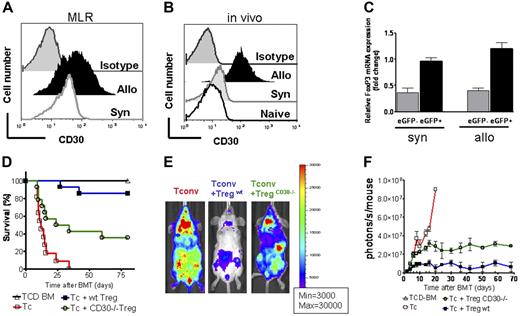
To investigate the surface expression of CD30 on Treg cells after alloantigen exposure, we used an MHC class I and II mismatch BMT model in which Treg cells are capable of protecting from aGvHD lethality.2 The mean fluorescence intensity for CD30 expression increased significantly more during allogeneic compared with syngeneic antigen stimulation in vitro (Figure 1A) and in vivo (Figure 1B), suggesting a potential role for CD30 in Treg-cell costimulation during alloantigen exposure. Increased CD30 expression on Treg cells over baseline was observed after 24 hours of alloantigen exposure, with a peak at 96 hours and a 15% decline at 144 hours (data not shown). Until that time point, more than 95% of the analyzed cells were FoxP3 positive.
Surface expression of CD30 increases upon allogeneic stimulation in vitro and in vivo. CD30 deficiency affects Treg-mediated GvHD protection. (A) CD4+CD25high FoxP3+ cells (H-2b) were coincubated with CD11c+ APCs derived from syngeneic C57B/6 mice (gray open histogram, MFI: 67.1) or allogeneic Balb/c mice (black solid histogram, MFI: 209.2) for 96 hours. The gray solid histogram represents cells stained with isotype control Ab. Cells were gated on CD4+FoxP3+. (B) eGFP-labeled Treg cells (CD4+CD25highH-2b) were isolated from secondary lymphoid organs on day 4 after transfer into C57B/6 mice (syngeneic, light gray histogram) or Balb/c mice (allogeneic, black solid histogram). Recipients were given 5 × 10⁶ TCD-BM (H-2b) after lethal irradiation with 800 cGy. Additionally, 2.5 × 10⁵ eGFP+ Treg cells (day 0) plus 5 × 10⁵ CD4+/CD8+ (1:4) T cells (day +2) were given (both H-2b). Histograms represent the mean fluorescence intensity for cells that are gated on eGFP+ cells. The gray open histogram and the dark gray filled histogram represent naive Treg cells (unmanipulated from 6-week-old mice) and isotype control–stained Treg cells, respectively. CD30 expression is increased after allogeneic compared with syngeneic antigen exposure (MFI black solid versus light gray histogram, P = .003). (C) Relative Foxp3 mRNA expression level was determined by quantitative real-time PCR in eGFP-positive versus -negative cells. The eGFP+ population represents the transferred Treg cells after syngeneic or allogeneic transplantation as indicated below the bar pairs. Expression was normalized to GAPDH. The negative controls are donor derived (CD4H-2b) eGFP− cells (gray bars). Error bars indicate SD from the mean. (D) Survival of recipients given 5 × 10⁶ TCD-BM cells (H-2b) after lethal irradiation with 800 cGy. Additionally, 2.5 × 10⁵ wt or CD30−/− Treg cells (day 0) plus 5 × 10⁵ CD4+/CD8+ (1:4) T cells (day +2) were given (both H-2b). Survival of mice receiving TCD-BM (▵, n = 15), with Tconv cells (□, n = 15), with Tconv cells and wt Treg cells (▪, n = 14), and with Tconv cells and CD30−/− Treg cells (○, n = 14). Survival of Balb/c recipients is significantly lower when Treg cells lack CD30 compared with wt Treg cells (○ versus ▪, P = .002). (E) Proliferation of luc+ Tconv cells (left column) is reduced by cotransfer of Treg cells (middle column) and to a lesser extent by Treg cells derived from CD30−/− donors (right column) as depicted for day 7 after BMT. (F) Signal intensity of luc+ Tconv cells as quantified in photons per mouse per second over total body area for the indicated group. TCD-BM (▵, n = 15), with Tconv cells (□, n = 15), with Tconv cells and wt Treg cells (▪, n = 14), and with Tconv cells and CD30−/− Treg cells (○, n = 14). Signal intensity is significantly higher in animals receiving CD30−/− Treg cells compared with wt Treg cells (○ versus ▪, P = .007, days 6-25). Data are pooled from 2 representative experiments.
Surface expression of CD30 increases upon allogeneic stimulation in vitro and in vivo. CD30 deficiency affects Treg-mediated GvHD protection. (A) CD4+CD25high FoxP3+ cells (H-2b) were coincubated with CD11c+ APCs derived from syngeneic C57B/6 mice (gray open histogram, MFI: 67.1) or allogeneic Balb/c mice (black solid histogram, MFI: 209.2) for 96 hours. The gray solid histogram represents cells stained with isotype control Ab. Cells were gated on CD4+FoxP3+. (B) eGFP-labeled Treg cells (CD4+CD25highH-2b) were isolated from secondary lymphoid organs on day 4 after transfer into C57B/6 mice (syngeneic, light gray histogram) or Balb/c mice (allogeneic, black solid histogram). Recipients were given 5 × 10⁶ TCD-BM (H-2b) after lethal irradiation with 800 cGy. Additionally, 2.5 × 10⁵ eGFP+ Treg cells (day 0) plus 5 × 10⁵ CD4+/CD8+ (1:4) T cells (day +2) were given (both H-2b). Histograms represent the mean fluorescence intensity for cells that are gated on eGFP+ cells. The gray open histogram and the dark gray filled histogram represent naive Treg cells (unmanipulated from 6-week-old mice) and isotype control–stained Treg cells, respectively. CD30 expression is increased after allogeneic compared with syngeneic antigen exposure (MFI black solid versus light gray histogram, P = .003). (C) Relative Foxp3 mRNA expression level was determined by quantitative real-time PCR in eGFP-positive versus -negative cells. The eGFP+ population represents the transferred Treg cells after syngeneic or allogeneic transplantation as indicated below the bar pairs. Expression was normalized to GAPDH. The negative controls are donor derived (CD4H-2b) eGFP− cells (gray bars). Error bars indicate SD from the mean. (D) Survival of recipients given 5 × 10⁶ TCD-BM cells (H-2b) after lethal irradiation with 800 cGy. Additionally, 2.5 × 10⁵ wt or CD30−/− Treg cells (day 0) plus 5 × 10⁵ CD4+/CD8+ (1:4) T cells (day +2) were given (both H-2b). Survival of mice receiving TCD-BM (▵, n = 15), with Tconv cells (□, n = 15), with Tconv cells and wt Treg cells (▪, n = 14), and with Tconv cells and CD30−/− Treg cells (○, n = 14). Survival of Balb/c recipients is significantly lower when Treg cells lack CD30 compared with wt Treg cells (○ versus ▪, P = .002). (E) Proliferation of luc+ Tconv cells (left column) is reduced by cotransfer of Treg cells (middle column) and to a lesser extent by Treg cells derived from CD30−/− donors (right column) as depicted for day 7 after BMT. (F) Signal intensity of luc+ Tconv cells as quantified in photons per mouse per second over total body area for the indicated group. TCD-BM (▵, n = 15), with Tconv cells (□, n = 15), with Tconv cells and wt Treg cells (▪, n = 14), and with Tconv cells and CD30−/− Treg cells (○, n = 14). Signal intensity is significantly higher in animals receiving CD30−/− Treg cells compared with wt Treg cells (○ versus ▪, P = .007, days 6-25). Data are pooled from 2 representative experiments.
In vivo up-regulation of CD30 expression was not caused by bystander activation due to the proinflammatory environment after irradiation-induced tissue damage since syngeneic BM transplant recipients displayed only moderate CD30 up-regulation (Figure 1B). To rule out that the increased CD30 expression was due to an overgrowth of Tconv cells in vivo, we measured Foxp3 expression levels by quantitative real-time PCR within the eGFP-positive Treg-cell population that was isolated from lymphoid tissues of the BM transplant recipients (Figure 1C). High levels of Foxp3 expression were observed in both syngeneic and allogeneic eGFP+ Treg-cell populations after BMT (Figure 1C).
To test the hypothesis that CD30 expression is important for Treg-cell function during allogeneic BMT, we compared CD30-deficient (CD30−/−) with wild-type Treg-cell donors with respect to aGvHD protection. Balb/c recipients given TCD-BM (C57B/6) alone appeared healthy. At the end of the observation period (day 80), 100% of the animals in the TCD-BM group were alive and without clinical signs of aGvHD (Figure 1D).
Mice that received Tconv cells along with TCD-BM developed severe aGvHD signs including reduced activity, hunched posture, ruffled fur, diarrhea, and weight loss, and all animals died within 35 days after BMT. Treg-cell cotransfer reduced aGvHD-related death after BMT and more than 80% survived (Figure 1D). Treg cells isolated from CD30−/− animals maintained some degree of suppression but were significantly less effective (P = .002) compared with wild-type Treg cells (Figure 1D).
Proliferation of luc+ Tconv cells (Figure 1E, left column) was inhibited by cotransfer of Treg cells derived from wt donors (Figure 1E middle column) but significantly less when derived from CD30−/− donors (Figure 1E, right column). Quantification of the signal derived from luciferase transgenic T cells demonstrated a reduced expansion in the presence of wt Treg cells (Figure 1F). Signal intensity was significantly higher if CD30−/− Treg cells compared with wt Treg cells were used (P = .007), indicating a less potent inhibition of Tconv-cell proliferation (Figure 1F).
Early blockade of the CD30/CD153 interaction reduces Treg-mediated suppression of aGvHD
To address the question of when CD30 signaling is critical for Treg-cell function, we investigated the impact of CD30L (CD153) blockade at different time periods using another previously reported major mismatch BMT model (FVB/N→Balb/c).29 We chose this model since expansion of luciferase transgenic (luc+) CD4+ and CD8+ Tconv cells as well as Treg cells can be assessed noninvasively by in vivo BLI.31 We have found that Treg cells are most effective when given prior to Tconv cells,43 which allows for their homing to aGvHD initiation sites, followed by activation and proliferation. Therefore, this model was particularly suitable to assess the relevance of the CD30/CD153 interaction on Treg-cell homing, activation, and expansion. To block the CD30/CD153 interaction in the early phase of Treg-cell expansion, CD153 blocking antibody was injected on days −2, 0, 2, and 4.
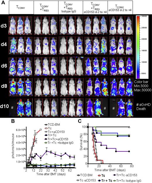
Mice that received TCD-BM plus luc+ Tconv cells displayed vigorous Tconv-cell expansion in contrast to animals that received Tconv cells and wt Treg cells (Figure 2A). By day 8, Tconv cells were distributed throughout the animal and accumulated in skin, liver, lymph nodes, and gastrointestinal tract (GIT) (Figure 2A, first column). Animals receiving Treg cells and anti-CD153 blocking antibody showed only slightly reduced expansion of Tconv cells compared with the group that received Tconv cells alone, but significantly higher signal intensity compared with groups receiving Treg cells alone or Treg cells plus isotype IgG in serial whole-body BLI (Figure 2A-B). Animals receiving Treg cells along with the CD153 blocking antibody had significantly reduced overall survival compared with Treg cells plus PBS and Treg cells plus isotype IgG (P < .001) (Figure 2C). Blockade of CD153 did not interfere with GvHD lethality in animals receiving Tconv cells (Figure 2C).
Treg-cell suppressor function is reduced when CD153 (CD30L) is blocked in the early phase after adoptive transfer. Expansion of luc+ donor Tconv cells in animals receiving Tconv cells alone or Tconv cells and Treg cells alone or in conjunction with isotype control Ab (rat IgG2b) or with anti-CD153 blocking Ab administered on days −2, 0, 2, and 4 as shown for 3 representative animals at days 3, 4, 6, 8, and 10 after BMT (A) and as quantified in emitted photons over total body area at serial time points after BMT (B). (A) Proliferation of luc+ Tconv cells (first column) is reduced by cotransfer of Treg cells (second column). While isotype IgG Ab does not interfere with Treg-mediated suppression (third column), addition of anti-CD153 blocking Ab reduces the suppressive effect of Treg cells (fourth column). The anti-CD153 blocking Ab has no impact on expansion of luc+ CD4/CD8 Tconv cells when given as indicated in the early phase after BMT (fifth column). (B) TCD-BM (▵, n = 15), with Tconv cells (□, n = 15), with Tconv cells and Treg cells (▪, n = 10), with Tconv cells and Treg cells and isotype IgG (○, n = 10), with Tconv cells and Treg cells and anti-CD153 blocking Ab (•, n = 10), and with Tconv cells and anti-CD153 blocking Ab (⋄, n = 10). Signal intensity is significantly higher in animals receiving Tconv cells and Treg cells and anti-CD153 blocking Ab compared with isotype IgG (• versus ○, P < .01, days 6-14). Error bars indicate SD from the mean. (C) Survival of Balb/c recipients is significantly lower when combining Treg cells with anti-CD153 blocking Ab compared with isotype IgG (• versus ○, P < .001). Anti-CD153 blocking Ab does not improve survival compared with PBS injection when administered on day −2 to day +4 (⋄ versus □, NS). Survival data from 3 independent experiments are combined.
Treg-cell suppressor function is reduced when CD153 (CD30L) is blocked in the early phase after adoptive transfer. Expansion of luc+ donor Tconv cells in animals receiving Tconv cells alone or Tconv cells and Treg cells alone or in conjunction with isotype control Ab (rat IgG2b) or with anti-CD153 blocking Ab administered on days −2, 0, 2, and 4 as shown for 3 representative animals at days 3, 4, 6, 8, and 10 after BMT (A) and as quantified in emitted photons over total body area at serial time points after BMT (B). (A) Proliferation of luc+ Tconv cells (first column) is reduced by cotransfer of Treg cells (second column). While isotype IgG Ab does not interfere with Treg-mediated suppression (third column), addition of anti-CD153 blocking Ab reduces the suppressive effect of Treg cells (fourth column). The anti-CD153 blocking Ab has no impact on expansion of luc+ CD4/CD8 Tconv cells when given as indicated in the early phase after BMT (fifth column). (B) TCD-BM (▵, n = 15), with Tconv cells (□, n = 15), with Tconv cells and Treg cells (▪, n = 10), with Tconv cells and Treg cells and isotype IgG (○, n = 10), with Tconv cells and Treg cells and anti-CD153 blocking Ab (•, n = 10), and with Tconv cells and anti-CD153 blocking Ab (⋄, n = 10). Signal intensity is significantly higher in animals receiving Tconv cells and Treg cells and anti-CD153 blocking Ab compared with isotype IgG (• versus ○, P < .01, days 6-14). Error bars indicate SD from the mean. (C) Survival of Balb/c recipients is significantly lower when combining Treg cells with anti-CD153 blocking Ab compared with isotype IgG (• versus ○, P < .001). Anti-CD153 blocking Ab does not improve survival compared with PBS injection when administered on day −2 to day +4 (⋄ versus □, NS). Survival data from 3 independent experiments are combined.
Proinflammatory cytokine accumulation as a parameter of aGvHD damage and apoptosis of Tconv cells in the presence or absence of Treg cells
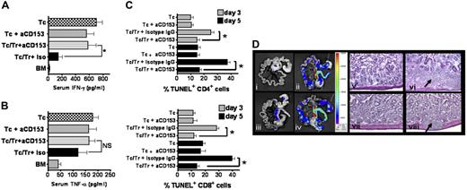
Serum IFN-γ and TNF-α levels were measured as an indicator of proinflammatory activity during the early phase of aGvHD. IFN-γ levels were significantly lower in animals that received Treg cells and Tconv cells plus isotype IgG compared with Treg cells and Tconv cells plus anti-CD153 blocking Ab from day −2 to day +4 (P < .05, Figure 3A). The same trend was observed for serum tumor necrosis factor (TNF-α), however it did not reach statistical significance (Figure 3B). These data indicate that CD153 blockade reduces the preventive effect of Treg cells on accumulation of proinflammatory cytokines after BMT. To investigate the relevance of apoptosis induction for Treg-cell function, we isolated CD4 and CD8 T cells from different lymphoid organs on day 5 following BMT. Cotransfer of Treg cells was associated with an increased apoptosis rate of CD4 and CD8 T cells compared with mice receiving only TCD-BM and Tconv cells or TCD-BM, Tconv cells, Treg cells, and anti-CD153 blocking Ab (Figure 3C). A comparable pattern with respect to the different amount of TUNEL+ CD4 and CD8 T cells was found on day 3 and day 5 (Figure 3C). Earlier time points were not investigated since Tconv cells were given on day 2 and therefore had only 24-hour contact with Treg cells on day 3. These data suggest that the impact of Treg cells on Tconv-cell expansion after BMT is in part mediated by apoptosis induction. To compare T-cell expansion in the GIT as a major aGvHD target organ, we performed ex vivo imaging (Figure 3D). Adoptive transfer of Treg cells reduced the ex vivo signal of luc+ Tconv cells in the GIT and the mesenteric lymph nodes (mLNs), an effect that was conserved in the presence of isotype IgG and reduced when CD153 was blocked (Figure 3D). Consistent with the BLI data, histologic examination of the GIT indicated the strongest T-cell infiltration in the bowel wall of animals receiving Tconv cells only or Tconv cells and Treg cells and anti-CD153 blocking Ab (Figure 3D).
Blockade of CD153 reverses Treg-mediated protection from accumulation of proinflammatory cytokines, apoptosis in T effector cells, and GvHD histopathology of the GIT. (A-B) Serum was collected on day 7 after transplantation from Balb/c recipients of TCD-BM plus Tconv cells (n = 17), TCD-BM plus Tconv cells and CD153 blocking Ab (n = 12), TCD-BM plus Tconv cells and Treg cells and CD153 blocking Ab (n = 18), TCD-BM plus Tconv cells and Treg cells and isotype Ab (n = 18), or TCD-BM alone (n = 17); *P < .05. Blockade of CD153 in the presence of Treg cells leads to increased IFN-γ (A) and TNF-α (B) levels that reach significance for IFN-γ when compared with Treg cells plus isotype IgG. Data are pooled from 3 independent experiments. (C) The diagrams present results from TUNEL staining of splenic donor-type (H-2kq) CD4 or CD8 cells isolated on day 3 or day 5 after BMT from Balb/c mice receiving TCD-BM plus Tconv cells, TCD-BM plus Tconv cells and CD153 blocking Ab (aCD153), TCD-BM plus Tconv cells and Treg cells and isotype IgG, and TCD-BM plus Tconv cells and Treg cells and CD153 blocking Ab, as indicated for the respective bar. Data are pooled from 3 independent experiments with a total of 9 mice per group (*P < .05). Error bars indicate SD from the mean. (D) The left panel (i-iv) depicts expansion of luc+ Tconv cells on day 8 after BMT in the GIT and in the mLNs as assessed by ex vivo BLI. (i) Background signal in the GIT of animals having received only TCD-BM without any luc+ cells. Proliferation of Tconv cells in the mLN and the GIT (ii) is reduced by cotransfer of Treg cells in the presence of isotype IgG (iii). Addition of anti-CD153 blocking Ab reduces the protective effect of Treg cells (iv). The right panel shows representative histologic sections of the bowel corresponding to the groups in the left panel. Significant T-cell infiltration (arrow) is found in animals receiving Tconv cells only (vi) or Tconv cells and Treg cells and anti-CD153 blocking Ab (viii).
Blockade of CD153 reverses Treg-mediated protection from accumulation of proinflammatory cytokines, apoptosis in T effector cells, and GvHD histopathology of the GIT. (A-B) Serum was collected on day 7 after transplantation from Balb/c recipients of TCD-BM plus Tconv cells (n = 17), TCD-BM plus Tconv cells and CD153 blocking Ab (n = 12), TCD-BM plus Tconv cells and Treg cells and CD153 blocking Ab (n = 18), TCD-BM plus Tconv cells and Treg cells and isotype Ab (n = 18), or TCD-BM alone (n = 17); *P < .05. Blockade of CD153 in the presence of Treg cells leads to increased IFN-γ (A) and TNF-α (B) levels that reach significance for IFN-γ when compared with Treg cells plus isotype IgG. Data are pooled from 3 independent experiments. (C) The diagrams present results from TUNEL staining of splenic donor-type (H-2kq) CD4 or CD8 cells isolated on day 3 or day 5 after BMT from Balb/c mice receiving TCD-BM plus Tconv cells, TCD-BM plus Tconv cells and CD153 blocking Ab (aCD153), TCD-BM plus Tconv cells and Treg cells and isotype IgG, and TCD-BM plus Tconv cells and Treg cells and CD153 blocking Ab, as indicated for the respective bar. Data are pooled from 3 independent experiments with a total of 9 mice per group (*P < .05). Error bars indicate SD from the mean. (D) The left panel (i-iv) depicts expansion of luc+ Tconv cells on day 8 after BMT in the GIT and in the mLNs as assessed by ex vivo BLI. (i) Background signal in the GIT of animals having received only TCD-BM without any luc+ cells. Proliferation of Tconv cells in the mLN and the GIT (ii) is reduced by cotransfer of Treg cells in the presence of isotype IgG (iii). Addition of anti-CD153 blocking Ab reduces the protective effect of Treg cells (iv). The right panel shows representative histologic sections of the bowel corresponding to the groups in the left panel. Significant T-cell infiltration (arrow) is found in animals receiving Tconv cells only (vi) or Tconv cells and Treg cells and anti-CD153 blocking Ab (viii).
Late CD153 blockade does not affect Treg-cell function
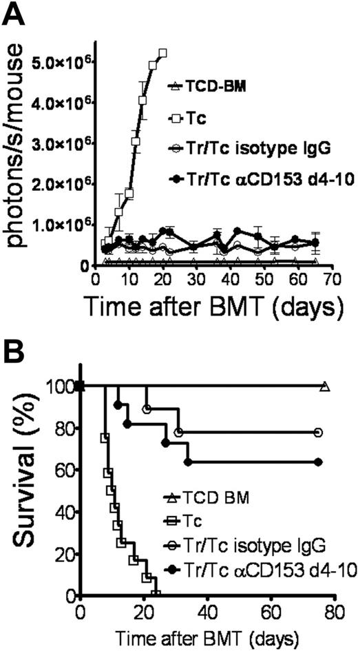
To assess the relevance of the CD30/CD153 interaction on Treg-cell function at a time point when initial homing, activation, and expansion had taken place, the CD153 blocking antibody was injected in the late phase after adoptive transfer, on days 4, 6, 8, and 10 following BMT. Tconv cells expanded in mice that received TCD-BM plus luc+ Tconv cells and caused the clinical signs of aGvHD. As expected, animals receiving Treg cells plus isotype IgG displayed a significantly reduced expansion of Tconv cells compared with the group that received Tconv cells alone in serial whole-body BLI (Figure 4A). Of importance, in vivo blockade of the CD30/CD153 interaction during the late phase did not impact Treg-cell function, indicating the relevance of the interaction in the early phase of Treg-cell activation and expansion (Figure 4A). There was no impact of the late CD153 blockade on Tconv-cell expansion, which makes a direct effect of the neutralizing CD153 Ab on Tconv-cell expansion unlikely (data not shown). Quantification of the photons derived from expanding Tconv cells over time demonstrated similar T-cell expansion among animals that received Treg cells plus isotype Ab or Treg cells plus late CD153 blockade (Figure 4A). Reduced expansion of luciferase-labeled Tconv cells in the presence of Treg cells translated into a survival advantage for the respective recipients that was not affected by the late blockade of the CD30-CD153 interaction (Figure 4B).
Treg-cell suppressor function is maintained when CD153 (CD30L) is blocked in the late phase after transplantation. Recipients underwent transplantation as described in “Materials and methods” for the FVB/N→Balb/c combination. To block the CD30/CD153 interaction in the late phase of Treg-cell expansion, the Abs (each 0.1 mg) were injected on days 4, 6, 8, and 10. Data from 3 independent experiments are combined. (A) TCD-BM (▵, n = 15), with Tconv cells (□, n = 15), with Tconv cells and Treg cells and isotype IgG (○, n = 12), and with Tconv cells and Treg cells and anti-CD153 blocking Ab (•, n = 12). Signal intensity is not significantly different in animals receiving Tconv cells and Treg cells and anti-CD153 blocking Ab compared with isotype IgG (• versus ○, NS). Error bars indicate SD from the mean. (B) TCD-BM (▵, n = 15), with Tconv cells (□, n = 15), with Tconv cells and Treg cells and isotype IgG (○, n = 12), and with Tconv cells and Treg cells and late anti-CD153 blocking Ab (•, n = 12).
Treg-cell suppressor function is maintained when CD153 (CD30L) is blocked in the late phase after transplantation. Recipients underwent transplantation as described in “Materials and methods” for the FVB/N→Balb/c combination. To block the CD30/CD153 interaction in the late phase of Treg-cell expansion, the Abs (each 0.1 mg) were injected on days 4, 6, 8, and 10. Data from 3 independent experiments are combined. (A) TCD-BM (▵, n = 15), with Tconv cells (□, n = 15), with Tconv cells and Treg cells and isotype IgG (○, n = 12), and with Tconv cells and Treg cells and anti-CD153 blocking Ab (•, n = 12). Signal intensity is not significantly different in animals receiving Tconv cells and Treg cells and anti-CD153 blocking Ab compared with isotype IgG (• versus ○, NS). Error bars indicate SD from the mean. (B) TCD-BM (▵, n = 15), with Tconv cells (□, n = 15), with Tconv cells and Treg cells and isotype IgG (○, n = 12), and with Tconv cells and Treg cells and late anti-CD153 blocking Ab (•, n = 12).
CD153 is up-regulated in a proinflammatory environment
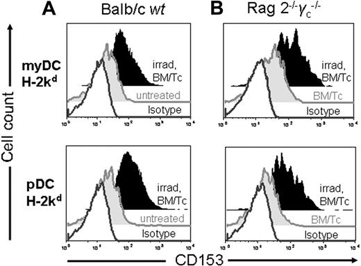
To quantify cell types that express CD153 in a proinflammatory environment, we analyzed spleen- and LN-derived antigen-presenting cells at different time points after BMT. On day 3 after BMT, the number of host-derived PI− myeloid DCs (myDCs) and plasmacytoid DCs (pDCs) isolated from the spleen were 4.3 × 10⁵ and 5 × 10⁴, respectively, for irradiated Balb/c animals and 1.4 × 10⁶ and 1.1 × 10⁵, respectively, for nonirradiated Balb/c animals. Besides expression on activated T cells and a small subset of B cells (data not shown), CD153 was found on recipient-type plasmacytoid and myeloid DCs (Figure 5A). Of interest, CD153 expression in untreated animals was significantly lower compared with irradiated recipients who underwent transplantation (Figure 5A). To rule out that the increase was related to the transferred cell populations, we used Rag2−/− γc−/− recipients that received TCD-BM and Tconv cells as described for the C57B/6→Balb/c combination, with or without irradiation (800 cGy). While there was no increase of CD153 expression over baseline in the nonirradiated Rag2−/− γc−/− recipients that received TCD-BM and Tconv cells, irradiated Rag2−/− γc−/− BM transplant recipients displayed significantly increased CD153 expression (Figure 5B) on day 3 after BMT.
Irradiation increases CD153 expression on myDCs and pDCs after BMT. The surface marker CD153 was measured by FACS analysis on different dendritic cell subsets in irradiated compared with nonirradiated wt Balb/c (A) or Rag2−/− γc−/− Balb/c (B) recipients. Where indicated, DCs were derived from animals that received TCD-BM and T cells as described for the C57B/6→Balb/c model. Recipient-type myeloid DCs (CD11c+CD11b+Gr-1−) or plasmacytoid DCs (CD11c+B220+CD11b−) derived from the spleen on day 3 after BMT display differential CD153 surface expression when isolated from untreated Balb/c mice or nonirradiated Rag2−/− γc−/− mice that received a transplant of TCD-BM and T cells. Grey open histogram: isotype IgG. CD153 expression on myDCs from irradiated compared with nonirradiated Balb/c mice (A) (MFI: 110 versus 24, P < .05) or Rag2−/− γc−/− mice (B) (MFI: 195 versus 42, P < .05). CD153 expression on pDCs from irradiated compared with nonirradiated Balb/c mice (A) (MFI: 182 versus 26, P < .05) or Rag2−/− γc−/− mice (B) (MFI: 137 versus 21, P < .05).
Irradiation increases CD153 expression on myDCs and pDCs after BMT. The surface marker CD153 was measured by FACS analysis on different dendritic cell subsets in irradiated compared with nonirradiated wt Balb/c (A) or Rag2−/− γc−/− Balb/c (B) recipients. Where indicated, DCs were derived from animals that received TCD-BM and T cells as described for the C57B/6→Balb/c model. Recipient-type myeloid DCs (CD11c+CD11b+Gr-1−) or plasmacytoid DCs (CD11c+B220+CD11b−) derived from the spleen on day 3 after BMT display differential CD153 surface expression when isolated from untreated Balb/c mice or nonirradiated Rag2−/− γc−/− mice that received a transplant of TCD-BM and T cells. Grey open histogram: isotype IgG. CD153 expression on myDCs from irradiated compared with nonirradiated Balb/c mice (A) (MFI: 110 versus 24, P < .05) or Rag2−/− γc−/− mice (B) (MFI: 195 versus 42, P < .05). CD153 expression on pDCs from irradiated compared with nonirradiated Balb/c mice (A) (MFI: 182 versus 26, P < .05) or Rag2−/− γc−/− mice (B) (MFI: 137 versus 21, P < .05).
Early-phase CD153 blocking is associated with significantly reduced expansion of adoptively transferred Foxp3+CD4+CD25+ Treg cells
In order to investigate the impact of CD153 blockade on Treg-cell expansion, we used Treg cells derived from luc+ animals, which allows for monitoring of in vivo trafficking and proliferation. Serial BLI showed that CD153 blocking did not abrogate migration of Treg cells to spleen, GIT, and cervical lymph nodes (Figure 6A). To correlate these results with the number of donor-derived Treg cells in secondary lymphoid organs, we quantified donor-derived (H-2q) CD4+Foxp3+ T cells from spleen, mLNs, iLNs, aLNs, and cLNs by immunofluorescence microscopic analysis at various time points after transplantation. Consistent with the expansion pattern, the frequency of donor-derived H-2kq+CD4+Foxp3+ cells 10 days following BMT, in the spleen, mLNs, and cLNs of Balb/c recipients having received Tconv cells and Treg cells in conjunction with early-phase anti-CD153 blocking Ab, was significantly lower compared with isotype IgG (Figure 6B). This difference was not observed in the axillary and inguinal LNs, however the number of H-2kq+CD4+Foxp3+ cells was low irrespective of treatment with anti-CD153 Ab (Figure 6B).
Expansion of luciferase-labeled Treg cells is reduced when CD153 is blocked early after transplantation. (A) Distribution and expansion of luc+ donor Treg cells in Balb/c mice (H-2kd) receiving Tconv cells and Treg cells (both H-2kq) and isotype IgG Ab or Tconv cells and Treg cells along with anti-CD153 blocking Ab as shown for representative animals on day 7 after BMT. Expansion of luc+ Treg cells (first animal) is reduced when the CD30/CD153 interaction is blocked in the early (middle animal) but not in the late (right animal) phase after adoptive transfer. Early blockade represents days −2, 0, 2, and 4, and late phase represents days 4, 6, 8, and 10 after BMT. (B) The number of donor-derived Treg cells (CD4+FoxP3+H-2Kq+) per high-power field was determined in 5 representative areas and plotted for the respective secondary lymphoid organ recovered from Balb/c recipients (n = 5) on day 10 after adoptive transfer of TCD-BM and Tconv cells plus Treg cells. *P < .05, animals receiving anti-CD153 Ab (blue bars) or isotype IgG (green bars) both in the early phase. (C) Whole-body photons derived from luc+ Treg cells expanding in Balb/c recipients at serial time points. TCD-BM (▵, n = 10), with Tconv cells and Treg cells and isotype IgG (□, n = 10), with Tconv cells and Treg cells plus early anti-CD153 blocking Ab (○, n = 10), and with Tconv cells and Treg cells plus late anti-CD153 blocking Ab (•, n = 10). Expansion of luc+ Treg cells is significantly reduced in the presence of the anti-CD153 blocking Ab during the early expansion phase compared with isotype IgG (○ versus □, P = .007) or late blockade (○ versus •, P = .008). Data are pooled from 3 independent experiments. (D) Treg cells (CD4+CD25highH-2kb+) from either wild-type (open bars) or CD30−/− (solid black bars) C57B/6 mice were incubated with CFSE-labeled CD4+CD25− T cells (H-2kb, Thy-1.1+) and γ-irradiated (30 Gy) APCs (CD11c+H-2kd+). To measure T-cell proliferation, Thy-1.1+ cells were analyzed by FACS after 72 hours. The bars represent the percentage of proliferating CD4+Thy 1.1+CFSE+ cells. One representative experiment of 3 is presented. (E) On days 5 and 7 after BMT, lymphoid organs were removed from recipients of wt or CD30−/− Treg cells and total cells per organ were counted in a single-cell suspension. Subsequent analysis of the resulting cell suspensions by FACS provided the percentages based on which the absolute cell numbers were calculated. *P < .05. Data are pooled from 2 independent experiments with 6 animals per group and time point. Error bars indicate SD from the mean.
Expansion of luciferase-labeled Treg cells is reduced when CD153 is blocked early after transplantation. (A) Distribution and expansion of luc+ donor Treg cells in Balb/c mice (H-2kd) receiving Tconv cells and Treg cells (both H-2kq) and isotype IgG Ab or Tconv cells and Treg cells along with anti-CD153 blocking Ab as shown for representative animals on day 7 after BMT. Expansion of luc+ Treg cells (first animal) is reduced when the CD30/CD153 interaction is blocked in the early (middle animal) but not in the late (right animal) phase after adoptive transfer. Early blockade represents days −2, 0, 2, and 4, and late phase represents days 4, 6, 8, and 10 after BMT. (B) The number of donor-derived Treg cells (CD4+FoxP3+H-2Kq+) per high-power field was determined in 5 representative areas and plotted for the respective secondary lymphoid organ recovered from Balb/c recipients (n = 5) on day 10 after adoptive transfer of TCD-BM and Tconv cells plus Treg cells. *P < .05, animals receiving anti-CD153 Ab (blue bars) or isotype IgG (green bars) both in the early phase. (C) Whole-body photons derived from luc+ Treg cells expanding in Balb/c recipients at serial time points. TCD-BM (▵, n = 10), with Tconv cells and Treg cells and isotype IgG (□, n = 10), with Tconv cells and Treg cells plus early anti-CD153 blocking Ab (○, n = 10), and with Tconv cells and Treg cells plus late anti-CD153 blocking Ab (•, n = 10). Expansion of luc+ Treg cells is significantly reduced in the presence of the anti-CD153 blocking Ab during the early expansion phase compared with isotype IgG (○ versus □, P = .007) or late blockade (○ versus •, P = .008). Data are pooled from 3 independent experiments. (D) Treg cells (CD4+CD25highH-2kb+) from either wild-type (open bars) or CD30−/− (solid black bars) C57B/6 mice were incubated with CFSE-labeled CD4+CD25− T cells (H-2kb, Thy-1.1+) and γ-irradiated (30 Gy) APCs (CD11c+H-2kd+). To measure T-cell proliferation, Thy-1.1+ cells were analyzed by FACS after 72 hours. The bars represent the percentage of proliferating CD4+Thy 1.1+CFSE+ cells. One representative experiment of 3 is presented. (E) On days 5 and 7 after BMT, lymphoid organs were removed from recipients of wt or CD30−/− Treg cells and total cells per organ were counted in a single-cell suspension. Subsequent analysis of the resulting cell suspensions by FACS provided the percentages based on which the absolute cell numbers were calculated. *P < .05. Data are pooled from 2 independent experiments with 6 animals per group and time point. Error bars indicate SD from the mean.
The expansion of the adoptively transferred luc+ Treg cells was significantly reduced in the presence of early-phase anti-CD153 blocking Ab compared with isotype IgG (P = .007) as measured by quantifying the total amount of photons per second per mouse (Figure 6C). Of importance, blockade of the CD30/CD153 during the late phase did not affect Treg-cell expansion compared with isotype IgG (Figure 6C). These data indicate a critical role of the CD30/CD153 interaction for Treg-cell activation and expansion, which occurs in the first several days following alloantigen exposure after BMT.
To probe the relevance of CD30 signaling for Treg cells in vitro, we used CD30−/− Treg cells as suppressors of alloantigen-driven Tconv-cell proliferation at different ratios. Tconv-cell proliferation was equally suppressed by wt compared with CD30−/− Treg cells (Figure 6D), indicating that in vitro suppressor function is not impacted by the lack of CD30. Quantification of total donor-type Treg-cell numbers on days 5 and 7 after BMT demonstrated a significant reduction in mesenteric LNs (day 5 and day 7), cervical LNs (day 7), and spleen (day 5 and day 7) when Treg cells were derived from CD30−/− compared with wt animals (Figure 6E). In concert with the in vitro data (Figure 6D), this observation supports the concept that CD30 deficiency leads to a numeric defect of Treg cells after allogeneic BMT.
Discussion
The protective effect of Treg cells in the setting of aGvHD has been well documented in murine minor and major BMT mismatch models.2–5 While L-selectin and CCR5 have been demonstrated to be critical for Treg-cell recruitment to aGvHD initiation sites,32–34 molecules on the Treg-cell surface that are relevant for controlling alloimmune responses remain to be defined. Several costimulatory pathways such as CD28/B7, OX40/OX40L, and 4-1BB/4-1BBL have all been shown to be dispensable for Treg-mediated suppression.34,35
The observation that CD30 is up-regulated on murine Treg cells upon alloantigen exposure in vitro and in vivo prompted us to further assess the functional role of the CD30/CD153 interaction in Treg-cell biology in BMT models of aGvHD.29 We found that Treg cells derived from mice genetically deficient in CD30 were significantly less potent in controlling aGvHD than similar cells from wt animals. These data indicated that CD30 is important for Treg-cell in vivo function during the aGvHD initiation process, which includes their effective expansion in secondary lymphoid organs.
To further address that Treg cells require CD30 signaling when in the process of protection from aGvHD, we blocked CD153 at different time periods. The time frames early versus late were chosen according to the homing pattern of conventional T cells that we previously described30 and because we found that during the first 4 days Treg cells home to lymphoid organs that may be involved in aGvHD priming. Early but not late blockade of the CD30/CD153 interaction abolished Treg-mediated aGvHD protection, accumulation of proinflammatory cytokines, and Tconv-cell apoptosis in vivo without interfering with Treg-cell migration to secondary lymphoid organs. In light of this finding and our observation that irradiation-induced inflammation impacts Treg-cell expansion (V.H.N., unpublished data, April 2006), we investigated the expression of CD153 on donor-type pDCs and myDCs that may provide costimulation for Treg-cell expansion after BMT. Of interest, we found CD153 to be up-regulated in the presence of a proinflammatory environment caused by irradiation but not when TCD-BM and Tconv cells were transferred into a lymphopenic Rag2−/− γc−/− host, indicating that the preparative regimen plays a critical role in Treg-cell expansion after BMT. Based on our observation that CD153 expression on APCs was up-regulated in the presence of a radiation-induced proinflammatory environment, it would be interesting to investigate the relevance of the CD30/CD153 interaction in models with less intense inflammation such as autoimmunity. The change of the APC population toward the donor type during days 4 to 8 may reduce the expansion of Treg cells and explain the continuous decline of the luc+ Treg-cell signal after day 7. Late CD30/CD153 interaction with donor-type APCs may be less important for Treg-cell expansion, and this may account in part for the observation that Treg-cell expansion cannot by impacted by anti-CD153 blockade in the late phase.
We avoided long-term blockade of the CD30/CD153 interaction in our model since this has been shown to reduce CD4+ T-cell–mediated aGvHD,36 which was our indicator for Treg-cell suppressor function in vivo. A recent study by Blazar et al demonstrated that the absence of CD30 on donor conventional CD4 T cells resulted in reduced aGvHD mortality in sublethally irradiated, MHC class II–only disparate bm12 recipients.36 A comparable observation was made when CD153 was blocked.36 The survival advantage could be overcome by increasing the number of CD4+ donor T cells, suggesting that CD30 deficiency or CD153 blockade caused a quantitative difference of donor CD4 T cells in the recipient.36 The authors concluded from these findings that the improved survival in anti-CD153 mAb-treated recipients may be due to impaired donor CD4+ T-cell homing to or expansion within aGvHD target organs, especially those of the gastrointestinal tract.36
Although the BMT model that was used36 differs from our model with respect to MHC mismatch, irradiation, T-cell dose, duration, and dosing of the CD153 blockade and adoptive Treg-cell transfer, both studies indicate that CD30 is involved in CD4+ T-cell expansion and/or survival in irradiated hosts. This finding is consistent with the observation that CD30−/− CD4+ T cells are unable to receive adequate survival signals from OX40L+ CD30L+ accessory cells in B-cell follicles, which results in a defect in secondary humoral responses in CD30−/− mice.37
Previous studies have shown that CD30 is critical for Treg cells in preventing allograft rejection,9 suggesting that CD30 is an effector molecule on Treg cells that mediates suppression possibly by inducing apoptosis of CD8+ memory T cells. We demonstrate the relevance of CD30 signaling for Treg-cell function in the BMT setting and found reduced apoptosis rates in both CD4+ and CD8+ donor-derived T cells when CD153 was blocked. These data indicate that the impact of Treg cells on Tconv-cell expansion after BMT is at least in part mediated by apoptosis induction. This observation is in line with recent studies indicating that besides inhibition of IL-2 mRNA transcription,38 apoptosis induction may account for some of the suppressive effect of Treg cells on Tconv cells11 and B cells.8
In comparison to the study by Dai et al,9 the presented data are the first to demonstrate the importance of CD30 signaling for Treg-cell function in a bone marrow transplantation model that differs significantly from allograft transplantation, in particular with respect to the transferred donor immune system and inflammation due to radiation-induced tissue damage. Also, our study delineates the importance of the timing of CD30 signaling, showing that the CD30/CD153 interaction is critical in the early phase after Treg-cell transfer. Further, the presented data provide evidence that CD30 signaling is important for expansion of Treg cells in response to allogeneic and inflammatory stimuli but is dispensable for cell-cell contact–mediated suppression in vitro.
Zhang et al described CD30 to be a unique marker on antigen-induced T cells with regulatory properties that prevented allograft rejection by inducing apoptosis of activated T cells.12 Elimination of autoreactive T cells by Treg cells has been described in a model of murine experimental encephalomyelitis,39 and the control of homeostatic T-cell expansion by Treg cells seems to be in part regulated by enhanced apoptosis of Tconv cells.40 However, our study cannot answer the question whether intact early CD30/CD153 signaling is required for the in vivo apoptosis induction itself or solely provides an activation and expansion signal for Treg cells. In the latter case, the reduced apoptosis frequency in the presence of anti-CD153 blockade would be secondary to a reduced Treg-cell number. The notion that CD30 is most relevant for Treg-cell expansion is supported by our data showing that CD30−/− Treg cells are suppressive in vitro, where equal numbers of Treg cells were used.
In the short term in vitro MLR system, CD30 deficiency did not reduce cell-cell contact–dependent Treg-cell suppressor function. This observation was complementary to the observation that CD30 signaling was important for Treg-cell expansion in vivo. In vitro, the activation and expansion defect of CD30−/− Treg cells can be overcome by using defined ratios of effector and regulatory T cells. Also, the in vitro assay cannot mimic the microenvironment in the irradiated BM transplant recipient. In particular, with respect to Treg-cell activation and expansion, significant differences between the in vitro versus in vivo dynamics of Treg cells have been reported.41 Therefore, the observation that CD30−/− Treg cells can suppress in vitro is consistent with the finding that the CD30 molecule is most critical for their activation and consecutive proliferation rather than for the exertion of their suppressor function.
Of interest, bioluminescence imaging disclosed that early blockade of CD153 significantly reduced Treg-cell expansion, indicating that CD30 signaling enhances the proliferation of alloantigen-exposed Treg cells. Consistent with this finding, bidirectional signaling via CD153 as a prerequisite for costimulatory signaling toward the CD30-expressing cell has been described.42
In conclusion, we have found that CD30 signaling in the early phase after BMT is critical for expansion and possibly induction of donor T-cell apoptosis by Treg cells. This study identifies CD30 signaling as a major pathway critical for immune regulation by Treg cells in controlling aGvHD as a pathological immune response.
Authorship
Contribution: R.Z. designed and performed research, analyzed data, and wrote the paper; V.H.N. helped to design experiments, performed research, and analyzed data; J.-Z.H. and E.Z. performed research and analyzed data; A.B. contributed new reagents, performed research, and analyzed data; M.B. performed research and analyzed data; C.H.C. designed research and contributed vital new reagents and analytic tools; and R.S.N. designed research and helped to write the paper.
Conflict of interest disclosure: The authors declare no competing financial interests.
Correspondence: Robert S. Negrin, Center for Clinical Science Research, 269 W Campus Dr, Rm 2205, Division of Bone Marrow Transplantation, Stanford University School of Medicine, Stanford, CA 94305; e-mail: negrs@stanford.edu.
The publication costs of this article were defrayed in part by page charge payment. Therefore, and solely to indicate this fact, this article is hereby marked “advertisement” in accordance with 18 USC section 1734.
Acknowledgments
This study was supported by National Institutes of Health grants (RO1 CA0800065 and P01 HL075462 both to R.S.N.); Dr Mildred-Scheel-Stiftung, Germany (R.Z.); National Institutes of Health grant K08 AI060888 (V.H.N.); American Society of Clinical Oncology Young Investigator Award (V.H.N.); American Society of Blood and Marrow Transplantation (J.-Z.H.); and Supergen Postdoctoral Fellowship from the Amy Strelzer-Manasevit Research Program (A.B.). This study was supported in part through the Small Animal Imaging Resource Program (SAIRP) of the National Cancer Institute (NCI; grant R24CA92862) and NCI InVivo Cellular Molecular Imaging Center (ICMIC) grant P50 CA114747. R.Z. is a recipient of a 2006 ASH Trainee Research Award.
The authors thank R. M. Wong for expert assistance with statistical analysis; J. Baker and J. Olson for excellent technical assistance; and S. Youssef and E. M. Hur for helpful discussion. We are also grateful to T. Mak (University of Toronto) for the CD30-deficient mice.

