Abstract
The leukocyte integrin Mac-1 (αMβ2) plays a pivotal role in inflammation and host defense. Upon leukocyte activation, Mac-1 undergoes a conformational change exposing interaction sites for multiple ligands. We aimed to generate single-chain antibodies (scFv's) directed against activation-specific Mac-1 ligand-binding sites. Using human scFv phage libraries, we developed subtractive strategies with depletion of phages binding to nonactivated Mac-1 and selection of phages binding to activated Mac-1, using monocytes as well as CHO cells transfected with native or mutated, activated Mac-1. Three scFv clones demonstrated exclusive binding to activated Mac-1. Mac-1 binding of the ligands fibrinogen, heparin, and ICAM-1, but not C3bi, was inhibited. Using alanine substitutions, the paratope was identified within the heavy chain HCDR3s of the scFv's. The epitope was localized to Lys245-Arg261 of the αM I-domain. In a pilot study with septicemic patients, we provide initial support for the use of these scFv's as markers of monocyte activation and as potential diagnostic tools. Potential therapeutic use was tested in adhesion assays under static and flow conditions demonstrating the selective blockade of activated monocytes only. Furthermore, scFv HCDR3–derived peptides retain selectivity for the activated integrin, providing a unique template for the potential development of inhibitors that are specific for the activated Mac-1.
Introduction
Leukocyte adhesion to the vessel wall or extracellular matrix is the basis of extravasation and transmigration of leukocytes and thus plays a key role in various biological processes, such as host defense, inflammation, and atherogenesis. In particular, members of 3 molecular families are involved: integrins, selectins, and immunoglobulin-like molecules.1 The ability of these adhesion molecules to react quickly and adequately in response to specific stimuli is a precondition for a functional regulation of these processes.1,2 This ability can be mediated either by quantitative changes in surface expression or by changes in receptor avidity or affinity. The latter is especially true for integrins.1,2 These complex heterodimeric, transmembrane receptors are characterized by the property to be “activated” by a rapid conformational change that results in an affinity change for their natural ligands.2-4 This conformational change is the final result of the so called inside-out signaling, which is initiated by a complex intracellular activation cascade that causes a currently poorly defined change in the conformation of the integrin's intracellular domain that is finally transferred to the integrin's extracellular domain.1,2,4
One of the main integrins expressed on the surface of neutrophils and monocytes is the integrin αMβ2 (CD11b/CD18; Mac-1). A unique feature of Mac-1 is its ability to bind a wide variety of ligands, including C3bi, intracellular adhesion molecule-1 (ICAM-1),5 fibrinogen,6 factor Xa,7,8 heparin,9,10 GPIbα,11 JAM-3,12 and lipoprotein (a).13 Mac-1 contains a so-called I-domain as the typical ligand-binding site. Whereas in humans only indirect data describing up-regulation of Mac-1 have been reported,14,15 several animal models suggest a direct involvement of Mac-1 in various pathophysiologic processes like inflammation,16,17 atherosclerosis,18 and ischemia.19,20
Clearly, a reliable marker for the conformational state of Mac-1 would thus be a useful tool for further investigation of the mechanisms involved in Mac-1 activation and for the evaluation of Mac-1 function in various clinical conditions. Furthermore, an activation-specific blocker of Mac-1 is a promising therapeutic agent, allowing a selective blockade of activated monocytes and neutrophils, which would in turn allow a specific inhibition of inflammatory processes without affecting the overall function of leukocytes.
For these reasons, we decided to generate antibodies that are specific for the activated integrin Mac-1. Using conventional hybridoma technology, Diamond et al have generated an anti–Mac-1 IgG antibody.21 However, this antibody only bound to a subset of activated Mac-1 receptors.21 We use the versatile technology of single-chain antibody phage display, which imitates the natural immunization process in an in vitro approach, to generate conformation-specific anti–Mac-1 single-chain antibodies (svFv's). Combining scFv phage display with depletion/selection panning strategies establishes a unique tool that allows the selection of antibodies that are specific for function-determining conformational states of Mac-1. Using this tool, we were able to generate human activation-specific scFv's against Mac-1. Furthermore, we have characterized the binding properties of these newly generated single-chain antibodies and investigated the potential use of these scFv's and their peptide derivatives as diagnostic tools and/or therapeutic agents.
Materials and methods
Monocyte isolation and recombinant cells
We took blood from patients diagnosed with severe sepsis and from an age- and sex-matched control group without any signs of inflammation for flow cytometric analysis of Mac-1 activation. Patients with sepsis were recruited from the intensive care unit of the Department of Internal Medicine at the University of Freiburg, Germany. The study has been approved by the ethics committee of the University of Freiburg. Informed consent was provided in accordance with the Declaration of Helsinki. Blood was collected by venipuncture from healthy volunteers and anticoagulated with citric acid. Monocytes were isolated by Ficoll gradient centrifugation and plastic adhesion and maintained in RPMI medium with 10% fetal calf serum. CHO cell lines were generated expressing recombinant Mac-1 either in a native or a mutant form with a GFFKR deletion of the α-subunit (CD11b) and cultured as described previously.22,23 ICAM-1–expressing CHO cells were a kind gift from A. Duperray (Grenoble, France).
Panning
All procedures were performed separately with a synthetic and a natural scFv-phage library.24 The first round of panning was performed on human monocytes. Initially, phages 1000-fold over the complexity of the starting libraries were added to 106 monocytes in modified Tyrode buffer and incubated for 2 hours at room temperature. Then, monocytes were sedimented by centrifugation (20 minutes at 1000g), and the supernatant was transferred to a fresh Falcon tube (BD Bioscience, San Jose, CA) and incubated with PMA-stimulated monocytes. After 1 hour of incubation at room temperature, monocytes were washed in modified HEPES buffer. Bound phages were eluted by incubation with 0.1 M glycine (pH 2.2) for 15 minutes, followed by neutralization with 2 M Tris HCl (pH 8). Log-phase XL-1–blue bacteria were infected with the phages and then plated on agar plates containing 50 mM glucose, 100 μg/mL ampicillin, and 20 μg/mL tetracycline. Infection with M13 KO7 helper phages and precipitation were performed as described previously.24 Then, 3 rounds on Mac-1–expressing CHO cells were performed according to the protocol described with the following modifications: for the depletion step, the phages from the previous round (1000-fold over the output number) were incubated with 2 × 10⁷ wild-type Mac-1–expressing CHO cells in modified Tyrode buffer; for selection, GFFKR-deleted, “activated” Mac-1–expressing CHO cells were used. Screening of clones by restriction analysis, sequencing, and purification was performed as described (Figures S1–S2, available on the Blood website; see the Supplemental Figures link at the top of the online article).24
Flow cytometry
Heparinized blood was stimulated for 15 minutes at 37°C with or without 100 ng/mL PMA and lysed with Lysing-Solution (BD Bioscience) following the manufacturer's protocol. Then, the purified scFv's were added at various concentrations and incubated for 10 minutes. For the detection of scFv binding, a monoclonal Alexa Fluor 488 anti–His-tag antibody (Qiagen, Valencia, CA) was added and incubated for 10 minutes at room temperature. An anti-CD14–PE (Immunotech, Marseille, France) double-staining was performed to gate monocytes.
For the sepsis study, we took peripheral blood from 18 patients who were diagnosed with severe sepsis as defined in a consensus document25 and from an age- and sex-matched control group without inflammation. Patients between 40 and 78 years old were recruited from the intensive care unit at the University of Freiburg, Germany. For statistical evaluation, the Mann-Whitney U test was applied (Prism v4.0; Graphpad Software, San Diego, CA).
Mac-1–expressing CHO cells were adjusted to 5 × 10⁶/mL and incubated with a PE-labeled anti-CD11b (2LPM19c; Dako, Carpinteria, CA), a FITC-labeled anti-CD18 (clone 7E4; Beckman, Hialeah, FL), and MAN-1 (each 10 μg/mL) for 15 minutes at room temperature. MAN-1 binding was detected with the secondary anti–His-tag antibody as described. Matched isotypes (Beckman) served as negative controls. CHO cells as well as monocytes were fixed with Cellfix and analyzed in a FACSCalibur (all from BD Bioscience).
Peptide synthesis
Solid-phase peptide synthesis was performed with sequences derived from the HCDR3 regions of MAS-1 and MAS-2 or the described sequence within the I-domain of αM on an Applied Biosystems 433A peptide synthesizer (Foster City, CA) by Fmoc strategy. For synthesis of cyclic peptides, a cystein residue was added at each end of the sequence. Peptides were purified by high-performance liquid chromatography (HPLC) on a Vydac (Hesperia, Columbia, MD) C18 reversed-phase preparatory column and characterized by analytical HPLC and matrix-assisted laser desorption/ionization mass spectrometry (MALDI-MS).
I-domain peptide ELISA
The peptide sequence KFGDPLGYEDVIPEADR (Lys245-Arg261 of the Mac-1 I-domain) was synthesized as described, conjugated to ovalbumin, and diluted in coating buffer (1.6g/L Na2CO3 and 3g/L NaHCO3 [pH 9.6]) to a concentration of 20 μg/mL. A 96-well plate (Nunc, Roskilde, Denmark) was coated overnight at 4°C. Subsequently, wells were washed with washing buffer (PBS with 0.05% Tween-20; Roth, Karlsruhe, Germany) and blocked with 1% BSA (Sigma, St Louis, MO). MAN-1 was added in various concentrations and incubated overnight at 4°C. After another 6 washing steps, anti–His-tag antibody (Merck, West Point, PA) and an antimouse antibody HRP conjugate (Pierce, Rockford, IL) were used for detection of MAN-1 binding in a TMB substrate reaction (Pierce). Absorption was measured in an enzyme-linked immunosorbent assay (ELISA) plate reader at 450 nm.
Adhesion assays
Adhesion assays were performed on fibrinogen, heparin, and C3bi. Plates (96-well) were incubated with 100 U/mL heparin (Braun, Melsungen, Germany), 20 μg/mL fibrinogen (Calbiochem, Temecula, CA) solution, or 10 μg/mL human C3bi (Calbiochem), all in PBS with calcium and magnesium (pH 7.2) overnight at 4°C. Evaluation of cell binding to C3bi was performed as described previously by Shimaoka et al.26,27 C3bi functionality after immobilization was assessed by ELISA. C3bi was immobilized in various concentrations on 96-well plates. The neoepitope was detected with a specific biotinylated anti-C3bi antibody (Quidel, San Diego, CA). Avidin-HRP (Bio-Rad, Hercules, CA) and TMB substrate (Pierce) were used to measure the binding in an ELISA plate reader at 450 nm. The results demonstrate a concentration-dependent immobilization of C3bi and confirm that the neoepitope remains intact upon immobilization (data not shown).
Fibrinogen-coated plates were blocked with 0.1% agarose, and heparin- and C3bi-coated plates were blocked with 1% BSA. CHO cells expressing activated Mac-1 and, as a negative control, nontransfected CHO cells were preincubated with or without blocking antibodies (10 μg/mL) for 10 minutes at room temperature. Anti-CD11b mAb 2LPM19c served as positive control, and an unspecific scFv served as negative control. Cells (100 000 per well) were added and incubated for 30 minutes at 37°C. Nonadherent cells were washed off. Cell adhesion was quantified as described elsewhere.28
To evaluate Mac-1–mediated adhesion to ICAM-1, a monolayer of ICAM-1–expressing CHO cells was used after blocking with 1% BSA. CHO cells expressing Mac-1 in the activated or nonactivated state, which were partially preincubated with blocking antibodies, were allowed to adhere for 30 minutes at 37°C. After 2 washing steps, adherent Mac-1 cells, which were still in the round, unspread state, were counted in 6 visual fields. Adhesion to a monolayer of non–ICAM-1–expressing CHO cells served as blank values.
Analysis of Mac-1–mediated cell adhesion under flow conditions
Adhesion of recombinant Mac-1–expressing CHO cells to a fibrinogen matrix under shear stress was assessed using a modified parallel-plate flow chamber assembly (GlycoTech, Gaithersburg, MD) described by Lawrence et al.29 Fibrinogen-coated coverslips were prepared as described previously.28 CHO cells were adjusted to 106/mL. Cells were preincubated with MAN-1 (20 μg/mL), with cyclic peptides either derived from MAS-1 or MAS-2 (10 or 100 μM), with the blocking anti-CD11b antibody (2LPM19c; 10 μg/mL), or with no addition for 15 minutes prior to perfusion. Adherent cells were counted after 3 minutes of venous flow and 1 minute after the application of arterial flow. Monocyte-matrix interactions were visualized using phase microscopy (Olympus IX71 with a 20×/0.4 numerical aperture objective; Olympus, Center Valley, PA) captured digitally (MDC-1004 camera; Imperx, Boca Raton, FL) at 30 frames per second with XCAP software version 2.2 (Epix, Buffalo Grove, IL) through various time points. Images were analyzed offline using Image Pro-Plus version 5.1 (Media Cybernetics, Silver Spring, MD).
Alanine substitution of the HCDR3
Alanine mutants were constructed with a Quick Change site-directed mutagenesis kit (Stratagene, La Jolla, CA) according to the manufacturer's instructions. The pHOG 21 plasmid DNA of the original scFv clones MAN-1, MAS-1, and MAS-2 were used as templates for mutagenesis polymerase chain reaction (PCR) with sense and antisense primers (primer sequences available in Table S1). The resultant alanine mutant cDNAs were verified to have the intended mutations, but no extraneous mutations.
Results
Generation of conformation-dependent single-chain antibodies by phage display
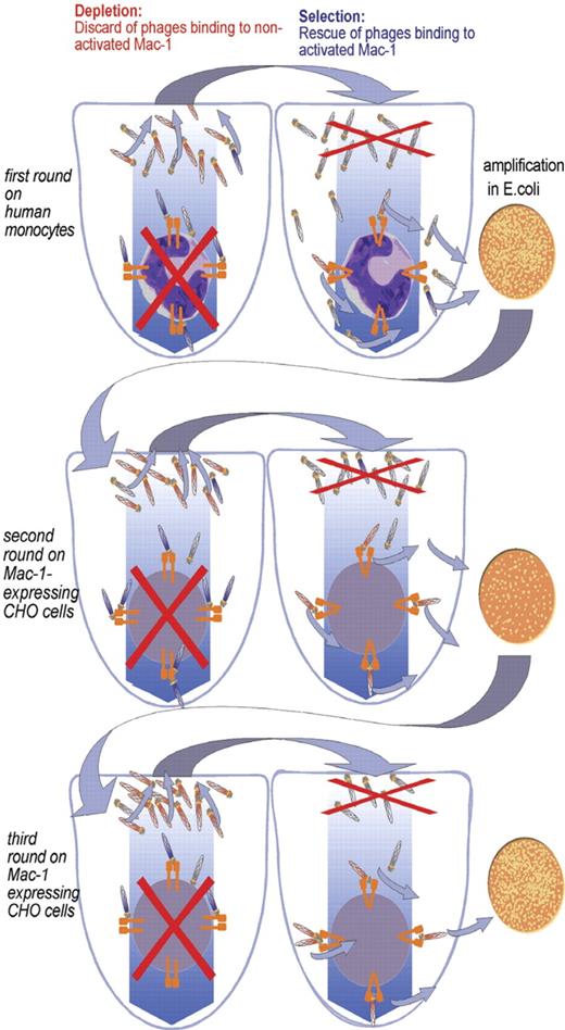
Starting materials were a natural and a synthetic phage library as described previously.24 For selection of phages directed against activation-specific epitopes of Mac-1, we developed a panning strategy with the following characteristics (Figure 1).(1) To wash off nonspecifically binding scFv's, a washing buffer with a relatively low pH (6.5) was used. (2) To direct the selection process to activated Mac-1, we presented this receptor with a distinct cell-surface background. Panning series were performed, with Mac-1 being expressed on 2 different cell types (monocytes and Mac-1–transfected CHO cells). (3) To deplete phages that bind to nonactivated receptors, we introduced a depletion step in our panning protocol, creating a subtractive selection strategy. The phage suspension was primarily incubated with nonactivated Mac-1 on monocytes as well as on CHO cells, and binding phages were discarded.
Subtractive panning (depletion/selection) strategy for the generation of conformation-specific single-chain antibodies. The first round was performed on human monocytes. A depletion step was performed in which phages bound to nonactivated Mac-1 or to the monocytes cell surface were separated by centrifugation and discarded. The supernatant was used for the next selection step in which the nonbinding phages in the supernatant were discarded and the binding phages were rescued and eluted. The obtained phages were then amplified in Escherichia coli and used for the next round. In the following 3 rounds (only 2 are pictured), the cell background was changed to Mac-1–expressing CHO cells to avoid enrichment of phages binding to activation-specific monocyte epitopes other than those on Mac-1.
Subtractive panning (depletion/selection) strategy for the generation of conformation-specific single-chain antibodies. The first round was performed on human monocytes. A depletion step was performed in which phages bound to nonactivated Mac-1 or to the monocytes cell surface were separated by centrifugation and discarded. The supernatant was used for the next selection step in which the nonbinding phages in the supernatant were discarded and the binding phages were rescued and eluted. The obtained phages were then amplified in Escherichia coli and used for the next round. In the following 3 rounds (only 2 are pictured), the cell background was changed to Mac-1–expressing CHO cells to avoid enrichment of phages binding to activation-specific monocyte epitopes other than those on Mac-1.
Altogether, we were able to enrich 1 natural (MAN-1 [Mac-1 activation-specific scFv/natural library]) and 2 synthetic (MAS-1 and MAS-2 [Mac-1 activation-specific scFv/synthetic library]) clones by differential panning. The full-length sequence of MAN-1, the HCDR3 sequences for MAS-1 and MAS-2, and production/purification methods are given in Figures S2 and S3.
Alanine scan of the HCDR3 region of MAN-1
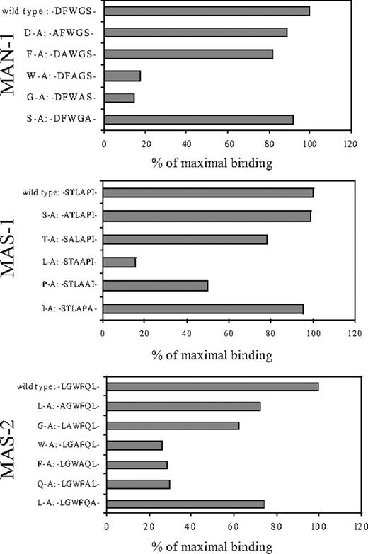
Since the CDR3 region of the heavy chain (HCDR3) is typically the major antigen-binding determinant of antibodies, we elucidated the role of the HCDR3 for epitope recognition of MAN-1, MAS-1, and MAS-2. Individual amino acids were substituted with alanine (alanine scan) by PCR. For MAN-1, the replacement of the first 2 and the last amino acids of the HCDR3 showed a small reduction in the binding ability of MAN-1 to activated Mac-1 (Figure 2). The substitution of the 2 central amino acids resulted in a significant loss in binding to Mac-1 (Figure 2). This emphasizes the important role of the HCDR3 and points to a specific role of the 2 central amino acids within the HCDR3. The importance of the centrally located tryptophan (W) is underlined by its appearance in the clone MAS-2. In an alanine scan of this clone, the replacement of this amino acid led to a loss of binding of the clone. For MAS-1, the centrally located leucin (L) seems to be the essential amino acid within the HCDR3. The mutations suggest that at least a component of the interaction of the scFv with Mac-1 is hydrophobic.
Alanine scan of the HCDR3 region of MAN-1, MAS-1, and MAS-2. The amino acids of the HCDR3, which were substituted by alanine, are given. The binding properties of the HCDR3 mutants were determined by flow cytometry on activated Mac-1 receptor in a whole-blood assay. Binding of the scFv's in flow cytometry was detected by an Alexa Fluor 488–labeled anti–His-tag antibody. Results are shown as percentage of the mean fluorescence of the original clones binding to activated Mac-1. Representative results out of at least 5 experiments are shown.
Alanine scan of the HCDR3 region of MAN-1, MAS-1, and MAS-2. The amino acids of the HCDR3, which were substituted by alanine, are given. The binding properties of the HCDR3 mutants were determined by flow cytometry on activated Mac-1 receptor in a whole-blood assay. Binding of the scFv's in flow cytometry was detected by an Alexa Fluor 488–labeled anti–His-tag antibody. Results are shown as percentage of the mean fluorescence of the original clones binding to activated Mac-1. Representative results out of at least 5 experiments are shown.
Flow cytometric proof of activation-specific binding to Mac-1
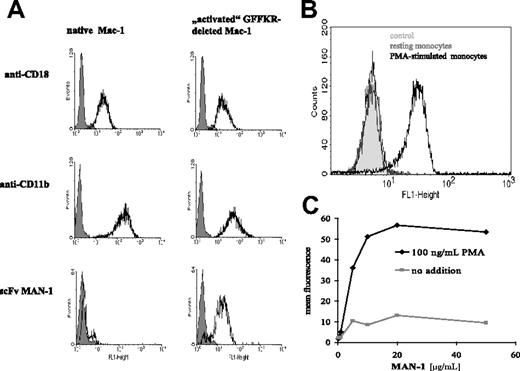
The binding of MAN-1 to Mac-1–expressing CHO cells was tested by flow cytometry (expression levels of CD11b and CD18 are shown in Figure 3A). MAN-1 demonstrated no binding to CHO cells expressing wild-type Mac-1 (Figure 3A). In contrast, MAN-1 strongly bound to CHO cells expressing the GFFKR-deleted and thus activated Mac-1 (Figure 3A). Monocytes obtained from healthy donors were used to prove activation-specific binding of MAN-1 to Mac-1. MAN-1 demonstrated a strong binding to PMA-activated but not to nonactivated monocytes (Figure 3B-C). Selective binding to the activated Mac-1 was maintained at concentrations beyond the saturation level as shown in the comparison of MAN-1 binding to PMA-activated and nonactivated monocytes at various concentrations of MAN-1 (Figure 3C). Similar activation-specific binding to Mac-1 was found with the clones MAS-1 and MAS-2 (data not shown).
Activation-specific binding of single-chain antibody MAN-1 in flow cytometry. (A) MAN-1 binding to Mac-1–expressing CHO cells. The expression level of the CHO cell line expressing the native and the GFFKR-deleted Mac-1 are evaluated with anti-CD11b and anti-CD18 mAbs (open curves) in comparison to matched isotype controls (shaded curves). (B) MAN-1 binding to PMA-activated but not to nonactivated human monocytes. (C) Binding of various concentrations of MAN-1 to PMA-activated monocytes in comparison to resting monocytes. The binding of MAN-1 in panels A, B, and C is detected with an Alexa Fluor 488–labeled anti–His-tag antibody and compared with the binding of an nonspecific single-chain antibody. Typical examples are shown, as routinely assessed after each scFv purification (n > 20).
Activation-specific binding of single-chain antibody MAN-1 in flow cytometry. (A) MAN-1 binding to Mac-1–expressing CHO cells. The expression level of the CHO cell line expressing the native and the GFFKR-deleted Mac-1 are evaluated with anti-CD11b and anti-CD18 mAbs (open curves) in comparison to matched isotype controls (shaded curves). (B) MAN-1 binding to PMA-activated but not to nonactivated human monocytes. (C) Binding of various concentrations of MAN-1 to PMA-activated monocytes in comparison to resting monocytes. The binding of MAN-1 in panels A, B, and C is detected with an Alexa Fluor 488–labeled anti–His-tag antibody and compared with the binding of an nonspecific single-chain antibody. Typical examples are shown, as routinely assessed after each scFv purification (n > 20).
Evaluation of potential MAN-1 cross-reactivity
Since the group of β2-integrins are structurally related and share common ligands, MAN-1 may bind to other β2-integrins besides Mac-1. In a flow cytometric assay, MAN-1 binding to activated monocytes is not inhibited by antibodies described to block the β2-integrins LFA-1 (αLβ2; CD11a/CD18), p150,95 (αXβ2; CD11c/CD18), or αDβ2 (CD11d/CD18) as demonstrated in Figure S4A. In contrast, MAN-1 binding is blocked by an anti–Mac-1 antibody in a concentration-dependent manner up to a full blockade, suggesting that MAN-1 binding is specific for the β2-integrin Mac-1 (Figure S4A). Potential cross-reactivity was further addressed by immunoprecipitation of Mac-1 from lysed, PMA-activated monocytes using MAN-1 as the precipitating antibody. Two bands were precipitated and detected by silver staining that fit to the molecular weight of the αM subunit with around 170 kDa, and the β2-subunit with around 95 kDa (data not shown). This suggests that there is no significant binding of MAN-1 to αX or αD, which would otherwise result in precipitates at 145 kDa and 125 kDa, respectively. Since the molecular weight of αL is described to be close to αM (approximately 170 kDa), the immunoprecipitation experiment cannot exclude binding of MAN-1 to LFA-1. For this reason, and to further support the finding that there is no significant binding to αX or αD, we performed Western blot analysis of MAN-1 precipitates of lysed, PMA-activated monocytes using anti-CD11a, anti-CD11b, anti-CD11c, and anti-CD11d antibodies. Whereas the monocyte lysate stained positive for all of the β2-integrins, the MAN-1 immunoprecipitate did not show an appropriate band for CD11a, CD11c, and CD11d. In contrast, an anti-CD11b antibody was clearly positive with both the monocyte lysate as well as the MAN-1 precipitate (Figure S4B). Thus, competition experiments of MAN-1 with blocking antibodies against all the members of the β2-integrin family as well as immunoprecipitations and Western blots suggest a selective binding of MAN-1 to the β2-integrin Mac-1.
Since 1 of the Mac-1 ligands, fibrinogen, also binds to the platelet integrin GPIIb/IIIa (αIIbβ3; CD41/CD61) we also evaluated potential cross-reactivity of MAN-1 with this integrin. For this purpose, we used recombinant CHO cell lines expressing native, nonactivated and GFFKR-deleted, activated GPIIb/IIIa as described.30-32 Whereas the activation-specific monoclonal antibody Pac-133 binds to the activated GPIIb/IIIa, MAN-1 does not bind to GPIIb/IIIa either in the activated or in the nonactivated state (Figure S4C).
Localization of MAN-1 binding to the Mac-1 I-domain
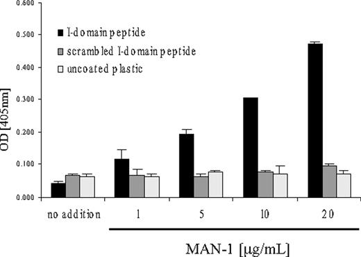
The I-domain is proposed to be the main activation-specific ligand binding site of Mac-1.1,3 An I-domain peptide, resembling the Lys245-Arg261 sequence of the Mac-1 I-domain, which represents the binding site of the Mac-1 ligand fibrinogen,34 was immobilized, and binding of MAN-1 was evaluated by ELISA. ScFv MAN-1 demonstrates a concentration dependant binding to the I-domain peptide compared with a control peptide, which was a scrambled (randomized) I-domain peptide sequence (Figure 4). This localizes the binding region for scFv MAN-1 to the I-domain-region Lys245-Arg261 of Mac-1.
MAN-1 binds to immobilized Mac-1 I-domain peptide. A scrambled I-domain peptide, which contained the same amino acids as the I-domain peptide in a randomized order, served as control. ScFv binding was detected with an anti–His-tag antibody and a secondary goat anti–mouse HRP antibody. Mean and standard deviation of triplicate experiments are given. A representative example of 4 experiments is given.
MAN-1 binds to immobilized Mac-1 I-domain peptide. A scrambled I-domain peptide, which contained the same amino acids as the I-domain peptide in a randomized order, served as control. ScFv binding was detected with an anti–His-tag antibody and a secondary goat anti–mouse HRP antibody. Mean and standard deviation of triplicate experiments are given. A representative example of 4 experiments is given.
Effects of MAN-1 on Mac-1–mediated adhesion under static conditions
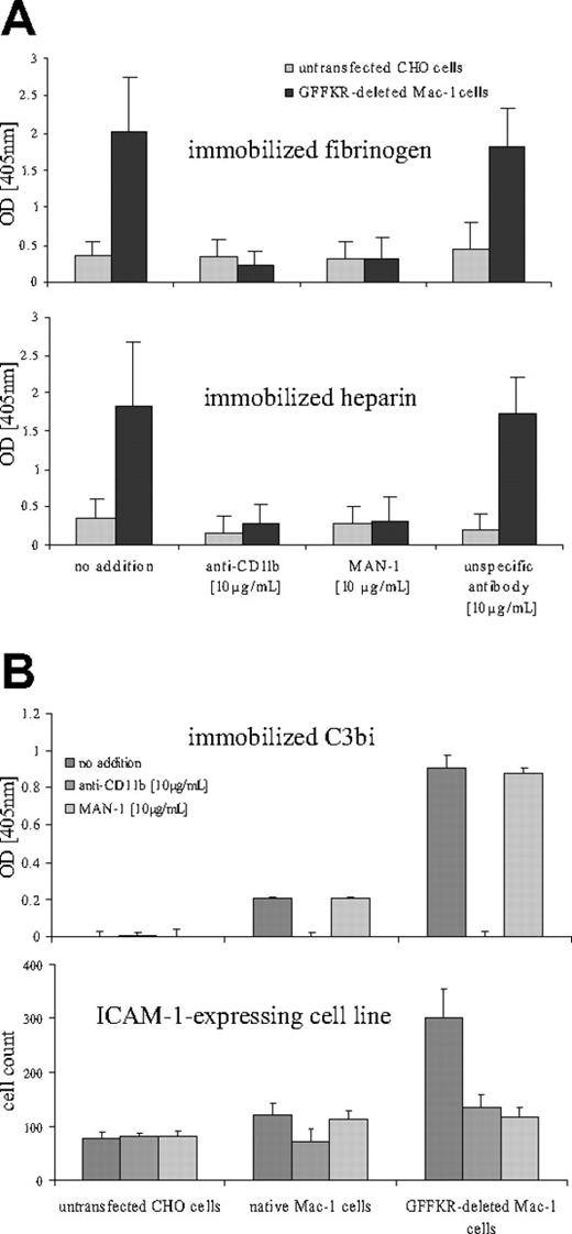
To evaluate the ability of MAN-1 to block ligand binding to Mac-1, adhesion assays on the Mac-1 ligands fibrinogen, heparin, and C3biwere performed. The adhesion of CHO cells, which express activated Mac-1, to both heparin and fibrinogen, could be blocked by MAN-1 to the same extent as by an anti-CD11b antibody (Figure 5A). These results underline the blocking ability of the MAN-1 antibody.
Single-chain antibody MAN-1 inhibits Mac-1 binding to the ligands heparin, fibrinogen, and ICAM-1, but not C3bi. (A) Inhibition of static adhesion of Mac-1–expressing CHO cells to immobilized fibrinogen and heparin by MAN-1. Adhesion of CHO cells transfected with the GFFKR-deleted Mac-1 to immobilized fibrinogen and heparin was evaluated. Nontransfected CHO cells served as negative control. Mean and standard deviation is given for triplicate experiments. Adherent cells were quantified with a phosphatase-substrate assay, and the absorbance was read at 405 nm. (B) Static adhesion of Mac-1–expressing CHO cells to ICAM-1–expressing CHO cells is inhibited by MAN-1, whereas adhesion to immobilized C3bi is not inhibited. Cells expressing the GFFKR-deleted, activated Mac-1 adhere more strongly to immobilized C3bi than nonactivated Mac-1 cells or a CHO cell control. Binding can be inhibited by an activation-nonspecific anti–Mac-1 antibody, but not by scFv MAN-1. Adherent cells were quantified with a phosphatase substrate assay, and absorbance was read at 405 nm. Mean and standard deviation is given for triplicate experiments. Adhesion of Mac-1–expressing CHO cells to immobilized ICAM-1–expressing CHO cells were counted based on their clearly distinguishable round shape on a flat monolayer of ICAM-1–expressing cells. Six visual fields were counted. Experiments were performed in triplicates. All static adhesion assays were performed at least 5 times. Representative results are shown.
Single-chain antibody MAN-1 inhibits Mac-1 binding to the ligands heparin, fibrinogen, and ICAM-1, but not C3bi. (A) Inhibition of static adhesion of Mac-1–expressing CHO cells to immobilized fibrinogen and heparin by MAN-1. Adhesion of CHO cells transfected with the GFFKR-deleted Mac-1 to immobilized fibrinogen and heparin was evaluated. Nontransfected CHO cells served as negative control. Mean and standard deviation is given for triplicate experiments. Adherent cells were quantified with a phosphatase-substrate assay, and the absorbance was read at 405 nm. (B) Static adhesion of Mac-1–expressing CHO cells to ICAM-1–expressing CHO cells is inhibited by MAN-1, whereas adhesion to immobilized C3bi is not inhibited. Cells expressing the GFFKR-deleted, activated Mac-1 adhere more strongly to immobilized C3bi than nonactivated Mac-1 cells or a CHO cell control. Binding can be inhibited by an activation-nonspecific anti–Mac-1 antibody, but not by scFv MAN-1. Adherent cells were quantified with a phosphatase substrate assay, and absorbance was read at 405 nm. Mean and standard deviation is given for triplicate experiments. Adhesion of Mac-1–expressing CHO cells to immobilized ICAM-1–expressing CHO cells were counted based on their clearly distinguishable round shape on a flat monolayer of ICAM-1–expressing cells. Six visual fields were counted. Experiments were performed in triplicates. All static adhesion assays were performed at least 5 times. Representative results are shown.
The binding of the activated Mac-1 to immobilized C3bi could only be blocked by the activation-unspecific anti-CD11b antibody, but not by MAN-1 (Figure 5B). Furthermore, flow cytometric measurements demonstrated that binding of soluble C3bi to PMA-activated monocytes is not inhibited by MAN-1, whereas an anti-CD11b antibody was able to block C3bi binding to activated monocytes (Figure S5).
In an ICAM-1 adhesion assay, MAN-1 was able to inhibit the binding of the activated Mac-1–transfected cell lines, but not the background binding of nonactivated Mac-1 to an ICAM-1–expressing cell line, as opposed to the unspecific anti-CD11b antibody, which inhibited both conformations (Figure 5B). Thus, MAN-1 inhibits ICAM-1 binding to activated Mac-1.
Inhibition of Mac-1–mediated adhesion by MAN-1 under flow conditions
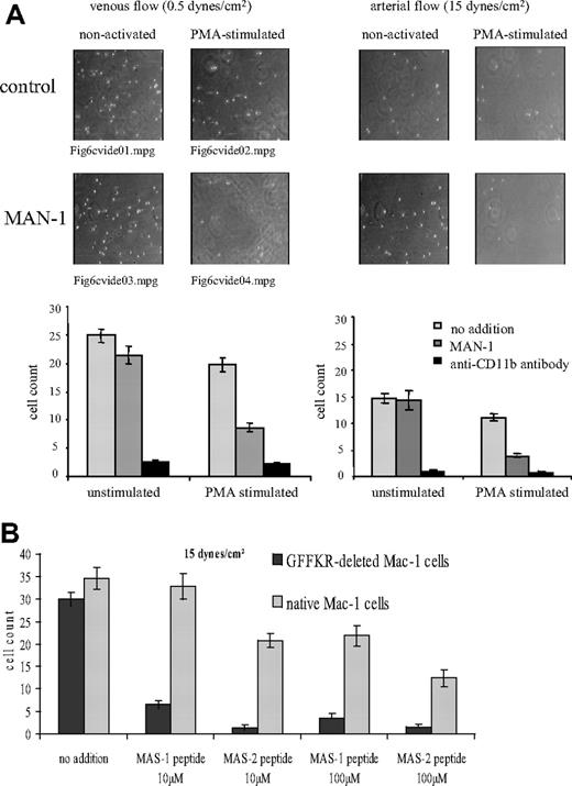
To evaluate the potential of MAN-1 as a therapeutic agent, we investigated the activation-specific blocking effect of MAN-1 under flow conditions. Based on previous data demonstrating selectin-independent adhesion of monocytes on fibrin/fibrinogen,35 we compared adhesion of activated and nonactivated monocytes on immobilized fibrinogen, which in contrast to soluble fibrinogen can mediate binding to the nonactivated Mac-1.36 Indeed, in flow experiments, monocytes can be captured by a fibrinogen matrix independent of their activity state (Figure 6A). Notably, PMA stimulation did not increase monocyte binding, which is consistent with a report demonstrating similar numbers of adherent monocytes with nonstimulated and stimulated monocytes under high shear rates (more than 0.5 dyne/cm2).37
Single-chain antibody MAN-1–derived peptides and circular MAS-1– and MAS-2–derived peptides specifically inhibit adhesion, which is mediated by the activated Mac-1 integrin. (A) MAN-1 inhibits adhesion of PMA-activated monocytes but not nonactivated monocytes to immobilized fibrinogen under flow conditions. An activation-nonspecific CD11b antibody (clone 2LPM19c) inhibits both activated and nonactivated monocytes. (B) Inhibition of adhesion of Mac-1–expressing CHO cells on immobilized fibrinogen by circular MAS-1– and MAS-2–derived peptides under flow conditions. For all flow experiments, means and standard deviations of adhering cells based on the counting of 5 visual fields are given. Representative examples of at least 6 experiments are demonstrated.
Single-chain antibody MAN-1–derived peptides and circular MAS-1– and MAS-2–derived peptides specifically inhibit adhesion, which is mediated by the activated Mac-1 integrin. (A) MAN-1 inhibits adhesion of PMA-activated monocytes but not nonactivated monocytes to immobilized fibrinogen under flow conditions. An activation-nonspecific CD11b antibody (clone 2LPM19c) inhibits both activated and nonactivated monocytes. (B) Inhibition of adhesion of Mac-1–expressing CHO cells on immobilized fibrinogen by circular MAS-1– and MAS-2–derived peptides under flow conditions. For all flow experiments, means and standard deviations of adhering cells based on the counting of 5 visual fields are given. Representative examples of at least 6 experiments are demonstrated.
This experiment was also performed with the CHO cells expressing native Mac-1 or GFFKR-deleted Mac-1 for adhesion on a fibrinogen matrix as described previously.28 MAN-1 selectively blocked adhesion of CHO cells that express the GFFKR-deleted and thereby activated Mac-1 (Figure S6).
The adhesion of monocytes to fibrinogen persisted under the arterial flow rate, which was applied at the end of the recording time. As we expected, only adhesion of the PMA-activated monocytes was inhibited by MAN-1. Adhesion of nonactivated monocytes was unaffected. Videos S1–S6, showing the monocyte flows, are available online, with the appropriate legends.
Inhibition of Mac-1–mediated adhesion by MAN-1–derived peptides under flow conditions
Since specificity of the scFv clones generated from the synthetic library is determined by the HCDR3 sequence, we evaluated whether activation-specific blockade can be achieved by peptides derived from the HCDR3s of MAS-1 and MAS-2. To increase stability, we synthesized cyclic peptides of the HCDR3 sequence. CHO cells expressing either the native or the activated form of Mac-1 were perfused at an arterial flow rate over a fibrinogen matrix. The MAS-1–derived peptides as well as the MAS-2–derived peptides blocked adhesion of CHO cells, which express the activated form of Mac-1, to a significantly higher extent than the CHO cells, which express the native form of Mac-1 (Figure 6B). This implies an activation-specific blockade of Mac-1 by the HCDR3-derived peptides.
MAN-1 as a diagnostic tool in patients with sepsis
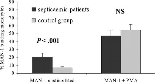
To demonstrate the feasibility of activation-specific anti–Mac-1 scFv's as diagnostic tools, we investigated 18 patients with severe sepsis. Compared with an age- and sex-matched control group without clinical or laboratory signs of inflammation, patients with sepsis demonstrated a significantly increased binding of MAN-1 to peripheral blood monocytes (Figure 7). Upon PMA stimulation, no significant difference in MAN-1 binding to monocytes could be observed between the 2 groups (Figure 7), implying that the stimulatory capacity of monocytes is preserved in both groups. However, in the patients with sepsis, monocytes are clearly preactivated. Thus, MAN-1 may represent a diagnostic tool for the detection of monocyte activation in sepsis.
MAN-1 binding as diagnostic marker for basal monocyte activation in patients with sepsis. MAN-1 binding to monocytes of 18 patients with severe sepsis compared with sex- and age-matched patients without any sign of inflammation as analyzed by whole-blood flow cytometry. MAN-1 binding without activation (basal activation) and after PMA stimulation are shown as a percentage of monocytes positive for MAN-1 as detected by an Alexa Fluor 488–labeled anti–His-tag antibody. Significance level P and NS for nonsignificant are given. Error bars depict SEM.
MAN-1 binding as diagnostic marker for basal monocyte activation in patients with sepsis. MAN-1 binding to monocytes of 18 patients with severe sepsis compared with sex- and age-matched patients without any sign of inflammation as analyzed by whole-blood flow cytometry. MAN-1 binding without activation (basal activation) and after PMA stimulation are shown as a percentage of monocytes positive for MAN-1 as detected by an Alexa Fluor 488–labeled anti–His-tag antibody. Significance level P and NS for nonsignificant are given. Error bars depict SEM.
Discussion
The aim of the present study was to generate human single-chain antibodies that are specific for the activated conformation of the leukocyte integrin Mac-1, and to demonstrate their potential as diagnostic tools and therapeutic agents. We developed a subtractive selection strategy allowing the isolation of specific scFv's from human scFv phage libraries. The functional properties of the generated scFv's using flow cytometry and cell adhesion assays under static as well as under flow conditions were characterized. We localized the binding epitope of a representative clone (MAN-1) to a 16–amino acid sequence of the αM I-domain. Using mutagenesis analysis of the heavy-chain CDR3s of 3 activation-specific single-chain antibodies, we could identify the paratope of the scFv's. In a pilot study with patients presenting with severe sepsis, we provide initial support for the use of our scFvs as a marker of monocyte activation and thereby as a potential diagnostic tool. The therapeutic potential of activation-specific Mac-1 blockade by the newly generated scFv's, and furthermore by small-molecular-weight HCDR3-derived peptides, was demonstrated in adhesion assays under static and flow conditions.
The procedure developed for the scFv selection is unique in its ability to select for conformation-specific antibodies. In contrast to classical panning strategies that are based on immobilized target molecules, we used a cell-based system allowing the display of complex, function-specific, transmembranous molecules consisting of multiple subunits. Furthermore, the phage display technology allows a subtractive approach, with depletion of phages that either bind nonspecifically or that bind to Mac-1 in its nonactivated state and selection of phages that bind to the activated Mac-1. To reduce nonspecific background binding, it was essential to change to a cell type that is distinct from human monocytes, but which expresses Mac-1 either in a nonactivated or an activated state. For this purpose, we used CHO cell lines that were either transfected with the native Mac-1 (for depletion) or with a mutated and thereby activated Mac-1 (for selection). The latter cell line has been developed in analogy to a cell-line model based on a GFFKR deletion in the integrin-α subunit that has been frequently used as a model for the activated GPIIb/IIIa (αIIbβ3).30-32 These Mac-1–expressing CHO cells bearing a deletion of the GFFKR region of the αM subunit demonstrate increased affinity to soluble ligands.22,23 The strategy to combine depletion and selection steps as well as the use of different cell backgrounds provides a unique specificity for a target molecule in defined conformational states, which can be used to target a wide variety of cell membrane proteins as well as protein complexes.
As an initial investigation of its diagnostic potential, the scFv MAN-1 was tested as a marker of sepsis. In particular in its early stages, this highly life-threatening clinical condition is difficult to diagnose due to the lack of conclusive laboratory parameters and to a wide variation in clinical appearance. The activation of monocytes/macrophages plays a pivotal role in the pathogenesis of sepsis and early diagnosis, and consequently, early treatment can indeed change the outcome of sepsis.38 Furthermore, it has been suggested that Mac-1 is a useful marker to diagnose sepsis.39 We hypothesized that the activation-specific anti–Mac-1 scFv can detect monocyte activation and thus can be used to diagnose sepsis. Indeed, in a pilot study comparing patients with severe sepsis with a sex- and age-matched control group without sepsis, the activation status of Mac-1 was significantly enhanced in patients with sepsis. Our data warrant further clinical studies to determine the suitability of activation-specific Mac-1 scFv's to contribute to the diagnosis of sepsis and other diseases with monocyte involvement. For instance, in Wegener granulomatosis, disease activity correlates with the extent of Mac-1 expression of monocytes.40 Mac-1 expression has been shown to correlate with the risk of restenosis after coronary angioplasty, to correlate with procoagulant activity after angioplasty in patients with acute myocardial infarction and to reflect the therapeutic effects of antiplatelet agents on monocyte activation after coronary stent implantation.41,42 Overall, in immune response–related diseases and in inflammation in general, activation-specific, anti–Mac-1 scFv's may provide new diagnostic opportunities.
Inhibition of Mac-1 is an attractive therapeutic strategy that may provide benefits in various diseases. For instance, neointimal thickening after arterial injury was significantly reduced by antibody blockade of Mac-1 and in Mac-1 knock-out mice.18 Rats treated with a blocking anti–Mac-1 antibody demonstrate reduced ischemic cell damage after transient cerebral artery occlusion,19 and Mac-1–deficient mice are less susceptible to cerebral ischemia/reperfusion injuries.20 Sepsis-induced lung infiltration of polymorphonuclear leukocytes (PMNs) was significantly reduced in a Mac-1 knock-out mouse model as well as in a transgenic mouse model in which NIF (neutrophil inhibitory factor), a blocking ligand of Mac-1, is overexpressed.16 In autoimmune diseases, Mac-1 blockade may offer a novel therapeutic approach. For example, in mice, autoimmune bullous pemphigoid could be prevented by antibody blockade of Mac-1 and by Mac-1 knock-out.17 For angiostatin, which has been shown to inhibit neovascularization, an antiadhesive/anti-inflammatory effect mediated by the blockade of Mac-1 has recently been demonstrated.43 In particular, in skin injury/inflammation, this mechanism may play an important role.43 Overall, an effective blockade of Mac-1 may allow targeting of chronic inflammatory processes in different pathologic settings.
The promising results with the activation-specific, anti–Mac-1 scFv's as selective inhibitors prompted us to initiate studies with the aim to develop small-molecular-weight inhibitors that might be further developed to orally active compounds. As a first step toward creating a peptide mimetic,44 we determined the paratopes of the single-chain antibodies by mutational analysis. In MAN-1 we identified 2 amino acids, tryptophane and glycine, within the CDR3 region of the heavy chain that are essential for activation-specific binding. The fact that the same amino acids are also found in MAS-2, 1 of the 2 clones of the synthetic single-chain library, underlines their structural importance. In scFv clone MAS-1, the exchange of a centrally localized leucine by alanine reduces the binding to background level. In all 3 scFv clones, the main paratope-forming amino acids are centrally located in the HCDR3 and are hydrophobic, providing structural hints for the design of small-molecular-weight inhibitors. As a second step, we produced and tested HCDR3-derived peptides. Indeed, peptides derived from the HCDR3 of MAS-1 and MAS-2 displayed highly activation-specific inhibition of Mac-1. The inhibitory potency of these peptides were similar to HCDR3-derived peptides, which were developed as lead compounds for the therapeutic blockade of anti-p185HER2/neu for patients with breast cancer.45 Notably, to our knowledge so far, all potent small molecule antagonists to I-domain–containing integrins are allosteric inhibitors,46 whereas the newly described HCDR3-derived peptides are based on a competitive inhibitory mechanism. Overall, we are able to define peptide sequences, which may serve as a basis for the development of orally active, activation-specific Mac-1 blockers.
We hypothesize that the specific blockade of activated Mac-1 provides advantages that translate into clinical benefits compared to the unselective blockade of Mac-1. One of the Mac-1 ligands that demonstrate a differential effect of activation-specific blockade is fibrinogen. In contrast to soluble fibrinogen, immobilized fibrinogen can mediate cell adhesion by binding to nonactivated Mac-1.36 As demonstrated, blockade by an activation-specific Mac-1 scFv leaves this Mac-1 function intact, whereas antibodies blocking the activated and the nonactivated Mac-1 inhibit Mac-1–mediated cell adhesion on immobilized fibrinogen under static and under flow conditions. Recently, the interaction between Mac-1 and the ligand fibrinogen has been described as powerful mediator of inflammation.36 Mice carrying a mutated P2C allele of fibrinogen, the major recognition site for the αM I-domain, showed a severely compromised host defense.36 ScFv MAN-1 is directed to the same site on Mac-1 as is fibrinogen, but in contrast to the fibrinogen mutant mice, cell adhesion to immobilized fibrinogen is still possible, and the compromise of the immune system may be less. Indeed, experiments with Mac-1 knock-out mice established a pivotal role of this integrin in host defense.47 Notably, the phagocytosis of bacteria (eg, Borrelia burgdorferi) can be mediated by Mac-1 in a nonactivated state either via C3bi or by direct interaction between B burgdorferi outer-surface protein and Mac-1.48,49 Furthermore, our results show that MAN-1 does not interfere with C3bi binding to the activated Mac-1 receptor. These results are consistent with the observation that the C3bi-binding region within the I-domain is not identical to the region for ICAM-1 and fibrinogen. Blocking antibodies that reduce binding to fibrinogen and ICAM-1 inhibit C3bi binding only slightly.50 Also, mutational analysis suggest a different binding site in Mac-1 for fibrinogen and C3bi.51 More recently, the binding region for C3bi within Mac-1 has been located and has been described to be within the peptide-sequence Asp398-Thr,451 which is not involved in fibrinogen binding and is distinct from the MAN-1 epitope.52 Overall, MAN-1 may not interfere with host defense mechanisms based on its activation-specific Mac-1 blockade and the selective epitope targeted by MAN-1.
In summary, we developed a depletion/selection strategy for human single-chain phage libraries with the aim to target an activation-specific epitope on Mac-1. We were able to generate conformation-specific single-chain antibodies that selectively bind to the activated integrin Mac-1. These single-chain antibodies and their peptide derivatives may be useful for the diagnosis of monocyte activation in disease and may be beneficial as therapeutic agents that selectively block activated leukocytes.
Authorship
Contribution: S.U.E. performed key experiments, designed research, analyzed data, and wrote the manuscript; M.S. designed research analyzed data, and wrote the manuscript; N.S. designed and performed research and analyzed data; J.S., N.B., and D.H. performed experiments; C.B. designed research; and K.P. designed research, analyzed, data and wrote the paper.
Conflict-of-interest disclosure: S.U.E., M.S., and K.P. are inventors in a patent application filed to protect the intellectual property of human activation-specific anti–Mac-1 antibodies and their derivatives. The remaining authors declare no competing financial interests.
S.U.E., M.S., and N.S. contributed equally to the article.
Correspondence: Karlheinz Peter, Baker Heart Research Institute, PO Box 6492, St Kilda Road Central, Melbourne, Victoria 8008, Australia; e-mail: karlheinz.peter@baker.edu.au.
The online version of this manuscript contains a data supplement.
An Inside Blood analysis of this article appears at the front of this article.
The publication costs of this article were defrayed in part by page charge payment. Therefore, and solely to indicate this fact, this article is hereby marked “advertisement” in accordance with 18 USC section 1734.
We are grateful to Drs J. Ylanne, J. Chin-Dusting, K. J. Woollard, C. Hagemeyer, and M. Udawela for advice and critical reading of the manuscript.
This work was supported by the National Health and Medical Research Council (NHMRC) and the National Heart Foundation of Australia.

