Abstract
Electrocardiographic abnormalities are common in Thalassemia Major patients as well as in animal models of iron overload. The relationship between these ECG abnormalities and the degree of cardiac iron overload has not been well characterized.
Hypothesis: Standard 12-lead ECG identifies preclinical cardiac iron deposition.
Methods: Fifty-four patients with Thalassemia Major underwent MRI quantification of cardiac iron within one month of a standard 12-lead ECG. Cardiac T2* measurements to quantify iron were performed using a validated multiecho gradient-echo sequence performed on a 1.5 Tesla General Electric CVi scanner. The PR, QRS, QT, and QTc durations as well as P-wave, T-wave and QRS axes and heart rates were measured. Normative data was derived from 20 control patients without detectable cardiac iron (T2* > 20 ms) and age-appropriate norms were created for each ECG criteria. Z-scores for the ECG parameters were then calculated for 34 patients having detectable cardiac iron (T2* < 20ms).
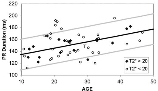
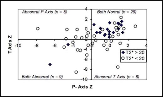
Results: ECG parameters varied significantly with age, with correlation coefficients ranging from 0.39 (QTc interval) to 0.66 (PR interval). Cardiac iron was associated with lower heart rates (Z=−0.44, p=0.06), QTc prolongation (Z=0.48, p=0.04) and leftward shift of the P and T wave axes [P-Axis (Z = −1.53, p<0.001); T-Axis (Z= −1.91, p<0.001)]. In total, 31 of 34 pts with cardiac iron had one or more ECG abnormalities (|Z|>2), compared to only 7 of 20 control pts (p<0.0001). Subjective ECG review was consistent with Z-score. The most common abnormalities associated with cardiac iron were non-specific ST-T wave changes (n = 15), sinus bradycardia (n=4), symmetric T-wave inversions (n = 4) and LVH (n = 4). The presence of any of these findings on ECG predicts detectable cardiac iron with a sensitivity and specificity of 68% and 85%. A combined metric of PR prolongation (Z>2), left shift of P or T axis (Z<–2), bradycardia, LVH, ST changes, or symmetric T-wave inversions produced a sensitivity of 94% and specificity of 75% for detectable cardiac iron.
Conclusions: Cardiac iron overload produces characteristic abnormalities in atrial conduction and ventricular repolarization. ECG may be an effective and inexpensive screening modality for cardiac iron overload.
Author notes
Disclosure:Consultancy: Dr Wood and Dr. Coates have served as consultants to Novartis. Research Funding: Dr. Woods and Dr. Coates have received research funding from Novartis and Apotex. Honoraria Information: Dr. Wood and Dr. Coates have received honoraria from Novartis and Apotex. Membership Information: Dr. Wood is an advisor to the Exjade Speaker’s Bureau and Dr. Coates is a member of the Exjade Speaker’s Bureau.


