Abstract
Abstract 3586
Poster Board III-523
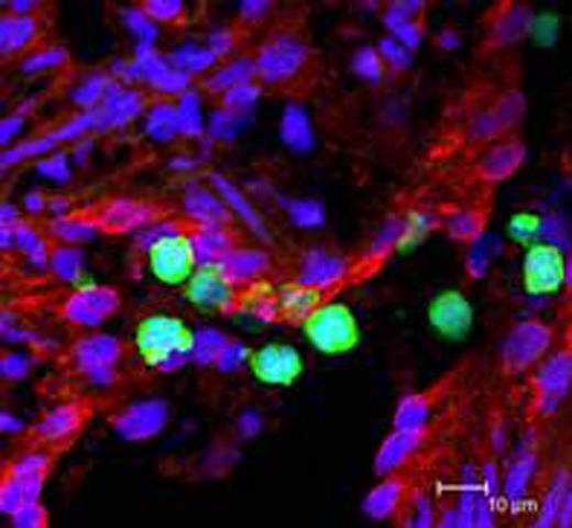
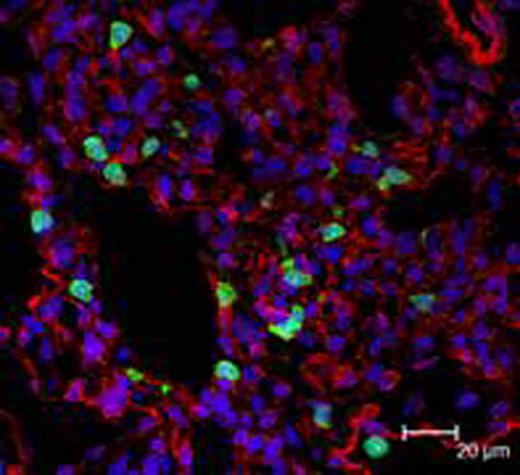
Shortly following G-CSF administration there is a dramatic yet transient disappearance of neutrophils from the circulation. To evaluate this disappearance, C57BL/6 LysM-EGFP knock-in male mice between 6-12 weeks of age were used. The LysM promoter/enhancer is specifically expressed in neutrophils and monocytes permitting evaluation of tissue distribution using EGFP marker localization by confocal microscopy. The ratio of EGFP+ leukocytes to background cellularity was determined in fixed volumes of tissue collected from the lung, liver, spleen, and kidney of mice either receiving saline (n=4) or at 15 minutes (n=6), 30 minutes (n=6), or 240 minutes (n=4) following 50ug/kg SQ rmG-CSF administration. No changes in ratio were observed in triplicate images of the liver or kidney of mice following G-CSF administration; however, there was a significant increase in EGFP+ leukocytes in the spleen and lung shortly following G-CSF administration. It increased from a control value of 6.3(2.6)% (n=12) to 15.9 (6.8)% (n=18) at 30 minutes in the lung and from 1.6 (0.7)% (n=12) to 8.3 (4.2)% (n=18) at 30 minutes in the spleen. While the value for the pulmonary images was less at 240 minutes (9.2 (5.1)%) the level of EGFP+ cells in the spleen continued to rise (12.3 (7.2)%). The increases were in neutrophils based on nuclear morphology. Thus the transient loss of neutrophils from the circulation following G-CSF administration appeared to be due to an accumulation of neutrophils within the pulmonary and splenic tissues. To confirm this observation, tissues were collected from three rhesus macaques 30 minutes following 10ug/kg SQ G-CSF. These tissues were compared to those of a control. Neutrophils were labeled with CD18 and a secondary antibody conjugated to FITC for quantification by confocal microscopy. As before, ratios of fluorescence were determined. The mean threshold intensity for CD18+ cells for the lung lobes for those animals receiving G-CSF (3.8 (3.0)%, n=54) was greater than control specimen lungs (1.5(1.1)%, n=18) and slightly increased for the spleen( from 4.1(3.7)% to 8.5 (6.3)%, n=9). Detailed confocal microscopy using a myeloid/histiocyte antigen clone Mac387 which labels neutrophils and monocytes, an endothelial marker anti-caveolin-1-Cy3 antibody, and a nuclear stain (To-Pro3) showed (see below, stained green, red, and blue, respectively) that the neutrophils were elongated, appeared activated, and were tightly adhered to the endothelium of the pulmonary vasculature. These results suggest that the transient neutropenia following G-CSF administration is associated with an accumulation of neutrophils within the pulmonary and splenic tissues.
No relevant conflicts of interest to declare.
Author notes
Asterisk with author names denotes non-ASH members.


