Abstract
Abstract 3576
Immunophenotype holds central role in the differential diagnosis of leukemic B-chronic lymphoproliferative disorders (B-CLPDs). However, select cases show overlapping characteristics and represent a diagnostic challenge. CD1d is an MHC-like molecule which presents glycolipids to the CD1d-restricted NKT cells, thus regulating innate and adaptive immune responses. Normal B-cells constitutively express CD1d, but its expression in B-CLPDs is currently unknown, with the exception of B-CLL in which there are only sparse and conflicting data. In the present study we assessed the diagnostic utility of CD1d expression in B-CLPDs and correlated the latter with various prognostic markers of B-CLL.
CD1d expression was measured by 4-color flow cytometry on peripheral blood (PB), bone marrow (BM) and lymph node (LN) samples from 147 patients with B-CLL, 24 with mantle cell lymphoma (MCL), 20 with lymphoplasmacytic lymphoma (LPL), 10 with splenic marginal zone lymphoma (SMZL), 14 with hairy cell leukemia (HCL) and 3 with atypical hairy cell leukemia (aHCL).The utility of CD1d as a diagnostic marker for B-CLL versus the rest B-CLPDs was evaluated with receiver operator characteristic (ROC) curve analysis. A cut off value of 40% was selected and subsequently sensitivity, specificity, and likelihood ratios (LR) were calculated. One-way ANOVA was used for the assessment of differences in CD1d mean fluorescence intensity (MFI) between B-CLPDs with very high CD1d expression. The association of CD1d expression with CD38, CD49d and IgHV status was tested with Pearson and Spearman correlation, respectively.
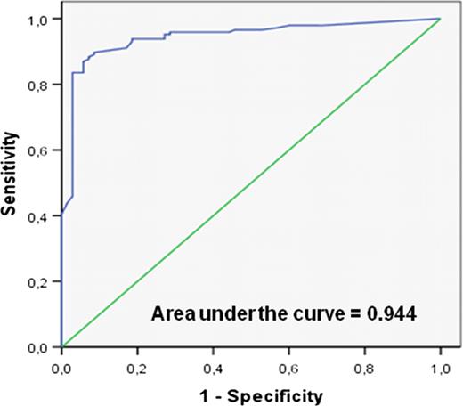
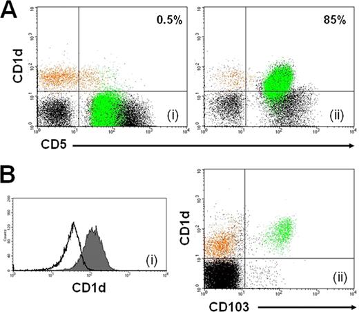
CD1d expression in B-CLPDs was as follows: B-CLL: 26%(range 0%-100%), MCL: 76%(15%-100%), LPL: 70%(53%-88%), SMZL: 96%(78%-100%), HCL: 100% and aHCL: 64%(51%-75)%. A CD1d expression of less than 40% was strongly indicative of B-CLL (LR: 26, specificity: 97%, sensitivity: 77%, figures 1 & 2A), as in all non B-CLL patients CD1d was > 40%. CD1d expression remained unaltered during either disease progression or relapse after chemotherapy in 17 patients with B-CLL that were assessed in various time points. Furthermore, CD1d was equally expressed in the BM, LN and PB of 6 patients with B-CLL, whereas it showed no significant correlation with CD38, CD49d and IgHV mutational status. Interestingly, in contrast to the generally low CD1d expression in B-CLL, all patients with trisomy 12 (n=5) expressed high CD1d levels (65-100%), in line with previous data reporting aberrant immunophenotypic features in trisomy 12. Also, HCL cells displayed significantly stronger intensity of CD1d expression (MFI: 196±26) compared to SMZL (MFI: 60±11, p=0.03), aHCL (MFI: 20.6±7, p=0.02) and normal B-cells (MFI: 62±5, p=0.03, figure 2B).
In conclusion, our findings suggest that CD1d is a useful immunophenotypic marker for the differential diagnosis of B-CLL versus other B-CLPDs, as it is significantly downregulated in the former and it remains unaffected by disease stage and treatment status. Additionally, CD1d expression could also help in the differential diagnosis of B-CLPDs with prominent splenomegaly and overlapping phenotypes.
ROC curve depicting the accuracy of CD1d expression of predicting the diagnosis of B-CLL in 218 patients with B-CLPDs.
ROC curve depicting the accuracy of CD1d expression of predicting the diagnosis of B-CLL in 218 patients with B-CLPDs.
CD1d expression in normal and clonal B-cells. (A) CD1d expression in a B-CLL (i) and a MCL (ii) patient. Normal B-cells are shown in orange and CD5+ neoplastic lymphocytes in green. (B) Stronger expression of CD1d in HCL (filled histogram in i, green color in ii) compared to SLVL (open histogram, i) and normal B cells (orange color, ii).
CD1d expression in normal and clonal B-cells. (A) CD1d expression in a B-CLL (i) and a MCL (ii) patient. Normal B-cells are shown in orange and CD5+ neoplastic lymphocytes in green. (B) Stronger expression of CD1d in HCL (filled histogram in i, green color in ii) compared to SLVL (open histogram, i) and normal B cells (orange color, ii).
No relevant conflicts of interest to declare.
Author notes
Asterisk with author names denotes non-ASH members.


