Abstract
Abstract 4263
Precise and effective measurements of iron overload in the liver, where iron deposition seems to be primarily noticeable (Noetzli LJ et al, Blood 2008), are important for the early diagnosis, treatment and follow-up of patients with hemochromatosis or hemosiderosi. T2* Magnetic resonance imaging (MRI) represents the most available noninvasive technique to assess hepatic iron content and shows a good correlation with biopsy results (Wood JC et al, Blood 2005). In the clinical practice, a single hepatic slice is acquired and T2* measurement is performed in a single region of interest (ROI) of the parenchyma (Pepe A et al, Eur J Haematol 2006). This approach may mask an heterogeneous iron distribution. Thus, the aims of this study were to set up a MRI acquisition technique for the detection of the iron burden in the whole liver and to evaluate the effectiveness of the single ROI technique.
Five transverse hepatic slices were acquired by a T2* gradient-echo sequence in 101 thalassemia major (TM) patients (48 males, mean age 29 ± 8 years, mean serum ferritin level 1413 ± 1209 ng/mL, mean pre-transfusion hemoglobin 9 ± 1 g/dl.) and 20 healthy subjects. The T2* value was calculated in a single ROI defined in the medium-hepatic slice. Moreover, the T2* value was extracted on each of the eight ROIs defined in the functionally independent segments according to Couinaud classification. The mean hepatic T2* value was obtained by averaging all segmental values.
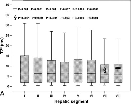
For patients the mean T2* values over segments VII and VIII were significantly lower than the other segments (P<0.0001) (figure 1). This pattern was substantially preserved in the two groups identified considering the T2* normal cut-off (Ramazzotti A. et al J Magn Reson Imaging 2009) (group A: single ROI T2* < 15.8 ms and group B: single ROI T2* ≥ 15.8 ms). All mean segmental T2* values as well as the mean hepatic T2* value were strongly correlated with the single ROI T2* value. After the application of a correction map based on T2* fluctuations in the healthy subjects, no significant differences were found in the segmental T2* values for the whole patient population (P=0.251), for both the previously defined groups (Group A: P = 0.073 and Group B: P=0.476) as well as for the Group A patients (N = 23) with chronic hepatitis (P=0.111).
Hepatic T2* variations are low and due to artifacts and measurement variability. The single ROI approach can be adopted in the clinical arena, taking care to avoid the susceptibility artifacts, occurring mainly in segments VII and VIII.
Segmental T2* variability in the whole patient population. Each box shows the median (black line), quartiles, and extreme values within a segment. The mean T2* values over the VII and VIII segments were significantly lower than the mean T2* values over the other segments. The P-value for each significant pairwise comparison is indicated
Segmental T2* variability in the whole patient population. Each box shows the median (black line), quartiles, and extreme values within a segment. The mean T2* values over the VII and VIII segments were significantly lower than the mean T2* values over the other segments. The P-value for each significant pairwise comparison is indicated
No relevant conflicts of interest to declare.
Author notes
Asterisk with author names denotes non-ASH members.


