Abstract
Abstract 462
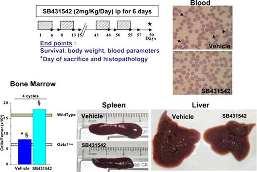
The marrow microenvironment in primary myelofibrosis and mouse models of the disease is characterized by increased levels of cytokines which regulate hematopoiesis including CXCL12, BMP4, VEGF and TGF-β. The observation that TGF-βnull myelofibrotic stem cells fail to transmit the disease by transplantation (Chagraoui et al, Blood 100:3495, 2002) has established an important role for TGF-β in disease development. Mice carrying the hypomorphic Gata1low mutation in which the enhancer that drives gene expression in megakaryocytes (MK) is deleted also develop myelofibrosis with age (Vannucchi et al, Blood 2002;100:1123). To clarify the role of TGF-β in development of myelofibrosis in the Gata1low mouse model, the levels of this factor in plasma and marrow of the mutant mice were measured. Gata1low mice express normal levels of TGF-β in plasma (1.8±0.7 vs 2.1±0.4 ng/mL) and levels of TGF-β mRNA (2.9±0.5 vs 1.5±0.3 arbitrary units, p<0.01) and protein (1.9±0.3 vs 0.86±0.2 ng/mL, p<0.01) only 2-times greater than normal in marrow. However, by immunoelectron microscopy, patchy TGF-β deposits associated with collagen fibers were observed in the marrow microenvironment of mutant mice (<3 vs >700 particles/field in wild-type and Gata1low marrow, respectively) indicating that fibrosis, by concentrating TGF-β locally, may contribute to disease progression also in Gata1low mice. To evaluate whether inhibition of TGF-β signaling would ameliorate myelofibrosis in this animal model, Gata1low mice were treated with SB431542 (C22H16N4O3, MW= 384.4), an inhibitor of TGF- β1/activin receptor-like kinases recently demonstrated to prevent renal fibrosis in mice (Petersen et al, Kidney Int 73:705, 2008.). Six males (7-9 months) and 6 females (12 months) were treated with SB431542 as described for renal fibrosis (see Figure). Equivalent numbers of mice treated with vehicle were used as control. Treatment was well tolerated (no deaths) and the SB43542-treated mice were easily recognized by being more active and with shinier coats. At the end of the 4th cycle, mice were sacrificed and analyzed for disease progression. The results were as follows:
SB43542-treatment did not affect hematocrit levels (43.2±1.2 vs 41.3±0.9 in SB43542- and vehicle-treated mice, respectively), increased platelets numbers [0.34(±0.03)×106/μL vs 0.2(±0.009)×106/μL, p<0.01] but platelets remained larger than normal, reduced white blood cell counts [5.2(±0.19)×103/μL vs 6.5(±0.3)×103/μL, p<0.01] and frequency of poikilocytes (1 every 4–5 fields vs >4/field). Also, progenitor cell trafficking was not reduced (CD34posCD117pos cells: 1.45 vs 1.2% colony forming cells: 10.8±2.4 vs 8.0±0.1 CFC/μL).
Treatment increased total cell number [18.0(±0.7) ×106 vs 8.2(±0.4) ×106/femur, p<0.01] and frequency of erythroid cells (20.5±2.5 vs 13.2±0.7%, p<0.01) but not of MK (40.2±5.7 vs 38.7±0.9%) in the femur. However, fibrosis and microvessel density were reduced (Gomori-Silver and CD34 staining). Increased Mallory staining of bones was observed but the femur became resistant to fracture, suggesting that overall bone structure improved.
SB43542-treatment reduced spleen weight (0.2±0.6 vs 0.45±0.05 gr, p<0.01) and cell numbers [265(±30)×106 vs 385(±5)×106 cells, p<0.01]. Therefore, although the frequency of erythroid cells and MK in the organ remained high, overall hematopoiesis in spleen was reduced.
SB43542-treatment restored the morphological appearance of the liver and reduced the frequency of MK (5.9±0.7 vs 20.4±4.7, p<0.01). These improvements were likely not due to an anti-inflammatory effect of the drug because parallel treatments with dexamethasone did not modify disease progression in Gata1low mice.
SB43542-treatment reduced the myelofibrotic traits expressed by Gata1low mice, confirming that increased TGF-β1 levels play an important role in disease manifestations in this animal model. We have previously published that Aplidin treatment restores the hematopoietic stem cell properties of Gata1low mice (Verrucci et al, J Cell Physiol,2010, May20, Epub ahead of print). The observation that SB43542-treatment primarily reduced microenvironmental abnormalities suggests that the two drugs may have synergistic effects in the treatment of myelofibrosis.
No relevant conflicts of interest to declare.
Author notes
Asterisk with author names denotes non-ASH members.


