Abstract
Vascular disruption induced by interactions between tumor-secreted permeability factors and adhesive proteins on endothelial cells facilitates metastasis. The role of tumor-secreted C-terminal fibrinogen-like domain of angiopoietin-like 4 (cANGPTL4) in vascular leakiness and metastasis is controversial because of the lack of understanding of how cANGPTL4 modulates vascular integrity. Here, we show that cANGPTL4 instigated the disruption of endothelial continuity by directly interacting with 3 novel binding partners, integrin α5β1, VE-cadherin, and claudin-5, in a temporally sequential manner, thus facilitating metastasis. We showed that cANGPTL4 binds and activates integrin α5β1-mediated Rac1/PAK signaling to weaken cell–cell contacts. cANGPTL4 subsequently associated with and declustered VE-cadherin and claudin-5, leading to endothelial disruption. Interfering with the formation of these cANGPTL4 complexes delayed vascular disruption. In vivo vascular permeability and metastatic assays performed using ANGPTL4-knockout and wild-type mice injected with either control or ANGPTL4-knockdown tumors confirmed that cANGPTL4 induced vascular leakiness and facilitated lung metastasis in mice. Thus, our findings elucidate how cANGPTL4 induces endothelial disruption. Our findings have direct implications for targeting cANGPTL4 to treat cancer and other vascular pathologies.
Introduction
Tumor metastasis is the main cause of mortality in cancer patients.1 It is determined largely by vasculature leakiness and the critical steps of intravasation and extravasation, which involve the directional migration of tumor cells across the disrupted endothelium. The endothelium provides a semipermeable boundary between the bloodstream and tumor. The paracellular permeability of the endothelium is mediated primarily by transmembrane endothelial adherens junction (AJ) and tight junction (TJ) proteins that are linked to the actin cytoskeleton,2 and connect adjacent endothelial cells (ECs).3 Thus, the disruption of major components of AJs and TJs, such as the intercellular VE-cadherin and claudin-5 clusters, leads to changes in the actin cytoskeleton, cell shape, and the activation of intracellular signaling pathways, which partly via β-catenin initiate gene transcription.4 Apart from AJs and TJs, integrins also strengthen EC–EC contacts and provide a scaffold for cytoskeletal reorganization during cell organization, proliferation, and migration.5 Specifically, the expression of integrin α5β1 regulates endothelial monolayer integrity.6 Inherently, metastasis requires communication between tumor cells and ECs that culminates in the disruption of EC–EC contacts and the degradation of the vascular basement membrane. However, the molecular mechanisms that facilitate such communication are poorly understood.
In the tumor microenvironment, interactions among tumor cells, their secreted factors, and the endothelium induce vascular permeability to aid tumor metastasis.5,7 These factors bind to cognate cellular receptors, setting off a cascade of downstream molecular signaling events that determine the outcome of malignancy.8,9 In this respect, angiopoietin-like 4 (ANGPTL4) protein has been implicated in cancer metastasis10 ; however, its precise role in vascular and cancer biology remains debatable. Earlier studies suggested that ANGPTL4 could prevent metastasis by inhibiting vascular leakiness.11,12 However, ANGPTL4 was identified as 1 of the most highly expressed genes in distant metastases13 and it is associated with breast cancer metastasis to the lungs.14,15 Our recent work revealed that tumors produced high levels of the C-terminal fibrinogen-like domain of ANGPTL4 (cANGPTL4). We also showed that cANGPTL4 stimulates integrin-mediated signaling to maintain an elevated, oncogenic O2−:H2O2 ratio to confer anoikis resistance to tumor cells via autocrine adhesion mimicry.16 Furthermore, we have demonstrated that through interaction with matrix proteins integrin β1 and integrin β5, cANGPTL4 facilitates cell migration and wound healing, both of which are processes highly reminiscent of metastasis.17,18 Despite the plethora of evidence implicating ANGPTL4 in cancer metastasis, the heterotypic role of ANGPTL4 in vascular integrity remains unclear.
Here, we show that tumor-derived cANGPTL4 instigated the disruption of endothelial continuity by directly interacting with integrin α5β1, VE-cadherin, and claudin-5 in a temporally sequential manner. Tumor-derived cANGPLT4 and recombinant human (rh)–cANGPTL4 increased vascular permeability in vitro and in vivo. Using ANGPTL4-knockout and wild-type mice injected with either control or ANGPTL4-knockdown tumors confirmed that cANGPTL4 induced vascular leakiness and facilitated lung metastasis in mice. cANGPTL4 binds to and activates integrin α5β1-mediated PAK/Rac signaling that weakened EC–EC contacts and increased vascular leakiness. cANGPTL4 associated with the adhesive domains of VE-cadherin and claudin-5, resulting in their declustering and internalization, translocation of β-catenin to the nucleus, and a leaky endothelium. These results identify cANGPTL4 as a novel upstream mediator of endothelium permeability and a potential target in the treatment of cancer and other vascular-related pathologies.
Methods
Antibodies
Antibodies were obtained from the following sources: PAK1 and pPAK1 (Ser199/Ser204; Cell Signaling Technology); integrin β1 (clone JB1A), α5β1 (clone JBS5), β3 (MAB2008), and c-Jun (all Millipore); CD29/activated integrin β1 (HUTS-21; BD Biosciences); β-tubulin, β-catenin (12F7), epidermal growth factor receptor (1005; all Santa Cruz Biotechnology); VE-cadherin [BV9] and claudin-5 (Abcam); CD31 (Dako Denmark A/S); occludin (Invitrogen); zona occludens-1 (ZO-1; Zymed Laboratories); and Tie 1, Tie 2, junctional adhesion molecule (JAM)-C, β-tubulin, horseradish peroxidase (HRP)–conjugated goat anti–mouse, HRP-conjugated goat anti–rabbit, and HRP-conjugated donkey anti–goat (Santa Cruz Biotechnology). Mouse monoclonal anti–human cANGPTL4 mAb4A11H5 and rabbit polyclonal anti–human cANGPTL4 were produced in-house.16 Secondary antibodies Alexa Fluor 488–conjugated goat anti–rabbit IgG and Alexa Fluor 594–conjugated goat anti–mouse IgG also were used (Invitrogen).
Cell cultures
Primary human microvascular endothelial cells (HMVECs; Lonza) were cultured in EndoGRO-MV-VEGF medium (Millipore) in a humidified atmosphere of 5% CO2 at 37°C. The culture surface was precoated with 0.1% gelatin in PBS. All other cell lines were cultured in DMEM supplemented with 10% FBS. Conditioned medium (CM) was obtained from 1 × 10⁵ tumor cells grown in 1 mL of serum-free medium for 3 days.
Transient transfections assay
Transfections of HMVECs were carried out as per the manufacturer's protocol (Promega) with either constitutive-active Rac1 G12V or dominant-negative Rac1 T17N.
Expression and purification of recombinant cANGPTL4 proteins
In vivo tumorigenicity and miles vascular permeability assay
Six-week-old BALB/c athymic female nude mice (20-22 g) were injected either cANGPTL4 (6 μg/mL), or 0.9% saline buffer was injected intradermally at adjacent locations on the back. To determine vascular permeability, tumor-bearing mice received an intravenous injection of Evans blue dye. Twenty minutes later, the mice were killed and Evans blue dye was extracted from the tumor and normal muscle as described in Miles vascular permeability assay.12 The extravasated dye was extracted using formamide, and the amount was quantified by measuring absorbance at 610 nm. To determine the tumor vascular volume, 2MDa FITC-conjugated dextran (10 mg/kg) also was injected intravenously for 20 minutes. Fluorescence readings were obtained using 3300 fluorospectrometer (NanoDrop).
For another set of experiments, mice were injected subcutaneously at the interscapular region with either 2 × 10⁶ or 8 × 10⁶ cells (A-5RT3CTRL or A-5RT3ΔANGPTL4). The same procedures were carried out in C57B/L6J wild-type and ANGPTL4-knockout mice using B16F10CTRL or B16F10ΔANGPTL4 cells. Injection sites were rotated to avoid site bias. The injected tumor cells were allowed to grow for 8 weeks.
In vivo metastasis assay
Wild-type and ANGPTL4-knockout C57BL/6J mice were injected intravenously with either 5 × 10⁵ B16F10CTRL or B16F10ΔANGPTL4 cells. For cANGPTL4 treatment, wild-type C57BL/6J mice intravenously injected with either 2 × 10⁶ B16F10CTRL or B16F10ΔANGPTL4 cells were treated with PBS or cANGPTL4 (3 mg/kg) thrice weekly. After 3 weeks, the mice were killed and the lungs were harvested for further analyses. Total metastatic burden were quantified by RT-PCR of melanin A. Total mRNA were extracted from lungs and reverse-transcribed as described previously.17,18 All animal studies were approved and carried out in compliance with the regulations of the Institutional Animal Care and Use Committee of Nanyang Technological University.
Proximity ligation assay
A proximity ligation assay (PLA) was carried out according to the manufacturer's protocol (Olink Bioscience) using indicated pairs of antibodies. Images were taken using an LSM 710 confocal microscope (Carl Zeiss) with a Plan-Apochromat 63×/1.4 oil differential interference contrast objective and ZEN 2009 software (Carl Zeiss). PLA signals were quantified using BlobFinder software (Centre for Image Analysis, Uppsala University).19
TER measurement
Transendothelial electrical resistance (TER) was measured using an electrical cell-substrate impedance sensing system (Applied BioPhysics). HMVECs were seeded on sterile 8-well gold-plated electrode arrays precoated with fibronectin at 2 × 10⁵ cells/well and allowed to adhere and spread for 4 hours at 37°C. Data from the electrical resistance experiments were obtained over the experimental time course at 5-minute intervals. Confluent HMVEC monolayers that had stable TERs for 1 hour preceding administration of indicated treatments were used. As cells adhered and spread over the microelectrode, TER increased, whereas cell retraction, rounding, or loss of adhesion was reflected by a decrease in TER. Resistance values for each microelectrode were normalized as the ratio of measured resistance to baseline resistance and are plotted as a function of time.
Internalization assay
Confluent HMVEC monolayers were treated with either PBS or cANGPTL4 (6 μg/mL) for 3 hours. At indicated time point, an internalization assays was carried out as described previously.17
Immunofluorescence staining
Disrupted tight junctions were visualized by immunofluorescence staining for ZO-1. HMVECs were fixed with 4% paraformaldehyde for 15 minutes, permeabilized with 0.2% Triton X-100 for 15 minutes, and blocked with 2% BSA containing 0.1% Triton X-100 in a humid chamber for 1 hour at room temperature. Cells were incubated overnight at 4°C with anti–human-ZO-1 antibodies (1:100) in 0.2% BSA. After 2 washes in PBS, cells were incubated for 1 hour at room temperature with Alexa 488–secondary antibodies (1:250). Cells were counterstained with Alexa 594–phalloidin for F-actin and 4,6-diamidino-2-phenylindole (DAPI) for nuclei. Immunostainings performed without primary antibodies served as negative controls. For β-catenin staining, cells were fixed with 4% paraformaldehyde containing 5% sucrose for 15 minutes, permeabilized with 0.5% Triton X-100 in PBS for 4 minutes, and blocked in 5% normal goat serum with 0.1% Triton X-100. Cells were incubated overnight at 4°C with anti–human β-catenin antibodies (1:200) in 3% normal goat serum, followed by Alexa 594-secondary antibodies. Images were acquired using an LSM710 confocal microscope (Carl Zeiss) with a 63×/1.40 oil objectives and AxioCan MRm camera (Carl Zeiss) and analyzed using ZEN 2009 software.
Surface plasmon resonance
Surface plasmon resonance (SPR) was carried out as described previously17,18 using a BIAcore 2000 system (BIAcore). Six concentrations (0.16, 0.32, 0.63, 1.25, 2.50, and 5.0μM) of recombinant integrin β1, first extracellular domain of VE-cadherin (ECD1), extracellular repeat 1 domain of VE-cadherin (EC1), occludin, or JAM-A were used. Integrin was expressed in Drosophila S2 cells,17 and various extracellular domains of VE-cadherin and claudin-5 were purchased from Abnova. Global fitting of the SPR data were performed as described previously to determine the KD value. Preinjection or preincubation with the indicated antibodies or preimmune IgG was performed to determine specific interactions. Each sensorgram was corrected by subtracting a sensorgram obtained from a reference flow cell with no immobilized protein. The Rmax value was determined to be 283.1 resonance units using anti-cANGTL4 antibodies against the immobilized cANGPTL4. Values are mean ± SD (n = 3).
Statistical analysis
Statistical analyses were performed using 2-tailed Mann-Whitney U tests with SPSS software (SPSS). All statistical tests were 2-sided. A P value of ≤ .05 is considered significant.
Results
Elevated cANGPTL4 expression in human tumors
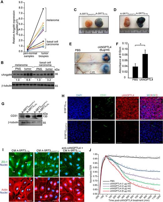
To examine the expression profile of cANGPTL4 in tumors, we performed immunofluorescence using anti-cANGPTL4 antibody on 2 human tumor tissue arrays that covered benign, malignant, and metastatic tumors originating from various anatomic sites. cANGPTL4 was elevated in all epithelial tumor samples compared with the corresponding normal tissue samples (supplemental Figure 1A-C, available on the Blood Web site; see the Supplemental Materials link at the top of the online article). Although fluorescence signals varied among different tumor types, they clearly showed that elevated cANGPTL4 was a common feature of tumors. Consistent with the observations in supplemental Figure 1A through C, cANGPTL4 protein level was elevated in all cancer cell lines compared with nontumorigenic lines (HaCaT and BJ; supplemental Figure 1D). Interestingly, we observed relatively higher cANGPTL4 levels in metastatic than in nonmetastatic cancer cell lines, suggesting a role of cANGPTl4 in tumor metastasis. To further corroborate the role of cANGPTL4 in metastasis clinically, we determined cANGPTL4 mRNA and protein levels in 6 human basal cell carcinoma (benign) and melanoma (metastatic) biopsies by quantitative real-time PCR and immunoblot analyses. We observed significant up-regulation of cANGPTL4 mRNA and protein levels in these epithelial cancers compared with their cognate peritumor normal samples. Notably, the melanomas expressed higher levels of cANGPTL4 mRNA and protein compared with benign basal cell carcinoma (Figure 1A-B). Similar trend was observed when comparing cANGPTL4 protein levels of fine-needle aspirates from 3 breast cancer patients to 4 human metastatic tumor lines (A-5RT3, MDA-MB231, MCF-7, and HT29) and 2 nontumorigenic lines (HaCaT and BJ). The mean cANGPTL4 protein concentrations were 12.4 μg/mL (fine-needle aspirate), 5.9 μg/mL (metastatic lines), and 0.8 μg/mL (nontumorigenic lines; supplemental Figure 1E). These data suggest that epithelial tumors secrete elevated levels of cANGPTL4, which is consistent with our recent report showing that tumor tissue expressed high levels of cANGPTL4 compared with basal level expression in the surrounding stroma.16 Altogether, our findings implicate a role for cANGPTL4 in cancer metastasis.
cANGPTL4 is elevated in metastatic tumors and disrupts endothelial junction integrity. (A-B) Expression levels of cANGPTL4 mRNA (A) and protein (B) in basal cell carcinoma (BCCs) and metastatic melanoma paired with peritumor normal tissues as determined by quantitative real-time PCR and immunoblot, respectively (n = 3). Data spots from same individual are linked by colored lines. Ribosomal protein L27 (L27) was used as a reference housekeeping gene. β-tubulin served as a loading and transfer control. (C-D) Evans blue dye extravasation induced by either A-5RT3CTRL– or A-5RT3ΔANGPTL4–induced tumors of different (C) or similar (D) tumor volume (A-5RT3CTRL, 1098 ± 422 mm3 vs A-5RT3ΔANGPTL4, 551 ± 135 mm3; P < .05; n = 6). (E-F) Evans blue dye extravasation induced by cANGPTL4 or PBS vehicle (G) and quantification (H) of extravasated Evans blue dye by measurement of absorbance at 610 nm. *P < .05. (G) Immunodetection of CD31 in A-5RT3CTRL– and A-5RT3ΔANGPTL4–induced tumors. (H) Immunofluorescence staining for CD31 and cANGPTL4 in B16F10CTRL- and B16F10ΔANGPTL4-induced tumors. Scale bar represents 100 μm. (I) Immunofluorescence staining for ZO-1 in a confluent HMVEC monolayer. Cells were treated with either CM from A-5RT3CTRL in the presence or absence of anti-cANGPTL4, from A-5RT3ΔANGPTL4 cells. HMVECs were counterstained with DAPI (blue) for nuclei and phalloidin (red) for actin stress fibers. Scale bar represents 40 μm. White arrows indicate endothelial junction lesions. (J) Transendothelial electrical resistance (TER) analysis of confluent monolayer HMVECs treated with the indicated amounts of cANGPTL4. PBS served as a vehicle control. Data (means ± SD) from 3 independent experiments.
cANGPTL4 is elevated in metastatic tumors and disrupts endothelial junction integrity. (A-B) Expression levels of cANGPTL4 mRNA (A) and protein (B) in basal cell carcinoma (BCCs) and metastatic melanoma paired with peritumor normal tissues as determined by quantitative real-time PCR and immunoblot, respectively (n = 3). Data spots from same individual are linked by colored lines. Ribosomal protein L27 (L27) was used as a reference housekeeping gene. β-tubulin served as a loading and transfer control. (C-D) Evans blue dye extravasation induced by either A-5RT3CTRL– or A-5RT3ΔANGPTL4–induced tumors of different (C) or similar (D) tumor volume (A-5RT3CTRL, 1098 ± 422 mm3 vs A-5RT3ΔANGPTL4, 551 ± 135 mm3; P < .05; n = 6). (E-F) Evans blue dye extravasation induced by cANGPTL4 or PBS vehicle (G) and quantification (H) of extravasated Evans blue dye by measurement of absorbance at 610 nm. *P < .05. (G) Immunodetection of CD31 in A-5RT3CTRL– and A-5RT3ΔANGPTL4–induced tumors. (H) Immunofluorescence staining for CD31 and cANGPTL4 in B16F10CTRL- and B16F10ΔANGPTL4-induced tumors. Scale bar represents 100 μm. (I) Immunofluorescence staining for ZO-1 in a confluent HMVEC monolayer. Cells were treated with either CM from A-5RT3CTRL in the presence or absence of anti-cANGPTL4, from A-5RT3ΔANGPTL4 cells. HMVECs were counterstained with DAPI (blue) for nuclei and phalloidin (red) for actin stress fibers. Scale bar represents 40 μm. White arrows indicate endothelial junction lesions. (J) Transendothelial electrical resistance (TER) analysis of confluent monolayer HMVECs treated with the indicated amounts of cANGPTL4. PBS served as a vehicle control. Data (means ± SD) from 3 independent experiments.
cANGPTL4 increased vascular leakiness in in vivo tumors
We next explored the role of cANGPTL4 in tumorigenesis. Metastatic human squamous cell carcinoma cell line A-5RT3 and murine melanoma B16F10 were transduced with either scrambled (control) small interfering (si)RNA (A-5RT3CTRL or B16F10CTRL) or siRNA against ANGPTL4 (A-5RT3ΔANGPTL4 or B16F10ΔANGPTL4; supplemental Figure 2A) to suppress endogenous ANGPTL4, as described previously.16,18 The injection of A-5RT3CTRL cells into immunodeficient mice induced large primary tumors at week 8, but A-5RT3ΔANGPTL4-induced tumors displayed a 30% lower tumor volume (A-5RT3CTRL, 1098 ± 422 mm3 vs A-5RT3ΔANGPTL4, 551 ± 135 mm3; P < .05; n = 6; Figure 1C). Miles assays revealed decreased vascular leakiness in A-5RT3ΔANGPTL4-induced tumors (∼ 5-fold lower), as evidenced by decreased extravasation of Evans blue dye (Figure 1D; Table 1). Differences in vascular volume among the tumor types were accounted for when determining tumor vascular permeability (Table 1). This differential vascular permeability was independent of the tumor volume, as observed in A-5RT3CTRL– and A-5RT3ΔANGPTL4–induced tumors (Figure 1D). Furthermore, none of the mice injected with A-5RT3ΔANGPTL4 developed distant metastases to the lymph nodes, whereas 83% of mice injected with A-5RT3CTRL showed metastases (supplemental Figure 2B). Notably, rh-cANGPLT4 was sufficient to induce vascular leakiness compared with the PBS control (rh-cANGPTL4, 0.084 ± 0.014 vs PBS, 0.04 ± 0.01; P < .05; n = 3; Figure 1E-F). Consistent to the increased vascular volume in A-5RT3CTRL– and B16F10CTRL-induced tumors, the expression of EC marker CD31 was elevated compared with their cognate ANGPTL4-deficient tumors (Figure 1G-H). Dual immunostaining for CD31 and cANGPTL4 also confirmed that epithelial tumors were the major producer of cANGPTL4, consistent with earlier findings using laser-captured microdissected samples (Figure 1H).16 Hence, these observations in vivo prompted us to investigate the molecular mechanism of cANGPTL4-induced endothelial permeability.
Confluent primary human microvascular ECs were cultured in 3-day-old CM from either A-5RT3CTRL or A-5RT3ΔANGPTL4 cells. EC junction integrity was disrupted when the cells were cultured in A-5RT3CTRL CM but not in A-5RT3ΔANGPTL4CM, as indicated by ZO-1 staining. Junction integrity was maintained in the presence of anti-cANGPTL4 monoclonal antibody, suggesting that tumor-secreted cANGPTL4 plays a major role in endothelial junction disruption (Figure 1I). We found that EC–EC junction integrity was disrupted when the cells were treated with only rh-cANGPTL4 (6 μg/mL) in the presence of either cycloheximide or actinomycin D, indicating that de novo protein synthesis and transcription were not required (supplemental Figure 2C). Treatment with cycloheximide, actinomycin D, or vehicle alone did not induce any vascular lesions (supplemental Figure 2C). This disruption was mirrored by a decrease in TER of the monolayer, suggesting a rapid and dose-dependent increase in paracellular permeability (Figure 1J). No change in TER was observed in the PBS control (Figure 1J). This effect was not because of apoptosis, even after 6 hours of rh-cANGPTL4 treatment (supplemental Figure 2D). Altogether, these results indicate that tumor-secreted cANGPTL4 disrupts endothelial integrity.
cANGPTL4 interacts with integrin α5β1, VE-cadherin, and claudin-5
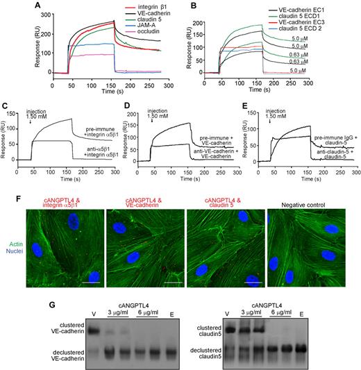
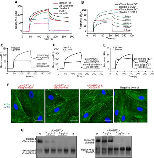
VE-cadherin and claudin-5 contribute to the control of paracellular permeability, whereas integrin α5β1, which is localized to EC–EC junctions, also mediates endothelial leakiness.4,6 Hence, we hypothesized that tumor-derived cANGPTL4 induces vascular leakiness and ultimately disrupts the endothelium barrier via direct interactions with specific junction proteins. SPR analysis revealed that cANGPTL4 interacted with VE-cadherin (KD = 1.12 × 10⁻⁷M), claudin-5 (KD = 5.87 × 10⁻⁸M), and integrin α5β1 (KD = 1.26 × 10⁻⁸M) but not with occludin or JAM-A (Figure 2A). More significantly, cANGPTL4 targeted the VE-cadherin EC1 domain and claudin-5 ECD1 domain that are responsible for homophilic interactions between adjacent ECs, suggesting that the interactions of cANGPTL4 with VE-cadherin and claudin-5 could play an important role in regulating vascular permeability (Figure 2B).20,21 No interactions were observed between cANGPTL4 and integrin β316,17 or other domains of VE-cadherin (EC3) and claudin-5 (second extracellular domain of claudin-5 [ECD2]), which also served as controls (Figure 2B). The observations of these interactions were corroborated by blocking with neutralizing antibodies against integrin α5β1 (Figure 2C), VE-cadherin (Figure 2D), claudin-5 (Figure 2E), and cANGPTL4 (supplemental Figure 3A-C). An in situ PLA detected complexes of integrin α5β1, claudin-5, or VE-cadherin with cANGPTL4 in both EC- (integrin, 22.50 ± 5.01; VE-cadherin, 16.43 ± 2.46; claudin-5, 12.35 ± 3.44 PLA spots/cell calculated from 250 cells) and A-5RT3CTRL–induced tumor biopsies (Figure 2F; supplemental Figure 3D), confirming that these interactions also exists in vivo. No PLA signals, that is, no interactions were observed between CD31 and cANGPTL4, claudin-5, VE-cadherin, or integrin α5β1 (supplemental Figure 3E).
cANGPTL4 interacts with integrin α5β1, VE-cadherin, and claudin-5. (A-E) Representative sensorgrams showing binding profiles between immobilized cANGPTL4 and integrin α5β1, VE-cadherin, claudin-5, occludin, and JAM-A (A); EC1, ECD1, VE-cadherin EC3, and claudin-5 ECD2 (B); integrin α5β1 (C), VE-cadherin (D), and claudin-5 (E) preblocked with either preimmune IgG or indicated cognate antibodies. (F) In situ PLA of cANGPTL4 and indicated binding partners. PLA signals are in red (integrin 22.50 ± 5.01 vs VE-cadherin 16.43 ± 2.46 vs claudin-5 12.35 ± 3.44 PLA spots/cell for 250 cells, n = 3). Cells were counterstained with Alexa 488-phallodin for actin stress fibers (green) and Hoechst dye for nuclei (blue). Negative control was performed in the absence of anti-ANGPTL4. Scale bar represents 40 μm. (G) Immunodetection of VE-cadherin and claudin-5 from membrane extract of HMVECs treated with cANGPTL4 (3 or 6 μg/mL) for 3 hours. Vehicle (v) is PBS, and E indicates treatment with 2.5mM EDTA in PBS.
cANGPTL4 interacts with integrin α5β1, VE-cadherin, and claudin-5. (A-E) Representative sensorgrams showing binding profiles between immobilized cANGPTL4 and integrin α5β1, VE-cadherin, claudin-5, occludin, and JAM-A (A); EC1, ECD1, VE-cadherin EC3, and claudin-5 ECD2 (B); integrin α5β1 (C), VE-cadherin (D), and claudin-5 (E) preblocked with either preimmune IgG or indicated cognate antibodies. (F) In situ PLA of cANGPTL4 and indicated binding partners. PLA signals are in red (integrin 22.50 ± 5.01 vs VE-cadherin 16.43 ± 2.46 vs claudin-5 12.35 ± 3.44 PLA spots/cell for 250 cells, n = 3). Cells were counterstained with Alexa 488-phallodin for actin stress fibers (green) and Hoechst dye for nuclei (blue). Negative control was performed in the absence of anti-ANGPTL4. Scale bar represents 40 μm. (G) Immunodetection of VE-cadherin and claudin-5 from membrane extract of HMVECs treated with cANGPTL4 (3 or 6 μg/mL) for 3 hours. Vehicle (v) is PBS, and E indicates treatment with 2.5mM EDTA in PBS.
cANGPTL4 interacted directly with the EC1 and ECD1 adhesive domains of VE-cadherin and claudin-5, respectively, and may lead to disruption of intercellular VE-cadherin and claudin-5 cluster formation.22 To examine this possibility, membrane fractions of confluent ECs treated with cANGPTL4 were extracted, resolved under native conditions, and probed with either anti–VE-cadherin or anti–claudin-5. cANGPTL4 treatment resulted in smaller protein bands compared with PBS (Figure 2G). Treatment with EDTA, which disrupts intercellular adhesion, served as a positive control for declustered VE-cadherin and claudin-5. These findings confirm that cANGPTL4 interacts with integrin α5β1, VE-cadherin, and claudin-5 to disrupt intercellular adhesion.
cANGPTL4 modulates endothelial integrity in a temporally sequential mechanism
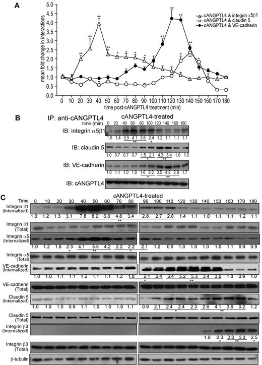
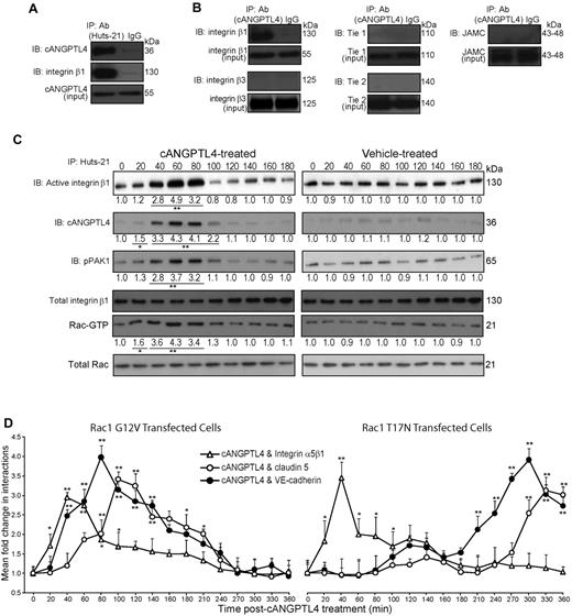
Next, we sought a mechanistic explanation for the effect of cANGPTL4 on the endothelium. We monitored the temporal interactions of cANGPTL4 and its 3 binding partners through kinetic PLA. A bimodal interaction profile of the cANGPLT4:integrin α5β1 complex was detected within 30-60 minutes, followed by cANGPTL4:VE-cadherin and cANGPTL4:claudin-5 at 120 minutes post-cANGPTL4 treatment (Figure 3A). In vitro coimmunoprecipitation of cANGPTL4 and its various interacting partners validated our PLA and SPR results (Figure 3B). Except for integrin α5β1, which showed elevated expression only 2 hours post-cANGPTL4 treatment, no change in protein level was observed for any other cANGPTL4-interacting partner, indicating that the bimodal cANGPTL4–protein interaction profiles did not result from increased protein expression (supplemental Figure 4A). We also showed that the initial cANGPTL4:integrin α5β1 interactions resulted in the complex being internalized into the cytoplasm at 40 minutes, followed by the internalization of the cANGPTL4:VE-cadherin and cANGPTL4:claudin-5 complexes between 120 and 150 minutes to result in the abrogation of the endothelial junctions (Figure 3C). This bimodal effect was not observed with the control treatment (supplemental Figure 4B). Notably, cANGPTL4:integrin α5β1 formation at 30-60 minutes coincided with a TER decrease, suggesting increased permeability (Figure 1K). Integrin β3, which does not interact with cANGPTL4, was internalized after 2 hours in both vehicle- and cANGPTL4-treated cells. Similar observations were reported in wild-type and ANGPTL4-knockdown keratinocytes,17 suggesting that this may be inherent to either integrin β3 or the experimental procedure. Thus, our results suggest that the initial interaction of cANGPTL4 with integrin α5β1 may be required to facilitate the subsequent interactions to mediate vascular disruption.
cANGPTL4 interacts with integrin α5β1, VE-cadherin, and claudin-5 in a bimodal manner. (A) In situ kinetic PLA detection of various complexes between cANGPTL4 and indicated binding partners in HMVECs treated with 6 μg/mL cANGPTL4. Values (means ± SD) represent mean fold change in the number of interactions compared with the zero time point, as determined from n = 3 independent experiments (600 HMVECs) using BlobFinder. Experiments were terminated when microscopic lesions were observed at 180 minutes after cANGPTL4 treatment. (B) Immunodetection of integrin α5β1, VE-cadherin, and claudin-5 in anti-cANGPTL4 immunoprecipitates from total protein lysates of HMVECs treated with 6 μg/mL rh-cANGPTL4. (C) Immunodetection of integrins β1, integrin α5, integrin β3, VE-cadherin, and claudin-5 from total protein lysate versus internalized fraction of HMVECs treated with cANGPTL4. Protein lysates were collected every 10 minutes (0-180 minutes). Values below each band represent the mean fold change in protein expression level compared with the cognate zero time point (n = 3). *P < .05; **P < .01.
cANGPTL4 interacts with integrin α5β1, VE-cadherin, and claudin-5 in a bimodal manner. (A) In situ kinetic PLA detection of various complexes between cANGPTL4 and indicated binding partners in HMVECs treated with 6 μg/mL cANGPTL4. Values (means ± SD) represent mean fold change in the number of interactions compared with the zero time point, as determined from n = 3 independent experiments (600 HMVECs) using BlobFinder. Experiments were terminated when microscopic lesions were observed at 180 minutes after cANGPTL4 treatment. (B) Immunodetection of integrin α5β1, VE-cadherin, and claudin-5 in anti-cANGPTL4 immunoprecipitates from total protein lysates of HMVECs treated with 6 μg/mL rh-cANGPTL4. (C) Immunodetection of integrins β1, integrin α5, integrin β3, VE-cadherin, and claudin-5 from total protein lysate versus internalized fraction of HMVECs treated with cANGPTL4. Protein lysates were collected every 10 minutes (0-180 minutes). Values below each band represent the mean fold change in protein expression level compared with the cognate zero time point (n = 3). *P < .05; **P < .01.
Inhibition of the cANGPTL4:integrin α5β1 complex delays cANGPTL4 interactions with VE-cadherin and claudin-5
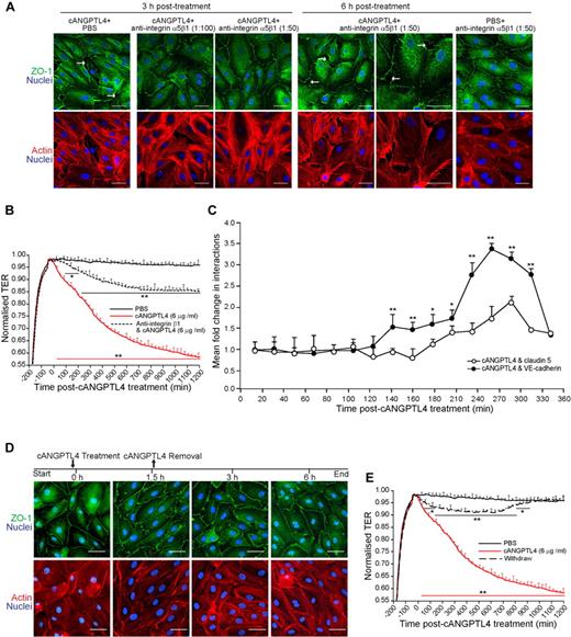
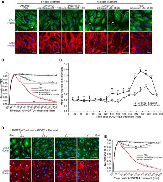
Antibody to integrin α5β1 increased endothelial leakiness but was insufficient to induce microscopic lesions at endothelial junctions.6 Thus, we sought to understand the impact of the upstream cANGPTL4:integrin α5β1 formation on the disruption of endothelial integrity. In the presence of monoclonal anti-integrin α5β1, cANGPTL4 required twice the length of time (6 hours) to induce lesions along the EC–EC contact compared with the cANGPTL4 alone (Figure 4A). Anti-integrin α5β1 blocked the interaction with cANGPTL4 (Figure 2E). ECs treated with anti-integrin α5β1 alone did not induce any lesions, even after 6 hours (Figure 4A). These results were corroborated by our TER data, which showed that the addition of anti-integrin α5β1 and cANGPTL4 (6 μg/mL) to ECs resulted in reduced endothelial permeability compared with ECs treated only with 6 μg/mL cANGPTL4 (Figure 4B). Accordingly, anti-integrin α5β1 caused a concomitant delay of ∼ 90 minutes in the interaction profile of cANGPTL4:VE-cadherin and cANGPTL4:claudin-5 compared with control (compare Figures 3A and 4C). The mean fold change (0.4 ± 0.12) in the interactions between cANGPTL4 and these 2 proteins remained similar, albeit with a nonsignificant decrease of ∼ 10% for VE-cadherin, suggesting that interactions with these 2 proteins are required for endothelial disruption (Figure 4C). Thus, we treated HMVECs with cANGPTL4 for 90 minutes, which allowed for cANGPTL4:integrin α5β1 interactions and initiated endothelial leakiness, followed by its removal. Immunostaining for ZO-1 revealed that cANGPTL4:integrin α5β1 interactions alone is not sufficient to induce endothelial disruption and that the later interactions are necessary for the observed disruption. (Figure 4D). Interestingly, we observed from our TER data that the removal of rh-cANGPTL4 promoted a “reversal effect” on endothelial permeability, conceivably because of the “re-zipping” of the EC–EC contacts, thus reinforcing the important of cANGPTL4:integrin α5β1complex formation in the entire process of endothelial disruption (Figure 4E). Hence, our results suggest that although integrin α5β1 is an essential player, its interaction with cANGPTL4 alone is insufficient to induce an endothelial disruption/lesion. The data suggest that the binding of cANGPTL4 to integrin α5β1 triggers internalization of the complex and “un-zipping” of the endothelial junctions to first promote endothelial leakiness. This facilitates the subsequent cANGPTL4 interactions with the adhesive domains of VE-cadherin and claudin-5 that weaken their homophilic contacts, resulting in their declustering and internalization and finally culminating in the disruption of EC–EC contacts.
Inhibition of cANGPTL4:interacting partner complex formation delays endothelial disruption. (A,B,D,E) Immunofluorescence staining for ZO-1 (A,D) and TER measurement (B,E) in a confluent HMVEC monolayer. Cells were treated with either 6 μg/mL cANGPTL4 in the presence of increasing anti-integrin β1 concentrations (1:100 and 1:50; A-B) or 6 μg/mL cANGPTL4 followed by removal of exogenous cANGPTL4 at 90 minutes (D,E). Treatments were for 3 or 6 hours. HMVECs were counterstained with DAPI (blue) for nuclei and phalloidin (red) for actin stress fibers. Scale bar represents 40 μm. White arrows indicate endothelial junction lesions. Data (means ± SD) from 3 independent experiments. *P < .05; **P < .01. (C) In situ kinetic PLA detection of cANGPTL4: indicated binding partner complexes in cANGPTL4-treated HMVECs in the presence of anti-integrin β1. Values (means ± SD) represent mean fold change in the number of interactions compared with the zero time point, as determined from n = 3 independent experiments (∼ 600 HMVECs) using BlobFinder. Experiments were terminated when microscopic lesions were observed (360 minutes after cANGPTL4 treatment).
Inhibition of cANGPTL4:interacting partner complex formation delays endothelial disruption. (A,B,D,E) Immunofluorescence staining for ZO-1 (A,D) and TER measurement (B,E) in a confluent HMVEC monolayer. Cells were treated with either 6 μg/mL cANGPTL4 in the presence of increasing anti-integrin β1 concentrations (1:100 and 1:50; A-B) or 6 μg/mL cANGPTL4 followed by removal of exogenous cANGPTL4 at 90 minutes (D,E). Treatments were for 3 or 6 hours. HMVECs were counterstained with DAPI (blue) for nuclei and phalloidin (red) for actin stress fibers. Scale bar represents 40 μm. White arrows indicate endothelial junction lesions. Data (means ± SD) from 3 independent experiments. *P < .05; **P < .01. (C) In situ kinetic PLA detection of cANGPTL4: indicated binding partner complexes in cANGPTL4-treated HMVECs in the presence of anti-integrin β1. Values (means ± SD) represent mean fold change in the number of interactions compared with the zero time point, as determined from n = 3 independent experiments (∼ 600 HMVECs) using BlobFinder. Experiments were terminated when microscopic lesions were observed (360 minutes after cANGPTL4 treatment).
cANGPTL4-induced disruption of the endothelium involves integrin-mediated PAK/Rac signaling
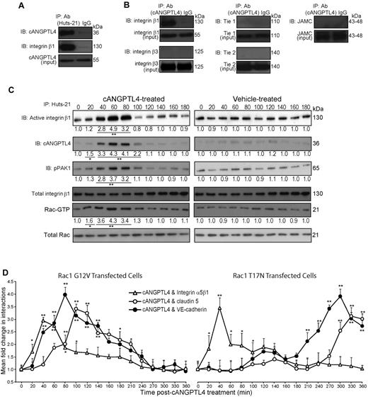
The pivotal role of integrin α5β1 in the process of EC–EC contact disruption lead us to examine the detailed molecular events resulting from the cANGPTL4:intergrin α5β1 complex formation. In vitro coimmunoprecipitation of cANGPTL4 with activated integrin β1 (antibody clone Huts-21) indicated that integrin was activated on association with cANGPTL4 (Figure 5A-B). No interactions were observed between cANGPTL4 and Tie1, Tie2, integrin β3, or the junction protein JAM-C (Figure 5B). Rac-GTP and PAK are downstream mediators of integrin signaling, whose roles in endothelial contractility and barrier function are well recognized.23,24 Our coimmunoprecipitation results showed that ANGPTL4-activated integrin β1 also stimulates the activation of Rac1 and phosphorylation of PAK, compared with PBS-treated ECs (Figure 5C). The increased cANGPTL4:integrin α5β1 interaction and activation of PAK/Rac1 signaling coincided with endothelial leakiness (Figure 1J), possibly by modulating integrin-mediated intercellular adhesion and contractility.23,25 To further underscore the role of Rac1, we transfected ECs with either constitutive active Rac1(G12V) or dominant-negative Rac1(T17N). Transfection of ECs with Rac1(G12V) accelerated the formation of cANGPTL4:VE-cadherin and cANGPTL4:claudin-5 complexes, whereas the expression of Rac1(T17N) delayed the complex delayed the complex formation (compare Figures 3A and 5D). This suggests that cANGPTL4 regulates endothelial integrity via the integrin α5β1-mediated Rac/PAK signaling axis that functions via signaling cross-talk to facilitate cANGPTL4 interactions with VE-cadherin and claudin-5, ultimately leading to vascular disruption.
cANGPTL4-activated integrin β1 mediates vascular permeability via activated Rac/PAK signaling axis. (A-B) Immunodetection of indicated proteins in anti–Huts-21 (A) and anti-cANGPTL4 (B) immunoprecipitates from total protein lysates of rh-cANGPTL4–treated HMVECs (6 μg/mL). IgG immunoprecipitates serves as control. (C) Immunodetection of indicated proteins from anti–Huts-21 immunoprecipitates of HMVECs treated with either vehicle (PBS) or rh-cANGPTL4. Total integrin β1, Rac1, and PAK served as controls. Protein lysates were collected every 20 minutes (0-180 minutes). Values below each band represent the mean fold change in protein expression level from 3 independent experiments compared with the cognate zero time point. *P < .05; **P < .01. (D) In situ kinetic PLA detection of cANGPTL4: indicated binding partner complexes in cANGPTL4-treated HMVECs transfected with either constitutive-active Rac1 G12V or dominant-negative Rac1 T17N. Values (means ± SD) represent mean fold change in the number of interactions compared with the zero time point, as determined from n = 3 independent experiments (∼ 600 HMVECs) using BlobFinder. *P < .05; **P < .01.
cANGPTL4-activated integrin β1 mediates vascular permeability via activated Rac/PAK signaling axis. (A-B) Immunodetection of indicated proteins in anti–Huts-21 (A) and anti-cANGPTL4 (B) immunoprecipitates from total protein lysates of rh-cANGPTL4–treated HMVECs (6 μg/mL). IgG immunoprecipitates serves as control. (C) Immunodetection of indicated proteins from anti–Huts-21 immunoprecipitates of HMVECs treated with either vehicle (PBS) or rh-cANGPTL4. Total integrin β1, Rac1, and PAK served as controls. Protein lysates were collected every 20 minutes (0-180 minutes). Values below each band represent the mean fold change in protein expression level from 3 independent experiments compared with the cognate zero time point. *P < .05; **P < .01. (D) In situ kinetic PLA detection of cANGPTL4: indicated binding partner complexes in cANGPTL4-treated HMVECs transfected with either constitutive-active Rac1 G12V or dominant-negative Rac1 T17N. Values (means ± SD) represent mean fold change in the number of interactions compared with the zero time point, as determined from n = 3 independent experiments (∼ 600 HMVECs) using BlobFinder. *P < .05; **P < .01.
cANGPTL4 modulates the nuclear translocation of β-catenin
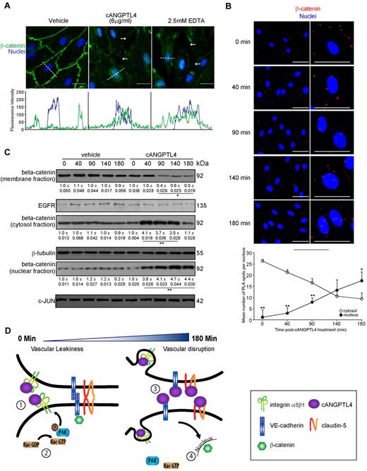
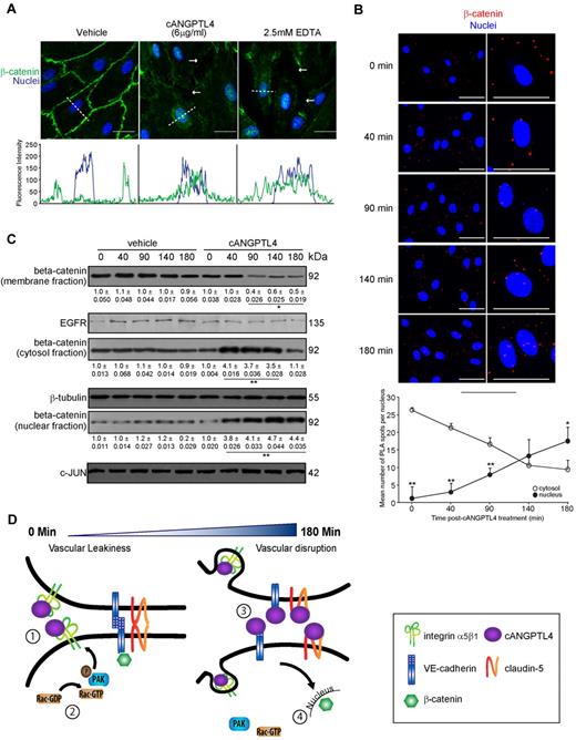
The perturbations and disassembly of VE-cadherin result in the translocation of membrane β-catenin to the nucleus.26,27 Hence, we asked whether cANGPTL4-induced declustering of VE-cadherin and claudin-5 resulted in nuclear translocation of β-catenin. In PBS-treated ECs, β-catenin formed a continuous lining along the cell margin as indicated by the immunofluorescence staining and intensity plot (Figure 6A; supplemental Figure 5A), whereas treatment with either cANGPTL4 (6 μg/mL for 3 hours) or 2.5mM EDTA (positive control) resulted in the disappearance of the β-catenin margin and accumulation in the nuclei, indicating cell–cell junction disruption (Figure 6A; supplemental Figure 5A). To better define the localization of β-catenin, we performed PLA that showed β-catenin in the nuclei at 180 minutes (β-catenin spots/nucleus increases from 1.2 ± 0.81 at 0 minutes to 17.5 ± 2.68 at 180 minutes, whereas those outside the nucleus decreases from 26.3 ± 3.29 minutes to 9.44 ± 3.98 minutes); this timing coincided with the declustering of VE-cadherin and claudin-5 (Figure 6B). Immunoblot analysis corroborated our PLA data (Figure 6C). Taken together, our data indicate that tumor-derived cANGPTL4 is a permeability-inducing factor capable of triggering endothelial disruption via sequential interactions with integrin α5β1, VE-cadherin, and claudin-5, resulting in the accumulation of nuclear β-catenin (Figure 6D).
cANGPTL4:interacting partner complex formation triggers the nuclear translocation of β-catenin. (A) Immunofluorescence staining for β-catenin (green) in an HMVEC monolayer treated with either 6 μg/mL cANGPTL4 or 2.5mM EDTA (as positive control) for 3 hours. HMVECs were counterstained with DAPI (blue) for nuclei. Scale bar represents 40 μm. White arrows indicate endothelial junction lesions associated with reduced β-catenin staining. Representative fluorescence intensity plot of β-catenin and DAPI, indicated by the white dotted line across the nuclei, were quantified using ZEN 2009 software (Carl Zeiss). (B) In situ PLA for β-catenin and quantification of the number of PLA spots per nuclei of cANGPTL4-treated HMVECs at the indicated time points. (0 minutes, 1.2 ± 0.81; 40 minutes, 3 ± 1.23; 90 minutes, 7.9 ± 1.93; 140 minutes, 13.3 ± 2.44; 180 minutes, 17.5 ± 2.68) PLA signals (red) revealed translocation of β-catenin into nuclei at 140 minutes after treatment (∼ 250 HMVECs). Cells were counterstained with Hoechst dye for nuclei (blue). Scale bar represents 40 μm. *P < .05; **P < .01. (C) Immunodetection of β-catenin in membrane, cytosol, and nuclei fractions of HMVECs treated with either vehicle (PBS) or cANGPTL4. Protein lysates were collected at the indicated times. Values (means ± SD) below each band represent the mean fold change in protein expression level compared with the cognate zero time point (n = 3). *P < .05; **P < .01. (D) Schematic diagram of cANGPTL4-mediated disruption of endothelial junctions. (1) cANGPTL4:integrin α5β1 formation (30-50 minutes) coincides with (2) the activation of Rac-GTP and pPAK in ECs (40-60 minutes) and with vascular leakiness (3); the interaction between cANGPTL4 with VE-cadherin and claudin-5 (120-140 minutes) disrupts intercellular contact formation, and stimulates (4) nuclear translocation of β-catenin (180 minutes).
cANGPTL4:interacting partner complex formation triggers the nuclear translocation of β-catenin. (A) Immunofluorescence staining for β-catenin (green) in an HMVEC monolayer treated with either 6 μg/mL cANGPTL4 or 2.5mM EDTA (as positive control) for 3 hours. HMVECs were counterstained with DAPI (blue) for nuclei. Scale bar represents 40 μm. White arrows indicate endothelial junction lesions associated with reduced β-catenin staining. Representative fluorescence intensity plot of β-catenin and DAPI, indicated by the white dotted line across the nuclei, were quantified using ZEN 2009 software (Carl Zeiss). (B) In situ PLA for β-catenin and quantification of the number of PLA spots per nuclei of cANGPTL4-treated HMVECs at the indicated time points. (0 minutes, 1.2 ± 0.81; 40 minutes, 3 ± 1.23; 90 minutes, 7.9 ± 1.93; 140 minutes, 13.3 ± 2.44; 180 minutes, 17.5 ± 2.68) PLA signals (red) revealed translocation of β-catenin into nuclei at 140 minutes after treatment (∼ 250 HMVECs). Cells were counterstained with Hoechst dye for nuclei (blue). Scale bar represents 40 μm. *P < .05; **P < .01. (C) Immunodetection of β-catenin in membrane, cytosol, and nuclei fractions of HMVECs treated with either vehicle (PBS) or cANGPTL4. Protein lysates were collected at the indicated times. Values (means ± SD) below each band represent the mean fold change in protein expression level compared with the cognate zero time point (n = 3). *P < .05; **P < .01. (D) Schematic diagram of cANGPTL4-mediated disruption of endothelial junctions. (1) cANGPTL4:integrin α5β1 formation (30-50 minutes) coincides with (2) the activation of Rac-GTP and pPAK in ECs (40-60 minutes) and with vascular leakiness (3); the interaction between cANGPTL4 with VE-cadherin and claudin-5 (120-140 minutes) disrupts intercellular contact formation, and stimulates (4) nuclear translocation of β-catenin (180 minutes).
cANGPTL4 promotes metastasis to the lungs in vivo
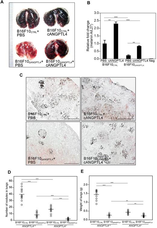
To further investigate the role of cANGPTL4 in the metastatic process and to substantiate its biologic relevance in cancer, we examined its effect on lung metastasis using 2 different mice models. Wild-type C57BL/6J mice (n = 12/group) were injected intravenously with either B16F10CTRL or B16F10ΔANGPTL4 cells (2 × 10⁶) and treated intravenously with either PBS or rh-cANGPTL4. Mice inoculated with B16F10ΔANGPTL4 had significantly fewer lung metastases compared with B16F10CTRL-injected mice. Intravenous administration of rh-cANGPTL4 enhanced lung metastases in both cases, as evidenced by real-time PCR analysis for melanin A, a marker of melanoma (Figure 7A-B). Cross-section staining of the lung tissues corroborated our in vivo data (Figure 7C; supplemental Figure 5B), underscoring the biologic metastatic potential of cANGPTL4 in tumor cells (Figure 7A-B). In addition, we examined lung metastases using wild-type (ANGPTL4+/+) and ANGPTL4-knockout (ANGPTL4−/−) mice injected with either 0.5 × 10⁵ B16F10CTRL or B16F10ΔANGPTL4. There was a significantly reduced number of tumor nodules in the lungs of ANGPTL4−/− compared with ANGPTL4+/+ mice, suggesting that metastasis of B16F10CTRL was impaired (Figure 7D-E). Altogether, our data indicate cANGPTL4 is a prometastatic tumor-derived factor, and they provide new insight into the mechanistic action of this protein in cancer.
cANGPTL4 facilitates metastasis. (A-C) Representative macroscopic images of lungs (A); relative expression of melanin A (B); and representative eosin-stained images of lung sections (C). Black nodules in panel C indicate intravasated melanoma (n = 5). Micrographs of lung sections were taken with MIRAX MIDI (Carl Zeiss) with a Plan-Apochromatic 20×/0.8 objective and MIRAX Scan software. Scale bar: represents 100 μm. (D-E) Number of nodules (D) and weights of lungs (E) from wild-type and ANGPTL4-knockout C57BL/6J mice intravenously injected with either 5 × 10⁵ B16F10CTRL or B16F10ΔANGPTL4 cells (n = 12). **P < .01; ***P < .001.
cANGPTL4 facilitates metastasis. (A-C) Representative macroscopic images of lungs (A); relative expression of melanin A (B); and representative eosin-stained images of lung sections (C). Black nodules in panel C indicate intravasated melanoma (n = 5). Micrographs of lung sections were taken with MIRAX MIDI (Carl Zeiss) with a Plan-Apochromatic 20×/0.8 objective and MIRAX Scan software. Scale bar: represents 100 μm. (D-E) Number of nodules (D) and weights of lungs (E) from wild-type and ANGPTL4-knockout C57BL/6J mice intravenously injected with either 5 × 10⁵ B16F10CTRL or B16F10ΔANGPTL4 cells (n = 12). **P < .01; ***P < .001.
Discussion
Metastasis requires dynamic tumor–endothelium communication through tumor-secreted vascular permeability factors to breach the endothelial barrier, thereby allowing the migration and invasiveness of the tumor cells across the endothelium. Hence, a better understanding of the mechanism by which such vascular permeability factors disrupt endothelial integrity could offer new therapeutic strategies. Recent studies have implicated a role for ANGPTL4 in metastasis,10 yet the mechanism by which ANGPTL4 modulates endothelial junction integrity remains unknown. Here, we provide insight into the heterotypic role of cANGPTL4 in vascular integrity. We show that cANGPTL4 disrupts intercellular adhesion via a novel integrin α5β1-mediated Rac/PAK signaling pathway and the declustering and internalization of VE-cadherin and claudin-5, resulting in nuclear accumulation of β-catenin. Using 2 different mouse models, our in vivo vascular permeability and metastatic assays confirmed that cANGPTL4 acts as a vascular permeability factor by inducing vascular leakiness and disruption. Interestingly, ANGPTL4-deficient tumors also showed decreased vascularization, suggesting that cANGPTL4 may facilitate metastasis through increased vascular permeability and volume.
EC–EC adhesion is mediated mainly by the α5β1 integrin and EC-specific adhesion complexes containing VE-cadherin and claudin-5.6,28 Our work shows that cANGPTL4 interacts with these 3 adhesive complexes in a sequential manner to disrupt the endothelial junctions. A major route by which integrins regulate cell–cell contacts is the Rac/PAK pathway, leading to modulation of EC contractility.23,29,30 We show that the initial interaction between cANGPTL4 and integrin α5β1 activates Rac and phosphorylates PAK, weakening cell–cell contacts and increasing endothelial leakiness. This facilitates cANGPTL4 access to the adhesive domains of VE-cadherin and claudin-5 to induce declustering of the adhesion molecules resulting in vascular disruption. We and others have shown that ANGPTL4 does not bind the angiopoietin receptor Tie1 or Tie2 to antagonize its action. Angiopoietin-1–mediated activation of Rac reduces vascular leak in the context of VEGF-induced vascular permeability. Thus, the role of Rac in vascular permeability is complex. The ligand and the cellular context that activate Rac will dictate its effect on vascular junction integrity.
The activation of Rac/PAK signaling trigger by cANGPTL4-bound integrin β1 is necessary to facilitate cANGPTL4 access to VE-cadherin and claudin-5 and to trigger vascular disruption. By preventing the association of cANGPTL4 with integrin β1 by anti-integrin β1, we observed delayed cANGPTL4:VE-cadherin and cANGPTL4:claudin-5 complex formation and a concomitant decreased vascular permeability. A similar delay in cANGPTL4:VE-cadherin and cANGPTL4:claudin-5 complex formation was detected in ECs expressing dominant-negative Rac1, without affecting the cANGPTL4:integrin β1 formation. This indicates that the Rac/PAK signaling acts downstream of integrin and facilitates cANGPTL4 interactions with VE-cadherin and claudin-5. Interestingly, by preventing the subsequent cANGPTL4 interaction with VE-cadherin and claudin-5, the initial increased in vascular permeability because of the activation of integrin-mediated Rac/PAK signaling did not propagated to observable vascular lesions. This confirms that Rac/PAK signaling is necessary to potentiate the vascular disruptive effect of cANGPTL4.
The interactions of cANGPTL4 with the VE-cadherin ED1 and claudin-5 EDC1 domains disrupt VE-cadherin and claudin-5 clustering. The EC intercellular clustering of VE-cadherin and claudin-5, mediated by their extracellular domains, is important for the maintenance of a semipermeable boundary between the bloodstream and the tumor.20,21,31 Our results also reveal a time-dependent cellular trafficking of β-catenin from the membrane to the nucleus in cANGPTL4-treated ECs, coinciding with endothelial disruption. The interactions between the cytoplasmic tail of VE-cadherin and the catenins are essential for the proper functioning of stable AJs.27 The nuclear accumulation of β-catenin in leaky endothelium and release of β-catenin from AJ junctions into the nucleus is consistent with the declustering of AJs.26,27,32 Moreover, the membrane-liberated β-catenin induces gene transcription that contributes to angiogenic responses of ECs after episodes of vascular leakage that promotes tumor metastasis.26,27,33
The role of ANGPTL4 in vascular leakiness, and thus metastasis, is controversial,10,12,34 and efforts to confirm it have been further hampered by the lack of understanding of the mechanistic action of ANGPTL4. We show that cANGPTL4 behaves as a vascular permeability factor and disrupts endothelial integrity via integrin α5β1-mediated Rac/PAK signaling and the declustering and internalization of VE-cadherin and claudin-5. The reason for the apparent discrepancy is unclear, but it may be because of the position of fusion tags in recombinant ANGPTL4. Studies that suggest an inhibitory role of ANGPTL4 in vascular leakiness have used a C-terminal fusion ANGPTL4, whereas our work and that of others who have presented evidence that ANGPTL4 is a prometastatic agent that induces endothelial permeability has used either an N-terminal fusion ANGPTL4 or untagged ANGPTL4. A dysfunctional endothelium usually presents as a leaky or disrupted vasculature and is characteristic of many pathologic conditions, including inflammatory edema, diabetes, atherosclerosis, and cancer metastasis.32,35 The disruption of EC–EC contacts is a major route exploited by primary tumors to promote metastasis. Our findings suggest that targeting cANGPTL4 to treat metastasis and vascular-related diseases could be a viable option.
The online version of the article contains a data supplement.
The publication costs of this article were defrayed in part by page charge payment. Therefore, and solely to indicate this fact, this article is hereby marked “advertisement” in accordance with 18 USC section 1734.
Acknowledgments
This work was supported by grants from the Ministry of Education, Singapore (ARC18/08); Nanyang Technological University, Singapore (RG127/05, RG82/07); and the Biomedical Research Council (10/1/22/19/644).
Authorship
Contribution: R.-L.H and Z.T. performed experiments, analyzed the results, and wrote the article; H.C.C., P.Z., M.J.T. C.K.T., C.R.I.L., and M.K.S. performed experiments; D.T.W.L. and S.M.T. performed experiments and contributed to discussion; S.K., J.L.D., and H.Y.L. analyzed the results, contributed to discussion, and edited the article; and N.S.T. analyzed the results, contributed to discussion, and reviewed and edited the article.
Conflict-of-interest disclosure: The authors declare no competing financial interests.
Correspondence: Nguan Soon Tan, School of Biological Sciences, Nanyang Technological University, 60 Nanyang Dr, Singapore 637551; e-mail: nstan@ntu.edu.sg.
References
Author notes
R.-L.H. and Z.T. contributed equally to this article.

