Abstract
Abstract 2587
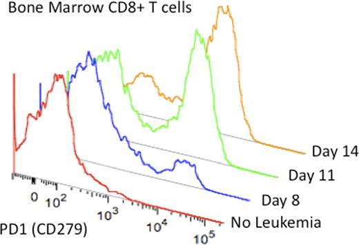
Acute lymphoblastic leukemia (ALL) is the most common pediatric malignancy and, despite tremendous success in therapy over the past 3 decades, remains a primary cause of cancer-related mortality in children. Enthusiasm for the use cellular immunotherapy for ALL has been tempered by the poor response to donor lymphocyte infusions following allogeneic hematopoietic stem cell transplantation. However, ALL blasts are susceptible to T cell and NK cell mediated lysis in vitro suggesting that poor response to in vivo immune interventions may be due to events occurring during the priming of the immune response. Using a murine model of precursor B cell ALL we examined the impact of leukemia progression on T cells in vivo. Methods: We developed a transplantable syngeneic model of pediatric ALL derived from transgeneic mice expressing human E2aPBX1, a recurring translocation present in 5% of pediatric leukemia (Bijl et al, Genes and Development, 2005). This murine line displays a precursor B cell phenotype and results in 100% lethality following injection of 100,000 cells (Qin et al, ASH, 2010). Using congenic (CD45.1) B6 recipients, we tracked the early progression of ALL in vivo and examined the T cells in the leukemia-containing compartments by flow cytometry and PCR. Results: Using congenic markers, ALL cells can be detected in bone marrow as early as 3 days following intravenous injection of 1,000,000 cells with a sensitivity of 0.01%. Spleen and lymph node involvement was seen later (10 days) followed by the detection of circulating blasts by 2 weeks. E2aPBX1 cells express variable levels of costimulatory molecules in vitro with no change in expression during in vivo progression. Notably, PDL1 and PDL2 are expressed both in vitro and in vivo at higher levels than on non-malignant precursor B cells in leukemia-bearing mice. Remarkably, although PD1+ T cells are not seen in the bone marrow of non-leukemia-bearing mice, PD1 expression on bone marrow T cells was markedly increased during progression such that 60–80% of all bone marrow CD4 and CD8 T cells were positive by 2 weeks following leukemia injection (figure). In addition to expression of PD1, these T cells also co-expressed Tim3, a phenotype associated with T cell exhaustion. Blockade of PD1 or PDL1 starting 3 days following leukemia injection had no impact on leukemia progression. However, combining PD1 blockade with the adoptive transfer of T cells from leukemia-primed donors resulted in improved survival compared to primed T cells alone (p=0.0004). Conclusions: Early progression of ALL results in the induction of PD1 and Tim3 on T cells in vivo. Combination of PD1 blockade plus adoptive T cell therapy results in therapeutic benefit suggesting that this axis may be an attractive target in ALL.
No relevant conflicts of interest to declare.
Author notes
Asterisk with author names denotes non-ASH members.


