Abstract
Abstract 4001
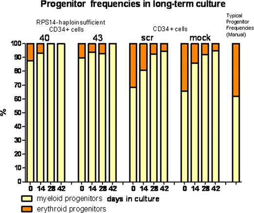
By means of a large RNA interference screen, Ebert et al. identified RPS14 as the target of del(5q), since haploinsufficiency of RPS14 in human hematopoietic stem cells (HSC) induces an erythroid differentiation defect, a characteristic feature of myelodysplastic syndromes (MDS) with isolated deletion in 5q (Nature451, 335–339 (2008)). However, to the best of our knowledge, these cells have not been observed for more than two weeks in culture. We have shown that telomere shortening and chromosomal instability play a crucial role, particularly during progression of MDS with del(5q) into acute myeloid leukemia (AML) (K Lange et al, Genes Chromosomes Cancer49, 260–269 (2010) and G Göhring et al, Leukemia, 2011 (in press)). Because of the limited proliferation of primary MDS cells in culture and because there is no appropriate MDS cell line, a good human cell culture model is urgently needed to investigate the relationship of telomere attrition and chromosomal instability during the progression of MDS with del(5q).
Therefore, it was our aim was to create a long-term culture (LTC) model to observe the effects of RPS14 haploinsufficiency on differentiation capacity, DNA repair, telomere shortening and chromosomal instability in human HSCs.
CD34+ cells were isolated from umbilical cord blood via magnetic cell separation. Knockdown of RPS14 to a level of 40 – 50% was performed via lentiviral transduction of shRNA vectors. As controls, in addition to untransduced mock cells, CD34+ cells were transduced with scrambled shRNA. LTC was performed on murine feeder layers (M2-10B4).
In short-term culture, as expected, knockdown of RPS14 led to an erythroid differentiation defect, decreased proliferative activity and an increased level of apoptosis. Cultivation on murine feeder layers enabled expansion of CD34+ cells for more than 6 weeks.
Generally, within 6 weeks RPS14 -deficient CD34+ cells showed a poorer proliferation than the control cells with a 214-fold versus a 6080-fold multiplication, respectively. Median numbers of γH2AX foci, indicators of DNA double-strand breaks, did not differ between RPS14-deficient and control cells with a median number of 9.4 and 8.2 foci/cell, respectively. Induction of γH2AC foci via Mitomycin C, a DNA cross-linking agent inducing double-strand breaks, did not reveal an altered DNA repair capacity with 14.3 and 11.6 foci/cell.
Telomere length decreased in RPS14- deficient cells as well as in control cells. Initially, telomeres showed a median length of about 12.5 kb, decreasing to a median length of 10.3 kb (range 8.4 – 11.4 kb) and 5.8 kb (range 5.7 – 8.9 kb) after 2 and 4 weeks, respectively, which afterwards elongated to a level of 8.7 kb (range 8.5 –8.9 kb) after 6 weeks. The mechanism underlying this primary shortening and subsequent re-elongation might be due to an up-regulation of telomerase or of the alternative lengthening of telomeres (ALT) mechanism. Cytogenetic investigations demonstrated an increase in chromosomal breakage in all cultures, pointing towards induction during LTC rather than due to RPS14 deficiency. Remarkably, chromosomal breakage and chromosome aberrations in single cells within the first 4 weeks of cultivation of hematopoietic stem cells transduced with scrambled shRNA was followed by clonal dominance of monosomy 7 after 6 weeks. This may be an effect caused by insertional mutagenesis. LM-PCR is currently under way to obtain insights into the affected insertion site(s). Monosomy 7 is one of the most frequent chromosome aberrations in MDS and frequently occurs as an additional aberration in MDS/AML with inversion 3q generating an MDS-EVI1 fusion transcript. Thus, it will be interesting to see whether monosomy 7 in our LTC model cooperates with EVI1 activation as in sporadic MDS and AML.
In conclusion, a LTC model on feeder layer cells seems to be an appropriate system to analyze hematopoietic stem cells and MDS in culture. However, cell culture artefacts inducing telomere shortening and chromosomal instability or insertional mutagenesis have to be taken into account and regular cytogenetic analyses should always be performed.
No relevant conflicts of interest to declare.
Author notes
Asterisk with author names denotes non-ASH members.


