Abstract
Abstract 4155
PET scan is increasingly used in the follow-up of lymphoma patients given allogeneic hematopoietic cell transplantation (Allo-HCT). However, whereas several studies addressed the question of the impact of PET positivity after autologous transplantation on transplantation outcomes, very few have been performed after allo-HCT. This is the aim of the current retrospective study.
We retrospectively analyzed data from 50 lymphoma patients who underwent an allo-HSCT after non-myeloablative conditioning between March 2000 and March 2009. The diagnoses were Hodgkin's lymphoma (n=8) and non-Hodgkin's lymphoma (n=42; 14 follicular, 11 mantle cell, 9 diffuse large B cell, 2 MALT, 1 Burkitt, 1 lymphocytic and 4 T-cell lymphomas). Patients were scheduled to benefit from a follow-up by PET scan on days 100, 180 and 365 and then yearly for a total of five years.
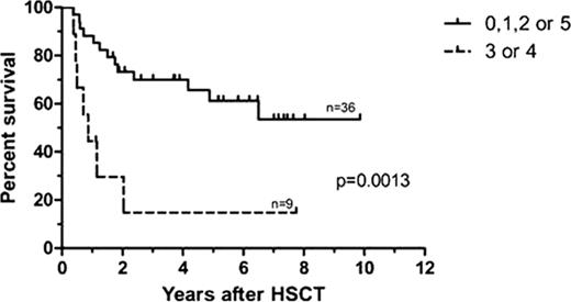
Day 100 PET scans were not performed in 5/50 patients (4 patients died before day 100, while another onewas in intensive care unit at that time).Among the remaining 45 patients, 20 (44.4%) presented hypermetabolic lesions, including 9 patients(20%) who had hypermetabolic lesions evocative of lymphoma.One-year OS (Figure 1) was higher in patients whose PET scan was negative or positivefor infectious/inflammatory reasons than for those with typical lymphoma lesions (85% vs 44%, p=0.0013). Among patients with day-100 PET positivity evocative of lymphoma, 7 patients died, 5 of them of their lymphoma, while 2 patients remained alive.
Overall survival according to the first PET scan on day 100 (0=complete response, 1= inflammatory/infectious, 2= probably inflammatory/infectious, 3=residual disease, 4=progression or development of new lymphomatous lesion, 5=suspicion of other neoplasia)
Overall survival according to the first PET scan on day 100 (0=complete response, 1= inflammatory/infectious, 2= probably inflammatory/infectious, 3=residual disease, 4=progression or development of new lymphomatous lesion, 5=suspicion of other neoplasia)
During further follow-up, twenty patients (44.4%) never presented hypermetabolic lesions after transplantation and 25 (55.6%) had at least one abnormal PET scan.
Among the 25 patients, only 11 (24.5%) had probable/proven neoplasia: 1 died with residual disease, 2 had residual lymphoma that went into remission after GVHR, 5 had biopsy-proven relapse, 1 had non-biopsy proven progression, 1 had lung cancer and 1 lung PTLD. The other 14 patients (31.1%) had suspicious lesions at one of the follow-up PET scans, but after further work-up, none of these lesions proved to be a relapse, and all disappeared afterwards. Biopsies were performed in 6 of these cases, including 2 lymph node (1 normal and 1 lymphoid hyperplasia), 2 lung (1 normal and 1 aspergillosis) and 2 GI (1 normal and 1GVHD) biopsies. For 6 patients, imaging studies (CT scan, MRI or echography) were normal or demonstrated infectious or inflammatory (including gut GVHD) disorders. The last 2 patients were thought to relapse based on both PET and CT scans, but refused biopsies, but their lesions regressed spontaneously.
In our study, a positive PET scan at day 100 after transplant is predictive of poorer OS. However, there is a noteworthyincidence of false-positive PET scans after non-myeloablativeallo-HCT. We therefore recommend that every suspicious lesion,and particularly in areas not previously involved by lymphoma, should be explored at least by CT scan and/or biopsy, before initiating any new treatment.
No relevant conflicts of interest to declare.
Author notes
Asterisk with author names denotes non-ASH members.


