Abstract
Abstract 1202
Transducin-like/Enhancer of Split 4 (TLE4), a homolog of the master developmental regulator in Drosophila, Groucho, is a co repressor previously described by our group as the putative tumor suppressor lost in a subset of t(8:21) AML patients also harboring 9q deletion. TLE4 has been found to be of fundamental importance in the repressive function of multiple highly conserved transcriptional complexes, including Hes1, Tcf/Lef, CSL, Runx1-3, and Pax5, all of which have been shown to be intimately connected to normal steady state hematopoiesis and deregulated in malignancy. Owing to its deletion in leukemia and its ability to confer functionality to a variety of signaling networks, our lab sought to investigate its role in hematopoiesis by generating a murine knockout model for Tle4.
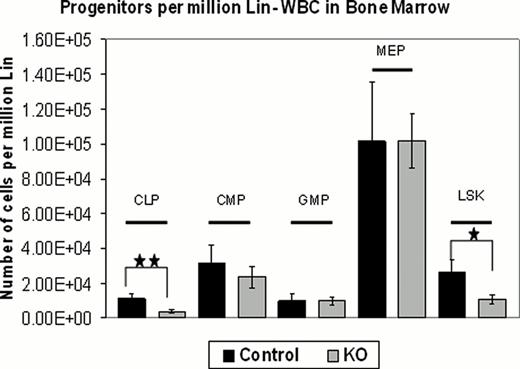
While normal at birth, within 2–3 weeks of age knockout (KO) animals become runted and eventually moribund by the time of weaning. Gross examination of the spleen and thymus in 3 week old animals reveals significant atrophy of these organs with severe abnormalities in normal tissue architecture. Tle4−/− mice lack clear demarcations between medullary and cortical regions of the thymus, in addition to having a significant increase in apoptosis seen via TUNEL staining. FACS analysis of the thymus shows CD4/CD8 double positive cells are heavily reduced in KOs, with an increase in both single positive mature populations. Additionally, there is a partial, but not complete, block in T cell progenitor development through TCR rearrangement (DN2-DN3 stages). Structural defects are seen in the spleen as well, with a failure in the formation of follicular zones. B cells are significantly reduced with an increase in CD11b+ cells in the spleen, in addition to a shift in the percentages of B1, B2, and marginal zone B cells in some animals. Histological examination of the long bones reveals dramatic abnormalities, both in hematopoietic and bone lineages. Tle4−/− mice exhibit a reduction in calcified bone, a dysplastic and reduced hypertrophic zone, and reabsorption of the trabecular region. These mice also have a gradual clearing of the marrow itself, with 2 week old animals first showing evidence of cytopenia that then progresses to full marrow failure by 3–3.5 weeks of age. The bone marrow exhibits a marked leukocytopenia, with a clear B lymphocytopenia as in the spleen. Furthermore, within the bone marrow, there is an increase in the number of CD4 and decrease in the number of CD8 T cells. ProB staging of developing B cells also trends towards a partial arrest in the transitions from the A through C fractions. When analyzed for progenitor populations, KO mice show severe reductions in the number of common lymphoid progenitors, while myeloid progenitors show no particular shifts in population frequency. Finally, there is a significant decrease in HSCs, defined as lin-Sca1+Ckit++ (LSK).
In order to assess whether the hematological phenotype observed in these mice are of a cell intrinsic or extrinsic nature, transplants were performed using embryonic day 13.5 fetal liver and 2 week old bone marrow. Analysis of recipient animals 16 weeks after transplant revealed leukocytopenia with a specific reduction in B cells in the blood, however the T cell and LSK defects seen in donor animals was not observed. Furthermore, when LSK CD34+ Flt3+ or – cells from KO or WT 2 week old animals were plated on OP9 cells over-expressing Deltex1 or GFP, there were no defects in T, B, or CD11b+ cell production.
The unique spectrum of phenotypic consequences concomitant to Tle4 deletion argues for a complex yet fundamental role in the maintenance of the hematopoietic niche. As illustrated by the ability to recapitulate some yet not all of the KO hematological aberrations in transplants, it appears that Tle4's involvement in lineage commitment is heterotypic in nature, an unsurprising finding considering its direct involvement in multiple highly conserved signaling pathways. Considering its principal role in those pathways, continued efforts to investigate the role the TLE family of corepressors play in the bone marrow niche may prove vital in reworking our paradigms for both normal and malignant hematopoiesis.
Tle4 deletion causes alterations in LSK and common lymphoid progenitor frequencies. (Controls n=7, KO n=8; *p<.032, **p<.01).
Tle4 deletion causes alterations in LSK and common lymphoid progenitor frequencies. (Controls n=7, KO n=8; *p<.032, **p<.01).
Representative Flow plots illustrating progenitor and LSK defects in Tle4−/− mice.
Representative Flow plots illustrating progenitor and LSK defects in Tle4−/− mice.
No relevant conflicts of interest to declare.
Author notes
Asterisk with author names denotes non-ASH members.


