Abstract
Although chronic myeloid leukemia (CML) originates from a primitive hematopoietic stem cell (HSC), it is the more differentiated progenitor cells that drive the expansion of the malignant clone. In addition, previous studies of chronic phase CML have shown that, despite the marked leukocytosis observed here, megakaryocyte-erythroid progenitors dominate the progenitor fraction. We sought to elucidate this by employing the new marker for leukemic stem cells, the human myeloid inhibitory C-type lectin-like receptor (hMICL), in the study of progenitor cell expansion in CML.
Bone marrow or peripheral blood stem cells were acquired from 11 normal donors and 31 CML patients at diagnosis in chronic phase and/or after 3-119 months of tyrosine kinase inhibitor (TKI) treatment. Cells were stained with fluorescent monoclonal antibodies and FACS sorted into HSCs (CD34+CD38-), hMICL+ progenitors (MpP; CD34+CD38+hMICL+), and hMICL- progenitors (MnP; CD34+CD38+hMICL-). Sorted cell subsets were subjected to growth in a 14-day methylcellulose assay and analyzed quantitatively for expression of the BCR-ABL fusion transcript.
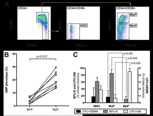
Immunological and functional properties of MpPs in normal donors. (A) Identification of MpPs, MnPs, and HSCs within the CD34+ compartment. (B) Cells with GMP phenotype (CD34+CD38+CD123lowCD45RA+) in MpP and MnP subsets. (C) Colony growth of bone marrow mononuclear cells (MNC) and sorted MpPs and MnPs in a 14-day methylcellulose assay. Error bars denote SDs.
Immunological and functional properties of MpPs in normal donors. (A) Identification of MpPs, MnPs, and HSCs within the CD34+ compartment. (B) Cells with GMP phenotype (CD34+CD38+CD123lowCD45RA+) in MpP and MnP subsets. (C) Colony growth of bone marrow mononuclear cells (MNC) and sorted MpPs and MnPs in a 14-day methylcellulose assay. Error bars denote SDs.
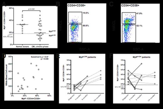
Human MICL expression in chronic phase CML patients. (A) Fraction of MpPs in normal donors and CML patients at diagnosis. (B) Typical immunological profiles of MpPLOW patients and (C) MpPHIGH patients. (D) Correlation between MpP fraction size and total white blood cell count at the time of diagnosis. (E) Development of MpP fraction size in individual patients after 3-6 months (solid lines) and after 12-119 months (dotted lines) of TKI treatment in MpPLOW patients and (F) MpPHIGH patients.
Human MICL expression in chronic phase CML patients. (A) Fraction of MpPs in normal donors and CML patients at diagnosis. (B) Typical immunological profiles of MpPLOW patients and (C) MpPHIGH patients. (D) Correlation between MpP fraction size and total white blood cell count at the time of diagnosis. (E) Development of MpP fraction size in individual patients after 3-6 months (solid lines) and after 12-119 months (dotted lines) of TKI treatment in MpPLOW patients and (F) MpPHIGH patients.
During the first 6 months of TKI treatment differing developments in MpP fraction size were observed for MpPLOW and MpPHIGH patients. While MpPLOW patients showed increasing MpP fractions during the first 6 months of treatment (fig 2E), 4/4 and 2/4 MpPHIGH patients displayed a decrease in MpPs at 3 and 6 months, respectively (fig 2F). Thus, in these patients, the majority of the Ph+ progenitor cells being cleared seemed to be GMPs.
In conclusion, our data demonstrate that hMICL is an early marker of granulocyte-macrophage differentiation, and provides a readily accessible approach to assessing the GMP population during TKI therapy in CML. Using the present approach we have uncovered a higher degree of variability in the composition of the progenitor compartment at diagnosis than previously reported, and shown that a significant proportion of the patients have expanded GMP populations. Ongoing studies are aimed at determining whether these patients may represent patients with a more advanced form of disease at the time of diagnosis.
Stentoft:Novartis: Consultancy, Financial support for relevant congress participation Other, Membership on an entity’s Board of Directors or advisory committees, Research Funding; Bristol-Myers-Squibb: Membership on an entity’s Board of Directors or advisory committees, Research Funding; Danish Regions: Membership on an entity’s Board of Directors or advisory committees. Hokland:Novartis: Research Funding.
Author notes
Asterisk with author names denotes non-ASH members.


