Key Points
The density of the interstitial collagen network increases in inflamed tissue, providing physical guidance to infiltrating neutrophils.
Neutrophil interstitial migration does not require the pericellular degradation of collagen fibers, but it is modulated by MMPs.
Abstract
In vitro studies suggest that leukocytes locomote in an ameboid fashion independently of pericellular proteolysis. Whether this motility pattern applies for leukocyte migration in inflamed tissue is still unknown. In vivo microscopy on the inflamed mouse cremaster muscle revealed that blockade of serine proteases or of matrix metalloproteinases (MMPs) significantly reduces intravascular accumulation and transmigration of neutrophils. Using a novel in vivo chemotaxis assay, perivenular microinjection of inflammatory mediators induced directional interstitial migration of neutrophils. Blockade of actin polymerization, but not of actomyosin contraction abolished neutrophil interstitial locomotion. Multiphoton laser scanning in vivo microscopy showed that the density of the interstitial collagen network increases in inflamed tissue, thereby providing physical guidance to infiltrating neutrophils. Although neutrophils locomote through the interstitium without pericellular collagen degradation, inhibition of MMPs, but not of serine proteases, diminished their polarization and interstitial locomotion. In this context, blockade of MMPs was found to modulate expression of adhesion/signaling molecules on neutrophils. Collectively, our data indicate that serine proteases are critical for neutrophil extravasation, whereas these enzymes are dispensable for neutrophil extravascular locomotion. By contrast, neutrophil interstitial migration strictly relies on actin polymerization and does not require the pericellular degradation of collagen fibers but is modulated by MMPs.
Introduction
Directed migration of leukocytes to the site of inflammation is a key event in the inflammatory response. In the last decades, the mechanisms regulating the initial steps of this process including intravascular rolling and adhesion as well as transendothelial migration of leukocytes have been studied in detail.1-4 After having passed the endothelial cell layer, the perivenular basement membrane, and the pericyte sheath, transmigrated leukocytes are thought to locomote through the interstitial tissue in an “ameboid” fashion, being largely independent of focal adhesions and pericellular proteolysis.4,5 The exact mechanisms underlying leukocyte migration within the inflamed interstitial tissue, however, are still unknown.
While multiphoton in vivo microscopy enabled investigations on lymphocyte and dendritic cell migration in lymphatic tissue,6-8 the majority of previously published studies on interstitial migration of inflammatory cells is based on in vitro studies in 2- and 3-dimensional systems or ex vivo assays. In these model systems, the complex architecture of the interstitial tissue, including its structural changes during inflammatory conditions9 as well as the significant phenotypic and functional alterations that leukocytes undergo during their extravasation,4,10 can hardly be mimicked. In this regard, the role of proteases in particular for leukocyte migration remains an issue of controversial debate.11-18
Over one third of all known proteolytic enzymes are serine proteases, which are characterized by the ability to hydrolyze the peptide bond of their substrates via a nucleophilic serine residue in the active site. Among these enzymes, plasmin is one of the most prominent members regarding its elementary function in the fibrinolytic system.19 In addition, there is increasing evidence that this protease is critically involved in various other physiological and pathophysiological processes such as the induction of receptor-mediated intracellular signaling pathways ultimately controlling cell adhesion and migration.20,21 Moreover, plasmin is able to activate various matrix metalloproteinases (MMPs), a class of proteolytic enzymes specialized on the degradation of the extracellular matrix, which is also supposed to contribute to the interstitial locomotion of cells.22 Using genetically altered mice or specific inhibitors, serine proteases, including plasmin14-16 as well as MMPs,12,17,18 have previously been implicated in leukocyte extravasation to the perivascular tissue by regulating interactions of these inflammatory cells with endothelial cells and the perivascular basement membrane. The role of these proteolytic enzymes for the subsequent migration of leukocytes through the inflamed interstitium remained largely unclear.
Here, we demonstrate that the density of the interstitial collagen network significantly increases upon onset of inflammation. These inflammatory remodeling events provide physical guidance to infiltrating neutrophils and allow these inflammatory cells to efficiently migrate through the interstitium. In this context, we show that directional interstitial locomotion of transmigrated neutrophils is strictly dependent on actin polymerization, but only marginally on actomyosin contraction, and does not require pericellular proteolysis. Although serine proteases including plasmin are critical for leukocyte extravasation, these proteolytic enzymes do not contribute to the extravascular motility of leukocytes. In contrast, MMPs promote directional interstitial migration of neutrophils through the interstitial tissue by regulating the surface expression of l-selectin and integrins on these inflammatory cells, corroborating these proteolytic enzymes as modulators of ameboidlike leukocyte motility.
Methods
Animals
Male C57BL/6 mice were purchased from Charles River (Sulzfeld, Germany). Male LysM-eGFP mice23 and CX3CR-1GFP/+ mice24 were generated as described previously and backcrossed on the C57BL/6 background for 6 to 10 generations. All experiments were performed using mice at the age of 10 to 12 weeks. Animals were housed under conventional conditions with free access to food and water. The experiments were performed according to German legislation for the protection of animals and approved by the local government authorities.
Analysis of leukocyte extravasation using the musculus cremaster assay
Surgical preparation.
Surgical preparation of the cremaster muscle was performed as originally described by Baez with minor modifications.25 Mice were anesthetized using 100 mg/kg ketamine and 10 mg/kg xylazine, administrated by intraperitoneal injection.
In vivo microscopy.
The setup for in vivo microscopy was centered around an Olympus BX 50 upright microscope (Olympus Microscopy, Hamburg, Germany), equipped for stroboscopic fluorescence epi-illumination microscopy as described previously.26
Experimental protocol.
Leukocyte recruitment to the cremaster muscle was induced by intrascrotal injection of platelet-activating factor (PAF) or CC chemokine ligand (CCL) 3/macrophage-inflammatory protein (MIP)–1α. After 3 hours, 5 vessel segments were randomly chosen, as described previously.26
Analysis of leukocyte interstitial migration using the musculus cremaster assay
In vivo microscopy.
The setup for in vivo microscopy was centered around an AxioTech-Vario 100 Microscope (Zeiss MicroImaging GmbH, Göttingen, Germany), equipped with a Colibiri LED light source (Zeiss MicroImaging GmbH) for fluorescence epi-illumination microscopy as described previously.26
Quantification of leukocyte migration parameters.
In vivo microscopy records were analyzed offline using the imaging software ImageJ (National Institutes of Health, Bethesda, MD) as described earlier.26
Experimental protocol.
Directional interstitial migration of leukocytes was induced in the cremaster muscle after perivenular microinjection (100 µm distance to the vessel under investigation) of 130 picoliters of PAF (100 nM) or CCL3/MIP-1α (250 nM). Microinjection was performed under visual control using a borosilicate micropipette connected to the injection system involving a semiautomatic micromanipulator (InjectMan NI 2, Eppendorf, Hamburg, Germany) and a microinjector (FemtoJet, Eppendorf, Hamburg, Germany) as described previously.27
Experimental groups.
In a first set of experiments, leukocyte migration to the peritoneal cavity was analyzed in control mice with an intraperitoneal injection of phosphate-buffered saline (PBS) as well as in mice receiving either tranexamic acid (TXA), ε-aminocaproic acid (EACA), the broad-spectrum serine protease inhibitor aprotinin, the broad-spectrum MMP inhibitor GM-6001, or drug vehicle undergoing stimulation with PAF or CCL3/MIP-1α (n = 4 each group).
In a second set of experiments, the single steps of the leukocyte extravasation process were analyzed in the cremaster muscle of control mice with an intrascrotal injection of PBS as well as of mice receiving TXA, EACA, aprotinin, GM-6001, or drug vehicle undergoing intrascrotal stimulation with PAF or CCL3/MIP-1α (n = 4 each group).
In a final set of experiments, directional interstitial migration of leukocytes was induced in the mouse cremaster muscle by perivenular microinjection of PAF or CCL3 and analyzed upon treatment with latrunculin B, blebbistatin, TXA, EACA, aprotinin, GM-6001, or corresponding drug vehicle (n = 4 each group).
Reagents.
The following inhibitors were used: aprotinin (100 000 KIU kg−1 intra-arterially [i.a.]; 5 minutes prior to onset of inflammation as a bolus and then as continuous intra-arterial infusion/superfusion of the cremaster muscle 100 000 KIU kg−1 h−1; Sigma-Aldrich, Deisenhofen, Germany) is a broad-spectrum serine-protease inhibitor. EACA (100 mg kg−1 i.a. 5 minutes prior to onset of inflammation as a bolus and then as continuous intra-arterial infusion/superfusion of the cremaster muscle 100 mg kg−1 h−1; Sigma-Aldrich) is a plasmin inhibitor; TXA (100 mg kg−1 i.a. 5 minutes prior to onset of inflammation as a bolus and then as continuous intra-arterial infusion/superfusion of the cremaster muscle 100 mg kg−1 h−1; Sigma-Aldrich) is a plasmin inhibitor. Latrunculin B (500 nM solution; Sigma Aldrich; as continuous superfusion of the cremaster muscle) is an inhibitor of actin-polymerization. Blebbistatin (500 nM solution; Sigma-Aldrich; as continuous superfusion of the cremaster muscle) is a selective inhibitor of nonmuscle myosin II. Control animals received equivalent volumes of corresponding drug vehicles.
Multiphoton in vivo microscopy
For in vivo 2-photon imaging, mice were anesthetized, and ears were fixed on a custom-built stage maintaining a physiologic temperature. Images were acquired with a TrimScope (LaVision Biotech, Goettingen, Germany) connected to an upright microscope with a 20× water immersion objective (Olympus America, Center Valley, PA).
Statistics
Data analysis was performed with a statistical software package (SigmaStat for Windows; Jandel Scientific, Erkrath, Germany). After testing normality of data (using the Shapiro–Wilk test), the 1-way analysis of variance test followed by the Dunnett (>2 groups) or the t test (2 groups) was used for the estimation of stochastic probability in intergroup comparisons. Mean values and standard errors of the means (SEMs) are given. P values < .05 were considered significant.
Results
Effect of aprotinin, TXA, EACA, and GM-6001 on leukocyte migration in experimental peritonitis
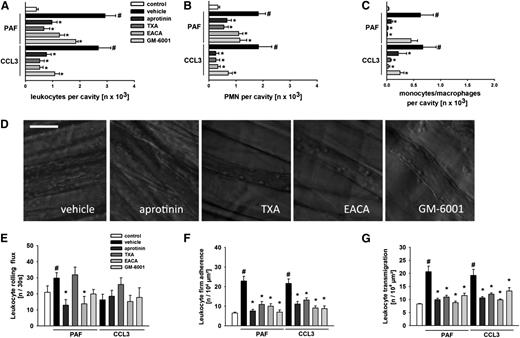
In the first set of experiments, the effect of the plasmin inhibitors TXA and EACA as well as of the broad-spectrum serine-protease inhibitor aprotinin and the broad-spectrum MMP inhibitor GM-6001 on leukocyte recruitment (Figure 1A) was analyzed in experimental peritonitis. Intraperitoneal injection of the lipid mediator PAF (a canonical neutrophil attractant) or the C-C motif chemokine CCL3 (a noncanonical neutrophil attractant) induced a significant increase in numbers of extravasated neutrophils (Figure 1B) and monocytes/macrophages (Figure 1C) in comparison with unstimulated controls. This increase was significantly reduced in animals receiving aprotinin, TXA, or EACA. Application of GM-6001 significantly diminished extravasation of neutrophils, and recruitment of monocytes/macrophages was significantly decreased upon stimulation with CCL3, but not in response to PAF.
Effect of aprotinin, TXA, EACA, and GM-6001 on leukocyte extravasation. Extravasation of total leukocytes (A), neutrophils (B), and monocytes/macrophages (C) to the peritoneal cavity was quantified 3 hours after intraperitoneal injection of PAF or CCL3 as detailed in the “Methods” section. (D) Representative in vivo transillumination microscopy images of inflamed cremasteric postcapillary venules in animals treated with aprotinin, TXA, EACA, GM-6001, or vehicle. Leukocyte rolling (E), firm adherence (F), and transmigration (G) were quantified 3 hours after intrascrotal injection of PAF or CCL3 by in vivo transillumination microscopy, as detailed in the “Methods” section. Panels show results for PBS-treated control mice as well as for mice receiving aprotinin, TXA, EACA, GM-6001, or drug vehicle (#P < .05 vs control; *P < .05 vs vehicle for n = 4 per group).
Effect of aprotinin, TXA, EACA, and GM-6001 on leukocyte extravasation. Extravasation of total leukocytes (A), neutrophils (B), and monocytes/macrophages (C) to the peritoneal cavity was quantified 3 hours after intraperitoneal injection of PAF or CCL3 as detailed in the “Methods” section. (D) Representative in vivo transillumination microscopy images of inflamed cremasteric postcapillary venules in animals treated with aprotinin, TXA, EACA, GM-6001, or vehicle. Leukocyte rolling (E), firm adherence (F), and transmigration (G) were quantified 3 hours after intrascrotal injection of PAF or CCL3 by in vivo transillumination microscopy, as detailed in the “Methods” section. Panels show results for PBS-treated control mice as well as for mice receiving aprotinin, TXA, EACA, GM-6001, or drug vehicle (#P < .05 vs control; *P < .05 vs vehicle for n = 4 per group).
Effect of aprotinin, TXA, EACA, and GM-6001 on leukocyte rolling, firm adherence, and transmigration
To further characterize the role of serine proteases and MMPs for each single step of the leukocyte extravasation process, we performed in vivo microscopy on the mouse cremaster muscle (Figure 1D). As it is well known, surgical preparation of the cremaster muscle induced leukocyte rolling in postcapillary venules. Stimulation with the lipid mediator PAF significantly enhanced the number of rolling leukocytes in comparison with unstimulated controls (Figure 1E). This elevation was significantly reduced in animals treated with aprotinin or EACA. Interestingly, leukocyte rolling was not significantly altered upon stimulation with the chemokine CCL3.
In unstimulated control animals, only a few leukocytes were found to be attached to the inner vessel wall of postcapillary venules. After 3 hours of stimulation with PAF or CCL3, the number of intravascularly firmly adherent leukocytes was significantly elevated (Figure 1F). This elevation was almost completely abolished in animals treated with aprotinin, EACA, TXA, or GM-6001.
Under control conditions, only a few leukocytes were observed within the perivenular interstitial tissue (Figure 1G). Intrascrotal injection of PAF or CCL3 induced a significant increase in numbers of transmigrated leukocytes. Similar to our findings for leukocyte firm adherence, this increase was almost completely abrogated in animals treated with aprotinin, EACA, TXA, or GM-6001.
Remodeling of the interstitial collagen network and interaction of interstitially migrating neutrophils with pericellular collagen fibers
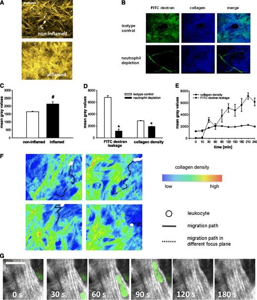
In a next set of experiments, interstitial collagen expression was analyzed by using the second harmonic generation signal in multiphoton in vivo microscopy on the mouse ear (Figure 2A). In response to PAF (Figure 2C), the average fluorescence intensity was significantly enhanced in comparison with PBS-treated controls. Moreover, swelling and increased contortion of single collagen fibers were observed upon onset of inflammation.
Interaction of neutrophils with interstitial collagen fibers. (A) Representative multiphoton in vivo microscopy images of interstitial collagen fibers (yellow) in the mouse ear 3 hours upon exposure to PBS or PAF. (B) Representative multiphoton in vivo microscopy images of FITC dextran leakage (green) and interstitial collagen fibers (blue) in the PAF-stimulated mouse ear of mice depleted from neutrophils or of control mice. Remodeling events within the interstitial tissue and FITC dextran leakage were quantified, as detailed in the “Methods” section. (C) Results for PBS-treated control mice as well as for mice stimulated with PAF (mean ± SEM for n = 3 per group; #P < .05 vs PBS). (D) Results for mice treated with a neutrophil-depleting anti-Ly6-G antibody or isotype control undergoing stimulation with PAF (mean ± SEM for n = 3 per group; *P < .05 vs isotype control). (E) Results for mice undergoing 240 minutes of stimulation with histamine (mean ± SEM for n = 3 per group). (F) Representative migration tracks (black) of neutrophils (white) within the interstitial tissue. Areas of low (blue), intermediate (yellow), and high (red) collagen density are indicated in pseudocolors (see supplemental Video 1A-D). (G) Representative serial images of an interstitially migrating neutrophil (green) encountering collagen fibers (white; also see supplemental Video 2). Scale bar = 20 μm.
Interaction of neutrophils with interstitial collagen fibers. (A) Representative multiphoton in vivo microscopy images of interstitial collagen fibers (yellow) in the mouse ear 3 hours upon exposure to PBS or PAF. (B) Representative multiphoton in vivo microscopy images of FITC dextran leakage (green) and interstitial collagen fibers (blue) in the PAF-stimulated mouse ear of mice depleted from neutrophils or of control mice. Remodeling events within the interstitial tissue and FITC dextran leakage were quantified, as detailed in the “Methods” section. (C) Results for PBS-treated control mice as well as for mice stimulated with PAF (mean ± SEM for n = 3 per group; #P < .05 vs PBS). (D) Results for mice treated with a neutrophil-depleting anti-Ly6-G antibody or isotype control undergoing stimulation with PAF (mean ± SEM for n = 3 per group; *P < .05 vs isotype control). (E) Results for mice undergoing 240 minutes of stimulation with histamine (mean ± SEM for n = 3 per group). (F) Representative migration tracks (black) of neutrophils (white) within the interstitial tissue. Areas of low (blue), intermediate (yellow), and high (red) collagen density are indicated in pseudocolors (see supplemental Video 1A-D). (G) Representative serial images of an interstitially migrating neutrophil (green) encountering collagen fibers (white; also see supplemental Video 2). Scale bar = 20 μm.
Because hydration has been found to induce shape changes of collagen fibers in vitro,28,29 leakage of the macromolecule fluorescein isothiocyanate (FITC) dextran and, concomitantly, interstitial collagen fiber density were analyzed in further experiments (Figure 2B). Depletion of neutrophils (which disrupt endothelial junctions during their extravasation) almost completely inhibited the PAF-elicited microvascular leakage as well as the subsequent inflammatory changes in interstitial collagen density (Figure 2C-D). Conversely, histamine-induced opening of endothelial junctions resulted in an immediate elevation in the leakage of FITC dextran into the perivascular tissue (Figure 2E), which was closely followed by the swelling of interstitial collagen fibers and a subsequent increase in the collagen fiber density of the perivascular tissue. These remodeling processes in the interstitium arose within the first 30 minutes after application of histamine, reached their peak values 60 minutes after the opening of endothelial junctions, and remained at this level for the entire observation period of 240 minutes.
In additional experiments, LysMeGFP mice (exhibiting fluorescence-labeled neutrophils) were used to visualize interactions between interstitially migrating neutrophils and collagen fibers. We observed that transmigrated neutrophils preferentially locomote in areas of the interstitial tissue with low collagen fiber density, thereby taking the path of least resistance (Figure 2F; supplemental Video 1A-B on the Blood website). When encountering areas of higher collagen fiber density, neutrophils immediately change their moving direction and continue their interstitial migration in areas of low collagen fiber density. When neutrophils get trapped between collagen fibers, these inflammatory cells slow down and push obstacles away to move forward rather than proteolytically degrade these extracellular matrix components (Figure 2G; supplemental Video 2).
Effect of latrunculin B and blebbistatin on directionality, velocity, and distance of interstitially migrating leukocytes
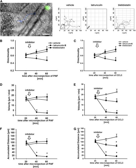
To further analyze the mechanisms underlying locomotion of transmigrated leukocytes in inflamed tissue, we used a novel in vivo chemotaxis assay (Figure 3A; supplemental Video 3). Perivenular microinjection of PAF or CCL3 induced directional migration of transmigrated leukocytes within the inflamed interstitial tissue. After superfusion of latrunculin B (which potently inhibits actin polymerization in interstitially migrating leukocytes; supplemental Figure 2), leukocytes immediately stopped to migrate within the interstitium, as indicated by significantly reduced migration directionality (Figure 3B-C) as well as almost completely abolished migration velocity (Figure 3D-E) and distance (Figure 3G) in comparison with vehicle-treated animals. In contrast, superfusion of blebbistatin (which blocks actomyosin contraction) did not significantly alter leukocyte interstitial migration parameters upon perivenular microinjection of PAF, while this compound slightly attenuated distance and velocity of interstitially migrating leukocytes upon perivenular microinjection of CCL3. Interestingly, blebbistatin selectively diminished PAF-elicited transmigration of neutrophils to the inflamed interstitium (a process which requires the actomyosin contraction-dependent squeezing of these inflammatory cells through endothelial junctions) without altering the preceding steps of the leukocyte extravasation cascade (supplemental Figure 3A-C).
Effect of latrunculin B and blebbistatin on leukocyte interstitial migration behavior. In vivo transillumination microscopy image of interstitially migrating leukocytes in the mouse cremaster muscle upon perivenular microinjection of PAF (injected together with green fluorescence–labeled microspheres for identification of the site of microinjection); the migration track of a single leukocyte is shown in blue (scale bar: 25 µm; also see supplemental Video 3). (A) Representative plots of migration tracks of transmigrated leukocytes in the cremasteric interstitial tissue of animals treated with latrunculin B, blebbistatin, or vehicle. Leukocyte interstitial migration behavior was analyzed after perivenular microinjection of PAF or CCL3, as detailed in the “Methods” section. Panels show results for directionality (B-C), velocity (D-E), and accumulated distance (F-G) of interstitially migrating leukocytes in animals treated with latrunculin B, blebbistatin, or vehicle (mean ± SEM for n = 4 per group; *P < .05 vs vehicle).
Effect of latrunculin B and blebbistatin on leukocyte interstitial migration behavior. In vivo transillumination microscopy image of interstitially migrating leukocytes in the mouse cremaster muscle upon perivenular microinjection of PAF (injected together with green fluorescence–labeled microspheres for identification of the site of microinjection); the migration track of a single leukocyte is shown in blue (scale bar: 25 µm; also see supplemental Video 3). (A) Representative plots of migration tracks of transmigrated leukocytes in the cremasteric interstitial tissue of animals treated with latrunculin B, blebbistatin, or vehicle. Leukocyte interstitial migration behavior was analyzed after perivenular microinjection of PAF or CCL3, as detailed in the “Methods” section. Panels show results for directionality (B-C), velocity (D-E), and accumulated distance (F-G) of interstitially migrating leukocytes in animals treated with latrunculin B, blebbistatin, or vehicle (mean ± SEM for n = 4 per group; *P < .05 vs vehicle).
Effect of aprotinin, TXA, EACA, and GM-6001 on migration directionality, velocity, and distance of interstitially migrating leukocytes
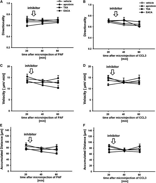
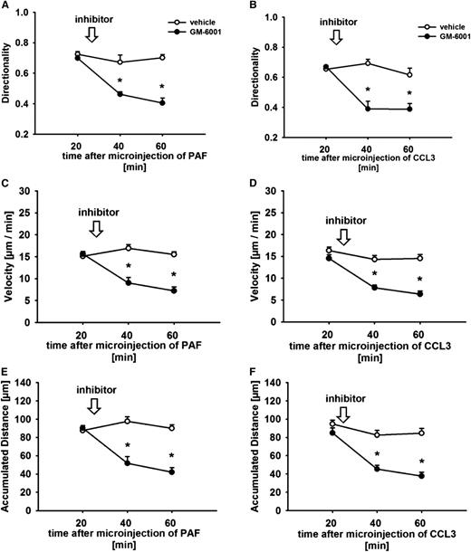
In further experiments, the effect of aprotinin, TXA, EACA, and GM-6001 on interstitial migration behavior of transmigrated leukocytes was analyzed. As shown above, perivenular application of PAF or CCL3 induced directional migration of leukocytes within the perivascular interstitial tissue of the cremaster muscle. While treatment with the serine protease inhibitors did not significantly alter migration behavior of interstitially locomoting leukocytes (Figure 4A-F), application of GM-6001 significantly reduced leukocyte interstitial migration directionality (Figure 5A-B), velocity (Figure 5C-D), and distance (Figure 5E-F) in comparison with vehicle-treated animals.
Effect of serine protease inhibitors on leukocyte interstitial migration behavior. Leukocyte interstitial migration behavior was analyzed after perivenular microinjection of PAF or CCL3 as detailed in the “Methods” section. Panels show results for directionality (A, B), velocity (C, D), and accumulated distance (E, F) of interstitially migrating leukocytes in animals treated with the serine protease inhibitors aprotinin, TXA, EACA, or vehicle (mean ± SEM for n = 4 per group).
Effect of serine protease inhibitors on leukocyte interstitial migration behavior. Leukocyte interstitial migration behavior was analyzed after perivenular microinjection of PAF or CCL3 as detailed in the “Methods” section. Panels show results for directionality (A, B), velocity (C, D), and accumulated distance (E, F) of interstitially migrating leukocytes in animals treated with the serine protease inhibitors aprotinin, TXA, EACA, or vehicle (mean ± SEM for n = 4 per group).
Effect of GM-6001 on leukocyte interstitial migration behavior. Leukocyte interstitial migration behavior was analyzed after perivenular microinjection of PAF or CCL3 as detailed in the “Methods” section. Panels show results for directionality (A, B), velocity (C, D), and accumulated distance (E, F) of interstitially migrating leukocytes in animals treated with the MMP inhibitor GM-6001 or vehicle (mean ± SEM for n = 4 per group; *P < .05 vs vehicle).
Effect of GM-6001 on leukocyte interstitial migration behavior. Leukocyte interstitial migration behavior was analyzed after perivenular microinjection of PAF or CCL3 as detailed in the “Methods” section. Panels show results for directionality (A, B), velocity (C, D), and accumulated distance (E, F) of interstitially migrating leukocytes in animals treated with the MMP inhibitor GM-6001 or vehicle (mean ± SEM for n = 4 per group; *P < .05 vs vehicle).
Effect of latrunculin B, blebbistatin, aprotinin, TXA, EACA, and GM-6001 on polarization of interstitially migrating leukocytes
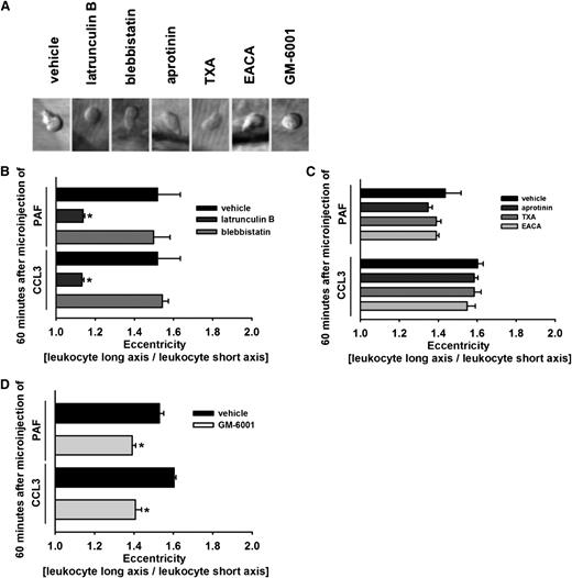
As a measure of polarization, eccentricity of interstitially locomoting leukocytes was analyzed by using in vivo microscopy (Figure 6A). In response to PAF or CCL3, interstitially migrating leukocytes were found to be polarized (values >1.2). Treatment with latrunculin B, but not with blebbistatin, completely abolished polarization of interstitially migrating leukocytes (Figure 6B) in comparison with vehicle-treated animals. Interestingly, aprotinin, TXA, or EACA did not significantly alter eccentricity of interstitially migrating leukocytes (Figure 6C), whereas in response to GM-6001, leukocyte polarization was significantly reduced (Figure 6D) in comparison with controls.
Effect of latrunculin B, blebbistatin, aprotinin, TXA, EACA, and GM-6001 on polarization of transmigrated leukocytes. (A) Representative in vivo transillumination microscopy images of transmigrated leukocytes in the cremasteric interstitial tissue of animals treated with latrunculin B, blebbistatin, aprotinin, TXA, EACA, GM-6001, or vehicle. As a measure of leukocyte polarization, eccentricity of transmigrated leukocytes was determined, as detailed in the “Methods” section. Panels show results for mice receiving (B) latrunculin B, blebbistatin, or drug vehicle, (C) aprotinin, TXA, EACA, or drug vehicle, and (D) GM-6001 or drug vehicle (mean ± SEM for n = 4 per group; *P < .05 vs vehicle).
Effect of latrunculin B, blebbistatin, aprotinin, TXA, EACA, and GM-6001 on polarization of transmigrated leukocytes. (A) Representative in vivo transillumination microscopy images of transmigrated leukocytes in the cremasteric interstitial tissue of animals treated with latrunculin B, blebbistatin, aprotinin, TXA, EACA, GM-6001, or vehicle. As a measure of leukocyte polarization, eccentricity of transmigrated leukocytes was determined, as detailed in the “Methods” section. Panels show results for mice receiving (B) latrunculin B, blebbistatin, or drug vehicle, (C) aprotinin, TXA, EACA, or drug vehicle, and (D) GM-6001 or drug vehicle (mean ± SEM for n = 4 per group; *P < .05 vs vehicle).
Systemic leukocyte counts and microhemodynamic parameters
Inner vessel diameters, blood flow velocities, and shear rates of analyzed postcapillary venules as well as systemic leukocyte counts were determined in order to assure intergroup comparability (supplemental Table 1). No significant differences were detected among all experimental groups.
Phenotyping of transmigrated leukocytes
To identify the phenotype of transmigrated leukocytes, we performed immunostaining for CD45 (common leukocyte antigen), Ly-6G (neutrophils), and F4/80 (monocytes/macrophages) of cremasteric tissue samples. In response to PAF or CCL3, over 85% of transmigrated leukocytes were positive for Ly-6G, and about 10% of transmigrated leukocytes were positive for F4/80. Moreover, within 120 minutes of stimulation with PAF or CCL3, (almost) exclusively GFP− leukocytes (representing neutrophils) transmigrated into the perivascular tissue of the cremaster muscle of CX3CR-1GFP/+ mice (exhibiting GFP+ monocytes/macrophages/NK cells; supplemental Figure 4).
Surface expression of proteases on neutrophils upon transmigration
Using flow cytometry, we sought to evaluate the surface expression profiles of plasmin(ogen) as well as of MMP-2, MMP-8, and MMP-9 on circulating and on transmigrated neutrophils (supplemental Figure 1) or monocytes (supplemental Table 3). In our experiments, we show that only a small proportion of circulating neutrophils and monocytes present plasmin(ogen) on their cell surface. Upon transmigration, the proportion of plasminogen-positive neutrophils and monocytes was significantly elevated. In addition, we found that almost all circulating neutrophils express MMP-2, −8, and −9. Upon transmigration, the expression of these MMPs was further enhanced. In contrast, only a small proportion of circulating monocytes was positive for MMP-2, MMP-8, or MMP-9. Upon transmigration, however, the proportion of MMP-expressing monocytes was significantly increased.
Effect of GM-6001 on surface expression of adhesion/signaling molecules
In the next set of experiments, the role of MMPs for surface expression of relevant adhesion/signaling molecules on neutrophils was analyzed by flow cytometry. Stimulation with PAF induced shedding of l-selectin from the surface of murine neutrophils, which was completely prevented upon application of GM-6001 (supplemental Table 2). In addition, GM-6001 significantly reduced the PAF-elicited elevation in surface expression of β1 integrins and further enhanced the PAF-elicited increase in surface expression of the β2 integrins Mac-1 and LFA-1. Moreover, we show that GM-6001 (A)—in contrast to the prototypical apoptosis stimulus staurosporine (B)—does not alter the viability of neutrophils isolated from the bone marrow (supplemental Table 4). Interestingly, surface expression of l-selectin or of integrins on murine monocytes was not significantly altered upon application of PAF (with or without addition of GM-6001 (supplemental Table 2).
Discussion
Directed migration of leukocytes to the site of inflammation is a hallmark of the inflammatory response. Previously, a diversity of adhesion and signaling molecules has been identified, specifically regulating the sequential steps of the leukocyte extravasation cascade. The mechanisms underlying the subsequent migration of leukocytes within the inflamed interstitial tissue, however, remained poorly understood.1-4 In this context, the functional relevance of proteases in particular is still an issue of controversial debate.11-18
In the present study, we sought to define whether and how serine proteases, which represent over one third of all known proteolytic enzymes, as well as MMPs, which form a class of enzymes specialized on the degradation of the extracellular matrix,22 are involved in leukocyte migration to the site of inflammation. Using a model of sterile peritonitis, we found that migration of neutrophils to the inflamed peritoneal cavity was effectively prevented by the broad-spectrum serine protease inhibitor aprotinin as well as by the broad-spectrum MMP inhibitor GM-6001. In addition, selective inhibition of the serine protease plasmin, which is considered to be an activator of MMPs, almost completely abolished peritoneal recruitment of neutrophils. These data clearly show that serine proteases and MMPs are essential for neutrophil recruitment to the site of inflammation.
Initially, proteolytic enzymes have been suggested to trigger leukocyte extravasation by facilitating the passage of these inflammatory cells between endothelial cells (via cleavage of junctional molecules) and through the perivenular basement membrane (by degradation of structural constituents). Using in vivo microscopy on the inflamed cremaster muscle, we were able to show that serine proteases and MMPs already contribute to intravascular accumulation of leukocytes. In previous reports, plasmin and other serine proteases are thought to interact with specific receptors,20,21 and MMPs modulate the functional properties of chemokines30-32 as well as regulate the expression of leukocyte adhesion/signaling molecules, thereby controlling intravascular leukocyte–endothelial cell interactions.33-35
On the route from the microvasculature to the site of inflammation, leukocytes are supposed to undergo dramatic functional and phenotypic changes. Here, we show that the proportion of plasmin(ogen)-bearing neutrophils as well as the expression levels of MMPs on neutrophils significantly increase upon transmigration. Similar surface changes during leukocyte diapedesis have been reported for integrins and other proteases.4,10 The functional relevance of these events for the subsequent migration of leukocytes within the interstitial tissue remained largely unclear.
The interstitium is a complex network of polysaccharides and proteins providing structural organization and physical support to tissues. Regarding cell motility, the interstitial tissue is suggested to be a requirement for as well as a physical barrier to locomotion of cells.30,31 According to our current understanding, leukocytes are thought to migrate within the interstitium in an ameboid fashion, being largely independent of focal adhesions and pericellular proteolysis.4,5 However, the majority of previously published studies on migration of inflammatory cells is based on in vitro and ex vivo assays. In these model systems, the complex architecture of the interstitial tissue as well as its structural changes in the inflammatory response9 or the dramatic phenotypic and functional alterations that leukocytes undergo during diapedesis4,10 can hardly be mimicked. Although multiphoton laser scanning in vivo microscopy enabled investigations on lymphocyte and dendritic cell migration,6-8 leukocyte interstitial migration in “nonlymphatic” tissues during inflammatory conditions is poorly understood.
As opposed to homeostatic lymphocyte trafficking in the lymph node, microvascular permeability immediately increases upon extravasation of neutrophils, resulting in a prompt influx of fluid into the inflamed interstitial tissue. On a microstructural level, hydration of collagen fibers significantly enhances their tortuosity as well as increases the volume of single collagen fibrils and of the extrafibrillar space.28,29 Using multiphoton laser scanning in vivo microscopy, we found that these inflammatory events are followed by collagen fiber swelling and, collectively, by a dramatic increase in the density of the interstitial collagen network. Interestingly, neutrophils preferentially migrate in “loose” areas of low collagen fiber density without any signs of collagen degradation. When encountering areas of higher collagen density, leukocytes gradually change their moving direction and continue to migrate by taking the path of least resistance. Consequently, these inflammatory changes in the interstitial collagen network are thought to limit the operating space of infiltrating neutrophils as well as to minimize distraction by opposing chemotactic cues, thereby enabling these inflammatory cells to efficiently swarm to the site of inflammation.
In this regard, neutrophils are thought to “flow” and “squeeze” through the interstitium as observed in basic in vitro and ex vivo model systems.36,37 For this ameboidlike locomotion pattern of leukocytes, the extension of the intracellular actin network at the moving front as well as the contraction of actomyosin at the cell rear are supposed to be the driving forces.36,37 Whether these processes are functionally relevant for the directed migration of leukocytes through the inflamed interstitial tissue is still unknown. Using a novel “3-Din vivo chemotaxis assay,”27 we found that interstitially migrating neutrophils immediately stopped to locomote upon inhibition of actin polymerization. Surprisingly, blockade of actomyosin contraction only marginally reduced migration velocity and distance of directionally moving neutrophils, suggesting that motility of these inflammatory cells within the inflamed interstitium is predominantly dependent on the extension of the intracellular actin network. According to previous in vitro data,37 actomyosin contraction might only be engaged when leukocytes have to pass areas of higher collagen density, allowing these inflammatory cells to rapidly and flexibly navigate through inflamed interstitial tissue.
With respect to these observations, we consequently hypothesized that proteases are dispensable for directed interstitial locomotion of leukocytes. In a further set of experiments, we were able to confirm that serine proteases are not required for neutrophil interstitial migration. These results are in agreement with previously published findings because broad-spectrum protease inhibition (which included several serine proteases and MMPs) did not affect motility of cultured leukocytes in a 3-dimensional collagen assay.11 Astonishingly, however, we found that blockade of MMP activity strongly reduced polarization and migration velocity of neutrophils locomoting within the inflamed interstitial tissue. In line with previous reports,33-35 we demonstrate that MMPs modulate expression of cell surface molecules (eg, integrins and l-selectin), which have recently been implicated in extravascular locomotion of neutrophils.38-40 Additionally, MMPs might change the functional properties of chemokines and their receptors in inflamed tissue by proteolytic processing, thereby modulating the effect of chemotactic cytokines on cell migration.30-32
In conclusion, our in vivo data indicate that in inflamed tissue the density of the interstitial collagen network significantly increases. These inflammatory remodeling events are thought to provide physical guidance to infiltrating neutrophils, allowing these inflammatory cells to efficiently migrate to the site of inflammation. The locomotion pattern of interstitially migrating leukocytes strictly relies on actin polymerization, but only marginally on actomyosin contraction, and does not require pericellular proteolysis. Although serine proteases including plasmin are critical for leukocyte extravasation, these proteolytic enzymes do not contribute to the extravascular motility of neutrophils. In contrast, MMPs promote both extravasation and directional interstitial migration of transmigrated neutrophils in inflamed tissue by regulating the surface expression of adhesion/signaling molecules on these leukocytes. Our findings provide fundamental insights into the principle mechanisms underlying the directed locomotion of neutrophils in the inflamed interstitial tissue and corroborate MMPs as modulators of ameboidlike leukocyte motility.
The online version of this article contains a data supplement.
The publication costs of this article were defrayed in part by page charge payment. Therefore, and solely to indicate this fact, this article is hereby marked “advertisement” in accordance with 18 USC section 1734.
Acknowledgments
The authors thank Gerhard Adams and Silvia Münzing for technical assistance.
This study was supported by Deutsche Forschungsgemeinschaft (DFG, SFB 914, to K.L., A.G.K., S.M., C.A.R, and F.K.) and Friedrich-Baur-Stiftung (to C.A.R.).
This study is part of the doctoral thesis of M.L.
Authorship
Contribution: M.L., B.U., K.S., A.E., M.M., D.P.-W., G.Z., M.P., M.R., and A.G.K. performed experiments and contributed to data analysis; and K.L., S.M., F.K., and C.A.R. designed experiments, contributed to data analysis, and wrote the manuscript.
Conflict-of-interest disclosure: The authors declare no competing financial interests.
Correspondence: Christoph Reichel, Department of Otorhinolaryngology, Head and Neck Surgery, Klinikum der Universität München, Marchioninistrasse 15, 81377 Munich, Germany; e-mail: christoph.reichel@med.uni-muenchen.de.

