Key Points
The human circadian system causes a morning peak in circulating levels of PAI-1, independent of any behavioral or environmental influences.
The circadian system determines to a large extent the PAI-1 rhythm observed during a regular sleep/wake cycle.
Abstract
Serious adverse cardiovascular events peak in the morning, possibly related to increased thrombosis in critical vessels. Plasminogen activator inhibitor-1 (PAI-1), which inhibits fibrinolysis, is a key circulating prothrombotic factor that rises in the morning in humans. We tested whether this morning peak in PAI-1 is caused by the internal circadian system or by behaviors that typically occur in the morning, such as altered posture and physical activity. Twelve healthy adults underwent a 2-week protocol that enabled the distinction of endogenous circadian effects from behavioral and environmental effects. The results demonstrated a robust circadian rhythm in circulating PAI-1 with a peak corresponding to ∼6:30 am. This rhythm in PAI-1 was 8-times larger than changes in PAI-1 induced by standardized behavioral stressors, including head-up tilt and 15-minute cycle exercise. If this large endogenous morning peak in PAI-1 persists in vulnerable individuals, it could help explain the morning peak in adverse cardiovascular events.
Introduction
The ability to clot blood can be life-saving after an injury, but thrombi within vessels can contribute to myocardial infarction, ischemic stroke, and sudden cardiac death.1-3 Ongoing fibrinolytic activity helps break down thrombi and maintain vessel patency and is largely managed by circulating tissue plasminogen activator, which converts plasminogen to plasmin.1-3 Fibrinolytic activity is significantly reduced in the morning due to an increase in plasminogen activator inhibitor-1 (PAI-1), the primary inhibitor of tissue plasminogen activator.1-3 Thus, increased PAI-1 and decreased fibrinolysis in the morning may increase the risk for development of occlusive thrombi and could help explain the morning peak in adverse cardiovascular events.1-4 To begin to understand potential underlying mechanisms in humans, we tested the degree to which the morning increase in PAI-1 is caused by a direct endogenous circadian rhythm in PAI-15-8 vs influences from the daily behavioral/environmental changes across the night and day (eg, rest/activity, fasting/feeding, dark/light, and ambient-temperature cycles).
Methods
We studied 12 healthy volunteers taking no medications (mean [range], 25.8 [20-42] years; body mass index, 23.6 [19.9-29.6] kg/m2; 6 women). Studies were approved by the Partners Human Research Committee, and participants gave written informed consent in accordance with the Declaration of Helsinki.
Participants were assessed throughout a 2-week laboratory protocol designed to desynchronize daily behavioral rhythms from internal circadian rhythms, while maintaining environmental factors constant. Participants had 2 baseline 24-hour days in normal room lighting conditions (∼90 lux wake episodes/0 lux sleep episodes), followed by a forced desynchrony protocol (FD) consisting of 12 standardized 20-hour “days” with controlled activity, posture, meals, sleep, room temperature, and light (<4 lux), as previously published.9-12 On the second baseline day and each 20-hour day, participants underwent a standardized test battery,12 which included a 15-minute 60° passive head-up tilt test and a 15-minute steady-state cycle ergometer test at 60% of maximal predicted heart rate. Core body temperature derived from a rectal thermistor was continuously recorded and used to determine circadian period and phase, with minimum core body temperature assigned as 0° (equivalent to ∼4:30 am in these participants).9 Blood samples were collected by intravenous catheter in EDTA tubes on ice, immediately spun down in a precooled centrifuge (4°C) for 10 min at 2200 rpm, and frozen at −80°C until assayed for PAI-1 antigen by enzyme-linked immunosorbent assay (R&D Systems, Minneapolis, MN). We note that PAI-1 antigen and PAI-1 activity are highly correlated.13 The normal daily pattern of plasma PAI-1 antigen was assessed from samples taken hourly across 24 hours on the second baseline day and night. Thereafter, the effect on PAI-1 of the endogenous circadian system, standardized activities, and their interaction was assessed from 4 samples taken across a range of activities on each of the 12 test batteries (so that all circadian phases were represented). The 4 standardized activities each day throughout the FD were supine rest (after 15 minutes lying on the tilt table), head-up tilt (10 minutes into the tilt), seated at rest (after 15-minute sitting on the ergometer), and mild cycle exercise (10 minutes into exercise). Endogenous circadian rhythms (2 harmonics) and behavioral effects (4 activities) were tested by cosinor mixed-model analysis of variance.
Results and discussion
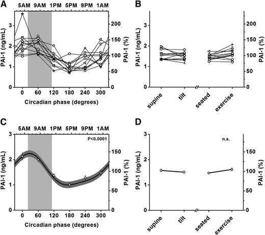
From group analysis of the FD data, there was a large amplitude endogenous circadian rhythm in PAI-1 with a peak at a circadian phase of 30°, corresponding to ∼6:30 am (∼1 hour prior to habitual wake time) and a trough at a phase corresponding to ∼3:30 pm (peak-to-trough amplitude = 1.24 ng/mL; 82% of mean; P < .0001; Figure 1A,C) with an increase from trough to peak of 124%. Although there was an effect in anticipation of the stress battery on the baseline day (see Figure 2B), throughout the subsequent FD there were no significant changes in PAI-1 across the sequence of standardized behaviors while controlling for circadian effects (Figure 1B,D) and no interaction between the effects of behaviors and circadian phase.
Presence of endogenous circadian rhythm (independent of behavioral effects) and absence of significant effect of behavioral stressors (independent of circadian effects) on PAI-1. PAI-1 had a significant endogenous circadian rhythm with the circadian peak corresponding to approximately 6:30 am, at the beginning of the vulnerable time of 6 am to noon (light gray bars in A,C). The behavioral states, including head up tilt and exercise, had no significant effect on PAI-1 (B,D). There were no statistical interactions between the circadian and behavioral effects. Panels A and B show individual data, while panels C and D show group analysis. Cosine models (black lines) and 95% confidence intervals (dark gray areas) are based on mixed-model analyses and use precise circadian phase data. To show that these models adequately fit the actual data, we also plot average data grouped into 60-circadian-degree windows with error bars representing standard error of the mean. Data are normalized according to each individual’s average across the FD protocol. Bottom x-axes (A,C), circadian phase with 0° indicating the timing of the fitted circadian core body temperature minimum (average ∼4:30 am in these participants); bottom x-axes (B,D), behavioral states; top x-axes (A,C), corresponding average clock time in these participants; right y-axes, percentage of each individual’s mean across the protocol; left y-axes, absolute values; light gray bars (A,C), most vulnerable period for adverse cardiovascular events observed in epidemiologic studies (∼6:00 am to noon). P values, significance of circadian effect from cosinor analyses.
Presence of endogenous circadian rhythm (independent of behavioral effects) and absence of significant effect of behavioral stressors (independent of circadian effects) on PAI-1. PAI-1 had a significant endogenous circadian rhythm with the circadian peak corresponding to approximately 6:30 am, at the beginning of the vulnerable time of 6 am to noon (light gray bars in A,C). The behavioral states, including head up tilt and exercise, had no significant effect on PAI-1 (B,D). There were no statistical interactions between the circadian and behavioral effects. Panels A and B show individual data, while panels C and D show group analysis. Cosine models (black lines) and 95% confidence intervals (dark gray areas) are based on mixed-model analyses and use precise circadian phase data. To show that these models adequately fit the actual data, we also plot average data grouped into 60-circadian-degree windows with error bars representing standard error of the mean. Data are normalized according to each individual’s average across the FD protocol. Bottom x-axes (A,C), circadian phase with 0° indicating the timing of the fitted circadian core body temperature minimum (average ∼4:30 am in these participants); bottom x-axes (B,D), behavioral states; top x-axes (A,C), corresponding average clock time in these participants; right y-axes, percentage of each individual’s mean across the protocol; left y-axes, absolute values; light gray bars (A,C), most vulnerable period for adverse cardiovascular events observed in epidemiologic studies (∼6:00 am to noon). P values, significance of circadian effect from cosinor analyses.
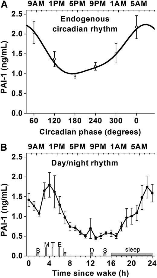
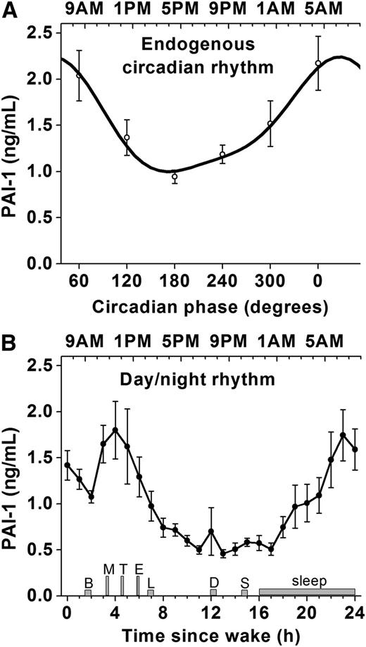
Comparison between the endogenous circadian rhythm in PAI-1 and day/night rhythm in PAI-1. Comparison between the endogenous circadian rhythm in PAI-1 assessed during the twelve 20-hour cycles of the FD protocol (A) and the day/night rhythm in PAI-1 assessed during the second 24-hour baseline day including sleep/wake, fasting/feeding, supine/upright, rest/activity, and dark/light cycle (B). Figures are aligned according to the approximate average corresponding clock time of the circadian profile (ie, 0 circadian degrees equates to ∼4:30 am and 3 hours prior to habitual scheduled awakening in these participants). In order to allow comparison of absolute values, subjects’ data were not normalized (in contrast to Figure 1). Timing of meals, sleep, and test battery (gray bars) are indicated for the baseline day (data for FD protocol were all collected during the test battery). B, breakfast; L, lunch; D, dinner; S, snack. M, mental stress test (10-minute computerized serial addition test); T, head-up tilt; E, exercise. Top x-axis (A), corresponding average clock time; top x-axis (B), average clock time. During the baseline day, participants were in semirecumbent posture until T, in varied postures throughout the remainder of the day, and remained lying down throughout the sleep episode. Error bars represent standard error of the mean.
Comparison between the endogenous circadian rhythm in PAI-1 and day/night rhythm in PAI-1. Comparison between the endogenous circadian rhythm in PAI-1 assessed during the twelve 20-hour cycles of the FD protocol (A) and the day/night rhythm in PAI-1 assessed during the second 24-hour baseline day including sleep/wake, fasting/feeding, supine/upright, rest/activity, and dark/light cycle (B). Figures are aligned according to the approximate average corresponding clock time of the circadian profile (ie, 0 circadian degrees equates to ∼4:30 am and 3 hours prior to habitual scheduled awakening in these participants). In order to allow comparison of absolute values, subjects’ data were not normalized (in contrast to Figure 1). Timing of meals, sleep, and test battery (gray bars) are indicated for the baseline day (data for FD protocol were all collected during the test battery). B, breakfast; L, lunch; D, dinner; S, snack. M, mental stress test (10-minute computerized serial addition test); T, head-up tilt; E, exercise. Top x-axis (A), corresponding average clock time; top x-axis (B), average clock time. During the baseline day, participants were in semirecumbent posture until T, in varied postures throughout the remainder of the day, and remained lying down throughout the sleep episode. Error bars represent standard error of the mean.
Figure 2 enables comparison of the effects on PAI-1 of the endogenous circadian cycle versus the effects of normal behavioral changes combined with the effects of the endogenous circadian cycle across the baseline day. Overall, there was great similarity between the baseline and circadian profiles of PAI-1 in terms of both the timing (<1 hour difference in peak) and absolute amplitude (<10% difference). The main differences were an overall average increase in PAI-1 from 1.00 ng/mL on the baseline day to 1.54 ng/mL during the 20-hour days) and a rapid increase of PAI-1 ∼3 hours after scheduled awakening on the baseline day, ie, after breakfast and likely in anticipation of the start of the very first stress test battery. Because samples in the FD were collected during the test battery and not before, we likely missed any such anticipation-triggered PAI-1 changes in the circadian analyses.
Previous studies have shown a significant daily rhythm in PAI-1 in humans, but the role of the endogenous circadian system vs behaviors was not established. Our main finding is that the endogenous circadian system appears to be the major contributor to the daily changes in PAI-1 seen during a normal sleep/wake cycle in humans. Although behavioral or environmental factors such as activity level could also influence PAI-1, our 15-minute mild stress tests likely did not capture the full range of PAI-1 changes that can be induced by behavior.14,15 This complexity is highlighted by a study that found a blunted PAI-1 rhythm in 2 blind individuals who were not synchronized to the 24-hour light/dark cycle.16 In animals, lesioning the central circadian pacemaker (suprachiasmatic nucleus) abolished the daily rhythm in PAI-117 but also abolished behavioral rhythmicity so it could not be determined if the PAI-1 rhythm is driven directly by the circadian system or indirectly through behavior. Nonetheless, recent genetic studies in animals have demonstrated that the PAI-1 promoter is under direct control of core clock genes5-8 and, in humans, a PAI-1 promoter polymorphism (4G5G) was found to increase morning PAI-1.18,19 Furthermore, a genome-wide association study identified a common variant in the core clock gene BMAL-1 (ARNTL) to be robustly associated with elevated morning PAI-1 levels.20 Building on this work, we have now discovered that there is a true endogenous circadian rhythm in circulating PAI-1 in humans, independent of the behavioral and environmental factors. Moreover, the circadian peak in PAI-1 at ∼6:30 am is at the start of the vulnerable window for adverse cardiovascular events (6 am to noon), as we hypothesized based on the possibility that the circadian system causes a prothrombotic state in the morning. One could speculate that evolutionary pressures shaped the circadian influences to favor blood clotting (ie, elevated PAI-1) at times of increased activity after sleep when risk of blood loss due to laceration injury was increased, eg, due to predator/prey/competitor encounters. On the other hand, this large endogenous circadian rhythm in prothrombotic PAI-1, if it persists in vulnerable individuals, could contribute to the increased morning peak in PAI-1 observed in these populations21,22 and to the morning peak in adverse cardiovascular events.1-3 We note that in a prospective study, individuals who later developed coronary heart disease had PAI-1 levels at baseline that were 31% higher than in case-control individuals after correcting for age, sex, and race,23 suggesting that the endogenous circadian modulation observed in the current study (82%) may be clinically meaningful.
We previously found that other cardiovascular risk factors are under direct circadian control, including platelet activation, count, and aggregability, plasma epinephrine and norepinephrine, plasma cortisol, systolic and diastolic blood pressure, heart rate, and vagal cardiac modulation.9-12 Of these factors, platelet activation and cortisol had endogenous circadian peaks, and plasma epinephrine the steepest circadian increase, during the vulnerable window for adverse cardiovascular events, around 9 am. Furthermore, in response to exercise, the largest circadian increase in epinephrine and largest circadian decrease in cardiac vagal modulation also occurred around 9 am. Together with these previous data, our current data suggest that the circadian system could contribute to the increased risk for cardiovascular events in the morning via a number of related cardiovascular mechanisms. Such mechanisms may have homeostatic advantage in most circumstances in anticipation of the expected behavioral changes in the morning, such as vagal withdrawal, sympathetic activation, and increased blood clotting. However, these same physiological adaptations could exacerbate existing risk factors in certain individuals, such as those with vulnerable atherosclerotic plaques in a coronary artery.4
There is an Inside Blood commentary on this article in this issue.
The publication costs of this article were defrayed in part by page charge payment. Therefore, and solely to indicate this fact, this article is hereby marked “advertisement” in accordance with 18 USC section 1734.
Acknowledgments
The authors thank Joanna Garcia and Carolina Smales for help with data processing and the staff of the Medical Chronobiology Program and Brigham and Women’s Hospital’s Center for Clinical Investigation for their help running the study. The authors also thank the participants for their participation in the study.
This research was supported by National Institutes of Health (NIH), Heart, Lung and Blood Institute grant R01-HL76409 (S.A.S.), a grant from the Harvard Catalyst Clinical Research Center (F.A.J.L.S.), NIH, National Center for Research Resources grant UL1-RR025758 (Harvard Clinical and Translational Science Center), NIH, Heart, Lung and Blood Institute grant P30-HL101299 (F.A.J.L.S.), and NIH, Heart, Lung and Blood Institute grant K24-HL076446 (S.A.S.).
Authorship
Contribution: F.A.J.L.S. and S.A.S. designed and performed research and wrote the paper; and F.A.J.L.S. analyzed data.
Conflict-of-interest disclosure: The authors declare no competing financial interests.
Correspondence: Frank A. J. L. Scheer, Division of Sleep Medicine, Brigham and Women’s Hospital, and Harvard Medical School, 221 Longwood Ave, Boston, MA 02115; e-mail: fscheer@rics.bwh.harvard.edu.


