Key Points
New onset of thrombosis is an independent risk factor for increased mortality in LA-positive individuals.
Life expectancy in our cohort of patients with LA was worse in comparison with an Austrian reference population.
Abstract
Data on the clinical course of lupus anticoagulant (LA)–positive individuals with or without thrombotic manifestations or pregnancy complications are limited. To investigate mortality rates and factors that might influence mortality, we conducted a prospective observational study of LA-positive individuals. In total, 151 patients (82% female) were followed for a median of 8.2 years; 30 of the patients (20%) developed 32 thromboembolic events (15 arterial and 17 venous events) and 20 patients (13%) died. In univariable analysis, new onset of thrombosis (hazard ratio [HR] = 8.76; 95% confidence interval [CI], 3.46-22.16) was associated with adverse survival. Thrombosis remained a strong adverse prognostic factor after multivariable adjustment for age and hypertension (HR = 5.95; 95% CI, 2.43-14.95). Concomitant autoimmune diseases, anticoagulant treatment at baseline, or positivity for anticardiolipin- or anti-β2-glycoprotein I antibodies were not associated with mortality. In a relative survival analysis, our cohort of LA positives showed a persistently worse survival in comparison with an age-, sex-, and study-inclusion-year–matched Austrian reference population. The cumulative relative survival was 95.0% (95% CI, 88.5-98.8) after 5 years and 87.7% (95% CI, 76.3-95.6) after 10 years. We conclude that occurrence of a thrombotic event is associated with higher mortality in patients with LA. Consequently, the prevention of thromboembolic events in LA positives might improve survival.
Introduction
Antiphospholipid antibodies (aPLAs), such as lupus anticoagulant (LA), anticardiolipin antibodies (aCLs), and antibodies against β2-glycoprotein I (anti-β2GPI), comprise a group of heterogeneous circulating autoantibodies that are directed against anionic phospholipids or affiliated plasma proteins and are frequently associated with a prothrombotic state.1 When associated with clinical manifestations, aPLAs are an essential component of the diagnosis of the antiphospholipid syndrome (APS).2 Still, aPLAs are also found in healthy individuals or are associated with infections without clinical manifestations of APS.3-6 So far, the pathogenesis of thrombotic manifestations in LA-positive patients is not completely understood and the clinical impact of aPLA positivity is as yet unclear.7
The occurrence of arterial and venous thromboembolism (TE) not only causes acute, potentially fatal organ damage, but may also compromise long-term organ functionality, possibly further aggravated by the association of aPLA positivity with autoimmune disorders, such as systemic lupus erythematodes (SLE) or other autoimmune diseases.8 So far, most investigations on the morbidity and mortality of aPLA-positive patients have focused on patients with SLE or patients already diagnosed with APS. In those cohorts, increased mortality has been reported for patients with APS.9 Still, the clinical significance of LA positivity, associated with incidence and recurrence rates of TE, and its impact on mortality have not been systematically analyzed as yet.
We conducted a prospective observational cohort study on persistently LA-positive individuals with and without a prior history of thrombosis or pregnancy complications: the Vienna Lupus Anticoagulant and Thrombosis Study (LATS). The aim of our study was to investigate overall mortality of our cohort of LA patients and to evaluate the impact of thrombosis on survival during the observation period.
Patients and methods
Starting from May 2001, adult patients who had repeatedly tested positive for LA within at least 6 or 12 weeks according to current recommendations2,10,11 were consecutively included in this prospective study after having given written informed consent. All included patients were asked to perform follow-up visits at the outpatient ward of our center every 6 months during the first 5 years, then at yearly intervals.
Data on previous and newly developed arterial and venous thromboembolic events, concomitant diseases, and medication were collected using a standardized questionnaire and chart review. All registered events had to be symptomatic and diagnosed with standardized methods (duplex sonography or phlebography for venous thrombosis, computed tomography [CT] or ventilation-perfusion lung scan for pulmonary embolism [PE], coronary arteriography for myocardial infarction, and CT scan or magnetic resonance imaging [MRI], CT/MRI angiography, or transcranial duplex sonography for stroke; transient ischemic attacks were diagnosed on the basis of the typical clinical presentation). All events occurring during the follow-up period were separately evaluated and adjudicated.
A blood sample was drawn at inclusion and at every visit and the LA and aCL and anti-β2GPI antibodies were determined. In case a patient became LA negative, he/she was excluded from further analysis. If the patient missed a scheduled visit, the questionnaire was completed by telephone interview or through chart review. Documented diagnoses of SLE, lupus-like disease (LLD), or other autoimmune diseases were reevaluated by a specialized rheumatologist according to the American College of Rheumatology criteria and recorded correspondingly.12,13 Causes of death were identified by review of death certificates, autopsy records, or through personal contact with the family physician, hospitals, or nursing homes. For comparison, data on the mortality of the Austrian general population were obtained from the Austrian death registry. The ethics committee of the Medical University of Vienna in accordance with the Declaration of Helsinki approved the conduct of the study (EC no. 068/2001).
Blood sampling and sample preparation
Blood samples were drawn with a 21-gauge butterfly needle (Greiner Bio-One) into a Vacuette tube (Greiner Bio-One) containing trisodium citrate (9 parts of whole blood, 1 of trisodium citrate 3.8%). All samples were mixed adequately by gently inverting the tubes and were processed within 3 hours after venipuncture. Platelet-poor plasma was prepared by centrifugation at 2500g for 15 minutes at 15°C, followed by a second step of centrifugation of the harvested plasma under the same conditions.
Determination of LA
Diagnosis of LA followed the Scientific and Standardization Committee (SSC)/International Society on Thrombosis and Haemostasis (ISTH) recommendations.11,14 We used 2 different screening tests: a lupus-sensitive activated partial thromboplastin time and a diluted Russell viper venom time (dRVVT). In patients under therapy with vitamin K antagonists (VKAs), only the aPTT was used as screening assay. In the case of prolongation of 1 or both screening tests, samples were further analyzed and confirmatory tests were performed, as described in detail by Wenzel et al.15 When, during the observation period, the confirmatory test was not definitely positive, LA was still regarded as positive, when the Rosner index, calculated as 100 × (clotting times of the 1:1 mixture − normal plasma)/patient’s plasma was higher than 15.16 As confirmatory assays, the StaClot LA (Diagnostica Stago) and the dRVVT-LA confirm (Life Diagnostics) were used.
Determination of aCL and anti-β2GPI antibodies
Determinations of immunoglobulin G (IgG) and IgM antibodies against cardiolipin (aCL) and β2GPI were done with indirect solid-phase enzyme immunoassays. Between 2001 and September 2005 the Varelisa Cardiolipin test (Pharmacia) was performed semiautomatically using a Tecan Genesis liquid handling system (Tecan Group Ltd). Starting from October 2005, the Orgentec Cardiolipin and, starting from October 2006, the Orgentec β2GPI tests (both Orgentec) were used as standard routine assays on a fully automated BEP2000 Advance System (Siemens Healthcare Diagnostics). All assays were performed following the manufacturers’ instructions.
According to the updated Sapporo criteria,2 aCL IgG and aCL IgM were regarded positive if results were >40 GPL/MPL U/mL for both the Varelisa Cardiolipin and the Orgentec Cardiolipin test. For anti-β2GPI, IgG and IgM (Orgentec assays) results were positive if >8 U/mL (corresponding to the 99th percentile of healthy controls).
Statistical analysis
All statistical analyses were performed using the commercially available package Stata (Windows version 12.0; Stata Corp). Continuous variables were summarized with the median and the interquartile range, and categorical variables by absolute frequencies and percentages. The association between categorical variables was assessed using the Pearson χ2 test and logistic regression. The association between a continuous variable and a categorical variable was assessed using the Student t test (with correction for variance heterogeneity when appropriate). For all time-to-event analyses, death from any cause was considered the event of interest. To eliminate information bias regarding the ascertainment of survival and TE status at potentially different time points, we truncated the follow-up period on December 31, 2012. Median survival time was calculated using the reverse Kaplan-Meier method. Survival probabilities were estimated and graphed over time according to the Kaplan-Meier method. Differences in survivorship functions between 2 or more groups were assessed with the log-rank test. The association between new onset of TE and mortality was visualized using landmark analysis17 (with an a priori specified landmark at 3 years after baseline). In a sensitivity analysis, we used different landmark time points, which did not alter our findings regarding TE and survival (not shown). Univariable and multivariable Cox regression models were used to evaluate the influence of baseline parameters on overall survival. TE was modeled as a time-dependent variable. Two patients experienced TE and death on the same day. In these patients, we specified the occurrence of TE to have happened 1 day prior to death. In a sensitivity analysis, we excluded these 2 patients in order to assess whether they exert a disproportionate influence on the regression coefficient for TE on survival. Life tables of the Austrian population according to calendar year, sex, and age from 1947 to 2012 were obtained from the webpages of the Austrian National Office for Statistics (http://www.statistik.at/web_de/statistiken/bevoelkerung/sterbetafeln/index.html) and the Human Mortality Project Database (http://www.mortality.org). The relative survival of our cohort in comparison with the age-, sex-, and calendar-year–matched Austrian general population was calculated according to the Ederer II method.18 Two-sided P values <.05 were regarded as statistically significant.
Results
Patient characteristics
In total, 159 LA-positive patients were included in the study, of whom 2 patients were excluded due to LA negativity and 6 patients did not return for follow-up visits despite repeated invitations. Thus, 151 persistently LA-positive patients (female = 123; 82%) were included in this data analysis. Eleven patients (7%) turned LA negative and 17 (11%) were lost to follow-up during the observation period. In total, patients were observed for a median of 8.2 years (95% confidence interval [CI], 7.4-9.2 years) resulting in an observation time of 1040 patient years. Patients’ characteristics are shown in Table 1. At time of inclusion, 99 patients had a positive history of TE; 40 female patients had a history of pregnancy complications. In 27 patients, both pregnancy complications and TE had occurred. Antiphospholipid syndrome was diagnosed in 112 patients (74%),2 whereas 39 patients (25%) were LA positive without clinical manifestations. Forty-eight patients (32%) had a concomitant systemic autoimmune rheumatic disease (ARD). Furthermore, 2 patients had Sjögren syndrome, associated with autoimmune thyroiditis in 1 case, and 1 patient had autoimmune hepatitis. At inclusion, 71 patients were under oral anticoagulation (OAC) with VKAs, 14 patients were on therapy with low-molecular-weight heparin (LMWH), and 37 patients were treated with low-dose aspirin (LDA), whereas 54 patients received no anticoagulant therapy.
Occurrence of venous and arterial TE
During follow-up, 30 patients (20%) developed 32 thromboembolic events. Twenty-eight patients experienced only 1 event, whereas 2 patients suffered 2 events, respectively. Fourteen of the events (47%) occurred in the arterial vascular bed and included 7 cases of cerebral ischemia (stroke or transient ischemic attack [TIA], 23%), 5 cases of myocardial infarction (MI; 17%, 2 of these fatal), and peripheral arterial thrombosis in 2 patients (7%). The 16 venous events (53%) included 7 isolated deep vein thromboses (DVTs; 23%), 6 in a leg vein and 1 in an arm vein, and 7 cases of PE (23%), 1 of them associated with DVT. Further manifestations were ocular vein thrombosis and thrombosis of the adrenal vein, associated with hemorrhagic infarction of the adrenal gland19 in 1 patient each. Of the 2 patients with recurrent events during the observation period, 1 patient had recurrent venous TE, the other one had MI after cerebral ischemia. The newly developed event was the first thrombotic manifestation in 9 patients.
Of the 14 arterial events, 4 occurred during sufficient OAC with VKA (international normalized ratio [INR] 2.0-3.0); 1 occurred under insufficient OAC (INR <2.0) and LDA. Three patients took only antiplatelet agents (2 LDA, 1 clopidogrel) at the time of the arterial event. Two patients were under anticoagulant therapy with LMWH. Of these, 1 patient received LMWH instead of VKA because of a planned pregnancy and 1 patient received postsurgical prophylaxis with LMWH. A further 4 patients had no anticoagulant therapy at the time of the arterial event.
Eight of the 16 venous thrombotic events occurred during OAC with VKA, documented to be insufficient (INR <2.0) in 3 cases. Two patients took LDA at the time of the venous event. A further 2 patients were anticoagulated with antiplatelet agents (1 LDA and 1 clopidogrel) and LMWH at the time of the venous thrombosis, due to planned pregnancy in one and due to recurrent TE under oral anticoagulant treatment with VKA in the other case. Three patients did not receive any anticoagulant therapy at the time of venous thrombosis. In 1 patient, VKA treatment at time of the event was excluded, but no information on treatment with LDA or LMWH was obtained.
Occurrence of death
During the observation period, 20 patients (13%) died. Causes of death and the anticoagulant treatment at time of death are depicted in Table 2. Two patients died of a fatal MI, and 2 patients died shortly after cancer-associated PE. These deaths were regarded as definitely associated with TE. We regarded death as possibly related to TE in a further 6 patients. In 4 of them, cardiac arrest was the documented cause of death (2 patients with known ischemic cardiomyopathy), and in a further 2 patients sudden death had occurred. No autopsies were performed in these patients. Five patients died of fatal hemorrhagic complications. Fatal cerebral hemorrhage occurred in 4 patients: 2 had impaired coagulation due to liver cirrhosis and thrombocytopenia (1 of them additionally received VKA), 1 patient had a rupture of an arterial aneurysm under OAC with VKA (INR 3.5), and 1 patient had intracerebral hemorrhage under VKA at an INR of 2.5. In 1 patient, massive gastrointestinal bleeding was recorded as cause of death.
In the overall cohort, survival probabilities were estimated at 98.7% after 1 year (95% CI, 94.7-99.7) and decreased to 91.7% (95% CI, 85.4-95.3) after 5 years and 79.7% (95% CI, 69.2-87.0) after 10 years. Patients who died during follow-up were already of advanced age at study entry, but did not differ from survivors with respect to other demographic or laboratory parameters. Neither was survival affected by the diagnosis of SLE or LLD.
Predictors of survival
The rate of newly developed TE events, especially in the arterial vascular bed, was increased in patients who died during the observation period compared with the rate of new-onset TE in survivors (Table 3).
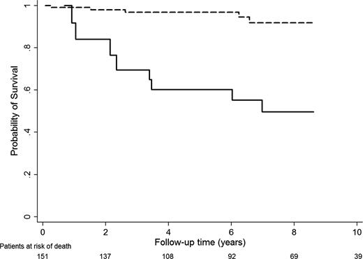
In a landmark analysis, patients with TE at 3 years of follow-up had a 5- and 10-year survival of 84.0% (95% CI, 49.9-95.7) and 49.7% (95% CI, 24.5-70.6), which was worse compared with patients without newly developed TE with survival rates of 98.0% (95% CI, 92.0-99.5) and 91.7% (95% CI, 79.7-96.7), respectively. Data are shown in Figure 1.
Survival according to thrombosis status (landmark set at 3 years after baseline). Survival rates are shown for patients free of a TE at 3 years of follow-up (dashed line) and patients with TE at 3 years of follow-up (solid line).
Survival according to thrombosis status (landmark set at 3 years after baseline). Survival rates are shown for patients free of a TE at 3 years of follow-up (dashed line) and patients with TE at 3 years of follow-up (solid line).
Results of univariable and multivariable analysis of predictors of survival are listed in Table 4. In univariable analysis of time to death, the occurrence of a TE event during follow-up, advanced age, hypertension, and diabetes at study entry were significant predictors of an adverse survival prognosis. New onset of arterial or venous TE remained a strong adverse prognostic factor even after multivariable adjustment for age and hypertension (hazard ratio [HR] = 5.95). The separate analysis of arterial and venous thrombosis identified the occurrence of arterial events as the strongest predictor of adverse survival prognosis in our cohort (HR = 10.6). Even after multivariable adjustment for age and hypertension, this impact on survival prevailed (HR = 5.7). To exclude a disproportionate influence of the 2 fatal MIs on the regression coefficients, the analysis was also performed after exclusion of these events (data not depicted in Table 4). New onset of arterial thrombosis was still a strong predictor of survival (HR = 7.3; 95% CI, 2.5-21.2; P < .001), which also remained significant after adjustment for age and hypertension (HR = 3.9; 95% CI,1.3-11.1; P = .01). The occurrence of newly developed venous events did not emerge as significant prognostic factor for survival, although the HR was 2.8 in multivariable adjustment for age and hypertension. We did not observe an effect of either a prior history of thrombosis or pregnancy complications, or oral anticoagulant therapy at study inclusion, or the diagnosis of concomitant autoimmune rheumatic disease on mortality. Positivity for anti-β2GPI IgG/IgM or aCL IgG/IgM antibodies or triple antibody positivity was not associated with survival after adjustment for age, occurrence of thrombosis, and hypertension. Diabetes at baseline and elevation of anti-β2GPI IgG were univariably but not multivariably associated with overall survival, which is likely due to positive confounding by age and hypertension. Importantly, patients with elevated anti-β2GPI IgG were younger (Student t test, P < .001) and had a lower presence of hypertension (χ2, P = .05) at baseline than patients with anti-β2GPI IgG below the cutoff. Conversely, patients with diabetes were older (Student t test, P < .001) and more likely to have hypertension (χ2, P < .001).
Survival of LA-positive patients in comparison with a matched Austrian population
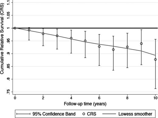
The survival of our cohort of LA-positive individuals was compared with that of an age-, sex-, and study-inclusion-year–matched Austrian population within a relative survival analysis framework. The cumulative Ederer II relative survival (CRS) indicated a consistently worse survival of our study cohort in comparison with the matched Austrian reference population (Figure 2). The CRSs after 1, 5, and 10 years of follow-up were 99.1% (95% CI, 95.3-100.3), 95.0% (95% CI, 88.5-98.8), and 87.7% (95% CI, 76.3-95.6), respectively. The upper confidence band of the 95% CI below 1 indicates a deterioration in survival of our cohort in comparison with the reference population.
Cumulative relative survival over time. The CRS is shown for the cohort of LA-positive individuals in comparison with an age-, sex-, and study-inclusion-year–matched Austrian reference population.
Cumulative relative survival over time. The CRS is shown for the cohort of LA-positive individuals in comparison with an age-, sex-, and study-inclusion-year–matched Austrian reference population.
Discussion
In our cohort of patients with persistent LA followed prospectively within the Vienna LATS, we could demonstrate that the occurrence of thromboembolic events in these patients is associated with an approximately sixfold increased risk of death. The association between occurrence of thrombosis and worse survival was independent of age, hypertension, sex, a positive history of thrombosis, anticoagulation at inclusion, concomitant autoimmune disease, and positivity for antibodies against cardiolipin (aCL) and β2GPI. Moreover, LA-positive patients had an increased mortality in comparison with a matched Austrian population.
Data from prospectively observed cohorts of patients with aPLAs are scarce and the patients that are included are heterogeneous. The largest group of patients published so far is from the Euro-Phospholipid Project.20 In that multicenter prospective cohort study, the authors collected data of 1000 patients and followed them for 10 years; 419 were lost to follow-up during the observation period. In contrast to our study, they also included patients with antibodies against cardiolipin or β2GPI only. Similar to our data, in the Euro-Phospholipid Project death was independent of anticoagulation, age, and concomitant autoimmune disease, which is somewhat unexpected, as one would surmise that anticoagulation would protect patients from experiencing fatal thrombotic events. Thromboembolic events definitely accounted for 20.0% of deaths in our population and 36.5% in the Euro-Phospholipid Project. Most probably the thrombotic tendency is heterogeneous in patients with APS. In most APS patients who have experienced thrombosis, long-term anticoagulant therapy is initiated; however, this does not eliminate the risk of death, especially not in those experiencing new thrombotic events. It may well be that closely monitored anticoagulant treatment might prevent a new onset of TE and therefore counteract an even higher increase in mortality. The fact that mortality was not associated with the history of thrombosis but with new occurrence of thrombosis during the observation period in our LA-positive patients suggests that, on the one hand, anticoagulation appears effective, but that on the other hand, history of thromboembolism or other markers (such as positivity for other aPLAs) might not precisely identify patients at the highest risk of death. Death was also independent of concomitant ARD in our study as well as in the Euro-Phospholipid cohort.20 To compensate for a possibly deteriorating effect, patients with ARD could undergo more frequent controls at specialized clinics; this measure might help to prevent increased ARD-related mortality.
In particular, arterial thromboses, such as stroke and TIA, myocardial infarction, and peripheral arterial thrombosis worsened survival in our cohort of LA-positive patients with an up to 10-fold increased risk of death. Interestingly, the rates of nonthrombosis-related death from hemorrhage (25%) or sepsis (15%) were surprisingly high in our cohort and in line with those reported by Cervera et al.20 In that cohort, 36.5% died of TE and a high rate of fatal infections (26.9%) was described. Thus, the reported increase in mortality cannot be solely interpreted as a consequence of fatal thrombotic events or the TE-associated deterioration of organ systems. Rather, our data indicate that more severe courses of disease might be associated with the occurrence of fatal or nonfatal arterial thrombosis during the observation period.
New onset of venous TE during the observation period was associated with a threefold increased risk of death. In studies in patients without APS and with primary manifestation of DVT, mortality after the acute phase was mainly increased due to cancer-associated deaths21,22 and survival was not impaired, if thrombotic manifestations triggered by cancer or aPLA positivity were excluded.23-25 This has also been confirmed in patients with hereditary thrombophilia with and without thrombosis. In a follow-up study of the European Prospective Cohort of Thrombophilia (EPCOT), no increased mortality in comparison with a control population was observed, not even in persons with thrombotic manifestations.23 As clearly shown by our data and others,20 patients with APS differ from these observations and have a considerably increased risk of long-term mortality.
The probability of survival in our cohort was 92% after 5 years and 80% after 10 years and was inferior to that reported by Cervera et al (94% after 5 years and 91% after 10 years).20 This difference might be due to the inclusion of LA-positive patients only, as LA is thought to carry the highest risk for thrombosis among all aPLAs.26,27 Furthermore, we also included patients without previous thrombotic manifestations and thus also without anticoagulant treatment. To compare the risk of death between LA-positive patients and the general population (ie, relative survival analysis), we matched our cohort of LA-positive individuals by age, sex, and date of diagnosis to the survival experience of an Austrian reference population. Interestingly, relative survival of our LA-positive patient cohort was significantly worse than the survival rate of the Austrian reference population. Given that the subgroup of LA-positive patients without newly developed TE had a relative favorable 10-year survival of 91.7%, this deterioration in relative survival appears to be attributable to the subgroup of LA patients who developed TE during follow-up. Again, these results support the hypothesis that LA-positive patients with TE may represent a specific subgroup of LA patients with a very high risk of death.
Seventy-seven percent of patients were receiving antithrombotic therapy at the time of the arterial or venous event. This leads to the conclusion that anticoagulant treatment might not sufficiently prevent thrombosis in LA-positive individuals.28-30 Treatment strategies for primary and secondary thromboprophylaxis in aPLA-positive patients and patients with APS are still a matter of debate.30-32 According to our data, thromboprophylaxis in LA-positive individuals should be carefully evaluated to prevent thrombosis and increased mortality.
With our observational design, we hardly can comment on the efficacy of anticoagulation to prevent recurrent thrombosis and death. Data that investigate the recurrence of venous TE in patients with APS in interventional studies are scarce and do not allow firm conclusions with regard to the need for long-term anticoagulation.33-35 In a systematic review, the risk ratio for recurrence in LA-positive patients was 2.82; however, this was not statistically significantly different in comparison with those without (95% CI, 0.83-9.64), and important methodologic limitations in most of the studies were noted.36 Two trials compared high dose vs conventional intensity of treatment with VKA and did not find a difference of recurrence rates.37,38 Data from interventional studies are urgently required to evaluate other anticoagulant regimens for the prevention of thrombotic events in this particular patient group.39 The choice of the anticoagulant needs to be carefully evaluated, as the occurrence of thrombosis and the associated deterioration in the survival prognosis do not seem to be sufficiently prevented by common treatment strategies. Clinical studies on the use of direct oral anticoagulants and/or immunosuppressive or immunomodulatory therapeutics are currently conducted.
Our study has several limitations, an important one being the relatively small number of patients. However, adult patients with persisting LA are rare and our observation period was long. With a median observation period of >8 years and 151 patients, we covered in total >1000 patient-years at risk of death. The inclusion of patients with LA only, but not of those with only aCL or anti-β2GPI antibodies, could also be interpreted as a limitation. However, LA is the strongest risk factor for thromboembolism26 and the diagnosis of the presence of LA follows strict guidelines. For other aPLAs, specifically for anti-β2GPI antibodies the cutoff for diagnosis of APS is an elevation above normal. There could be a bias in other studies, as still various assays lead to different results14,40 and false-positive results cannot be excluded. We are convinced that the single-center approach is a strength of our study, as only 11% of our LA-positive patients were not followed until the defined end of observation.
We conclude from our data that patients with LA are at an increased risk of death, especially those with new onset or recurrence of arterial thrombotic manifestations. Whether this risk can be decreased by a more rigid regime of anticoagulation or anticoagulants other than VKAs still remains to be investigated.
There is an Inside Blood Commentary on this article in this issue.
The publication costs of this article were defrayed in part by page charge payment. Therefore, and solely to indicate this fact, this article is hereby marked “advertisement” in accordance with 18 USC section 1734.
Acknowledgments
The authors thank Stefan Blueml (Department of Medicine III, Division for Rheumatology, Medical University of Vienna) for critical reevaluation of diagnoses of concomitant autoimmune rheumatic diseases and Tanja Altreiter (Department of Medicine I, Clinical Division of Hematology and Hemostaseology, Medical University of Vienna) for proofreading the manuscript.
J.G. has received financial support through the Bayer Hemophilia Clinical Training Award (http://www.bayer-hemophilia-awards.com). F.P. is supported by a studentship of the Austrian Science Fund (FWF-SFB54 “Inflammation & Thrombosis”).
Authorship
Contribution: C.Z., C.A., and I.P. designed the study; J.G., C.Z., C.A., I.P., and S.K. contributed patients; J.G., I.P., and S.K. collected and interpreted the data. F.P. analyzed the data; S.K., P.Q., and T.P. performed laboratory analyses; J.G., F.P., and I.P. wrote the manuscript; and all authors agreed with the manuscript’s content and approved its submission.
Conflict-of-interest disclosure: The authors declare no competing financial interests.
Correspondence: Ingrid Pabinger, Department of Medicine I, Clinical Division of Hematology and Hemostaseology, Medical University of Vienna, Waehringer Guertel 18-20, 1090 Vienna, Austria; e-mail: ingrid.pabinger@meduniwien.ac.at.


