Abstract

Introduction
While surveillance imaging does not appear to improve outcomes for patients with curable, aggressive non-Hodgkin lymphoma (NHL) achieving a complete remission (CR) after front-line induction therapy, there are limited data to guide the approach to surveillance in patients with mantle cell lymphoma (MCL). Despite the propensity for all patients to relapse after current induction therapies, the median progression-free survival (PFS) now exceeds 6 years in some series, suggesting that routine surveillance may not be of use early in the disease course. We evaluated the pattern of surveillance imaging at Emory University and described outcomes for patients who relapse after induction therapy based on method of relapse detection.
Methods
All patients with MCL evaluated at Emory with documentation of surveillance imaging post-induction were included. We evaluated each patient with regards to treatment response, presence of relapse, and method of relapse diagnosis in addition to baseline demographic, clinical, and treatment-related variables of interest. We then identified a subset of these patients who received their surveillance imaging in part or whole at Emory. We evaluated each Emory computed tomography (CT) or positron emission tomography (PET/CT) scan obtained and whether the scan was negative for relapse, suggestive of relapse, or equivocal. We also collected biopsy and further imaging data to identify which scans led to a final diagnosis of relapse. The cohort of patients was evaluated with descriptive statistics, and overall survival (OS) was analyzed both from time of original diagnosis and from time of relapse diagnosis using a Cox proportional hazard model. The cohort of surveillance images was evaluated using descriptive statistics regarding specificity and total burden of testing.
Results
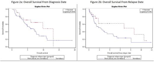
Of 136 included patients, 79 (58%) had a documented relapse during the observation period. Relapsed patients had a median surveillance period of 936 days from initial treatment date to relapse date, while patients with no documented relapse had a median follow-up of 1127 days from initial treatment to end of observation. Of the 79 patients with documented relapse, 23 (29%) were identified by routine surveillance imaging or other testing without clinical signs or symptoms, 44 (56%) were identified by clinical findings, and 12 (15%) had no documentation of method of relapse detection (Figure 1). Comparing patients diagnosed by routine surveillance versus clinical findings, there was no significant differences in OS from date of original diagnosis (HR = 0.65, p = 0.236) or from date of relapse (HR = 0.56, p = 0.121). (See Figures 2a and 2b).
For 95/136 patients identified as having part or all surveillance imaging performed at Emory, a total of 553 scans performed in the surveillance period were identified. Of these 553 scans, 349 (63%) were CT scans, 197 (36%) were PET/CT scans, 6 (1%) were MRI scans, and 1 (<1%) was an ultrasound. 51 (9%) scans were obtained as a result of a new clinically significant sign or symptom during the surveillance period, and 502 (91%) scans were obtained for surveillance only. Among the 502 surveillance scans, 44 (9%) were concerning for relapse, 451 (90%) were negative for relapse, and 5 (1%) were non-diagnostic. Among the 44 scans performed for surveillance concerning for relapse, 11 (25%) ultimately led to a confirmed relapse diagnosis. As a result, the positive predictive value of a positive surveillance scan is 25%, and the number of surveillance scans needed to detect one relapse is 46 scans.
Conclusions
Among 136 patients with some form of surveillance, 79 (58%) had a documented relapse, and 23 of these relapses (29%) were identified through surveillance testing. Those who had relapse diagnosed by surveillance had no significant difference in OS from time of diagnosis or time of relapse compared to patients identified by clinical indication, although a larger study may further delineate the impact of method of relapse detection. Among 502 surveillance scans performed on a subset of 95 of these patients at Emory, 11 (2%) resulted in confirmed relapse. Surveillance imaging in MCL should be utilized judiciously after discussions with patients and after consideration underlying risk factors. Future study of the role of surveillance imaging in MCL is needed.
Flowers:AbbVie: Research Funding; Pharmacyclics, LLC, an AbbVie Company: Research Funding; Roche: Consultancy, Research Funding; Acerta: Research Funding; TG Therapeutics: Research Funding; Millenium/Takeda: Research Funding; ECOG: Research Funding; NIH: Research Funding; Infinity: Research Funding; Mayo Clinic: Research Funding; Genentech: Consultancy, Research Funding; Gilead: Consultancy, Research Funding. Cohen:Pharmacyclics: Consultancy, Membership on an entity's Board of Directors or advisory committees; Celgene: Consultancy, Membership on an entity's Board of Directors or advisory committees; Novartis: Consultancy, Membership on an entity's Board of Directors or advisory committees, Research Funding; Millennium/Takeda: Consultancy, Membership on an entity's Board of Directors or advisory committees, Research Funding; Infinity: Consultancy, Membership on an entity's Board of Directors or advisory committees; Bristol-Myers Squibb: Research Funding; Seattle Genetics: Consultancy, Membership on an entity's Board of Directors or advisory committees, Research Funding.
Author notes
Asterisk with author names denotes non-ASH members.

This icon denotes a clinically relevant abstract


