Abstract

Iron, an essential micronutrient, plays an important role in cellular functions. To prevent deficiency, iron supplementation is universally recommended for preterm infants; nevertheless, assessment of newborn iron stores is not currently recommended. Both iron deficiency and iron excess early in life can have adverse effects on neurodevelopment and outcomes, and therefore sensitive and specific methods for evaluating iron status and determining optimal iron supplementation are essential. The current study aimed to evaluate iron status and iron-supplementation efficacy/toxicity in preterm infants using new laboratory methods, and to correlate iron status with clinical, nutritional, laboratory and therapeutic factors, and the amount of blood extracted and transfused.
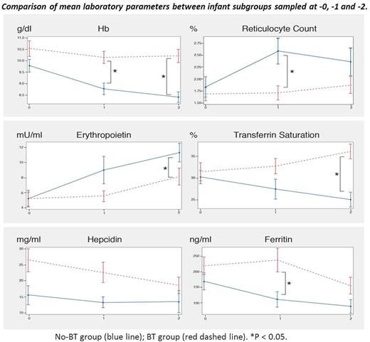
We evaluated 50 very low birth weight (VLBW) preterm infants treated under standard protocols at the Emek Medical Center NICU, 26 (54%) male. Iron supplementation was administered enterally from 4 wk of age (4 mg elemental iron/kg body weight daily). Laboratory studies included CBC, reticulocyte count, reticulocyte hemoglobin (Hb) content, iron, transferrin, transferrin saturation, ferritin, erythropoietin, hepcidin, CRP and non-transferrin-bound iron (NTBI) (Aferrix, Israel). Samples were obtained at 3 different times during hospitalization: (-0) before starting oral iron supplementation, (-1) on d 4-7 of supplementation, and (-2) after at least 2 wk of supplementation. To better understand iron metabolism, the analyzed preterm population was divided into subgroups for comparison: infants who did not require blood transfusion (No-BT) vs. those who received one or more transfusions (BT); infants who did not show abnormal NTBI levels (≤0.19 μmol/L; No-NTBI) vs. those with abnormal NTBI levels (≥ 0.2 μmol/L in one or more samples; NTBI).
Mean birth weight of the studied infants was 1264.5 ± 342 g, gestational age 29.1 ± 2.5 wk, age at hospital discharge 57.6 ± 20 d and weight at discharge, 2412 ± 421 g. The BT group included 35 (70%) infants. The mean birth weight was lower in the BT vs. No-BT group (1199 ± 351 g vs. 1416±273 g, P = 0.04). Mean red blood cell, Hb, and hematocrit were higher in the BT vs. No-BT group. Conversely, mean platelet count, reticulocyte count, and erythropoietin were higher in the No-BT vs. BT group, suggesting an erythropoietic response to lower Hb levels and utilization of iron through effective erythropoiesis. In the BT group, mean serum iron, ferritin, transferrin saturation and hepcidin were higher, whereas transferrin was lower than in the No-BT group. This reflects increased iron burden induced by the transfusions, making NTBI more likely to develop with potential toxicity. Regarding NTBI, 34 (68%) of the infants were in the No-NTBI group, and 16 (32%) showed increased NTBI levels. The Δiron (sum of iron from diet, supplementation and transfusion minus iron output via blood extractions) before starting supplementation was significantly higher in the NTBI vs. No-NTBI group. Mean transferrin saturation at -0 was higher, whereas transferrin at -2 was lower in the NTBI vs. No-NTBI group, demonstrating a higher iron burden in NTBI infants. The odds of developing NTBI rose 7.2% for each percent increment in transferrin saturation at -0 (O.R. 1.072, 95% CI, 1.005-1.143, P = 0.036), and a cutoff of 26% at -0 gave 80% sensitivity and 41% specificity for the presence of NTBI (AUC 0.68).
Thus VLBW infants show evidence of regulatory mechanisms for iron homeostasis and iron utilization for erythropoiesis; however, the protective responses are not developed enough to limit the appearance of circulating free iron with potential toxic effects. Neither adverse effects nor presence of NTBI were associated with enteral iron supplementation. Premature infants who received blood transfusions had a positive iron balance, higher Hb and ferritin levels, and reduced erythropoiesis. Consequently, these babies are more likely to develop free iron. Conventional biomarkers are effective for evaluating iron status in VLBW infants. Transferrin saturation seems to be the most reliable and "physiological" biomarker, and a potential predictor of abnormal NTBI levels. These findings support an individualized approach to iron supplementation based on the characteristics of the specific preterm infant while taking into consideration past need for blood transfusion and the infant's iron status.
No relevant conflicts of interest to declare.
Author notes
Asterisk with author names denotes non-ASH members.

This icon denotes a clinically relevant abstract


