Abstract
Background: Diabetic effects on blood rheology are important topics and have been extensively investigated. In particular, it has been known that diabetic RBCs exhibit significantly deteriorated deformability compared to healthy RBCs. Despite the importance of previously measured mechanical properties of diabetic RBCs, several problems remain to be elucidated. First, conventional techniques have difficulties in measuring intrinsic properties of individual cells. Detailed quantification of diabetic alteration in RBCs at the individual cell level is also hard to achieve. Accordingly, previous approaches do not allow the systematical correlative analyses to be performed on simultaneously measured various cell parameters. As a consequence, there are still no general agreements on the integrated effects of diabetic complications or hyperglycemia on individual human RBCs.
Methods: To effectively settle the arisen problems, we quantitatively and non-invasively measured the individual RBCs from diabetic and healthy blood donors to figure out the characteristics of diabetic RBCs by employing the quantitative phase imaging (QPI) technique. From the reconstructed three-dimensional (3-D) refractive index (RI) tomogram and the time-series of 2-D phase map of individual RBCs, the morphological (volume, surface area, and sphericity), biochemical (Hb concentration and Hb content), and mechanical (membrane fluctuation) parameters were retrieved. Systematic correlative analyses among measured six RBC parameters and independently determined HbA1c level, defined as the ratio of HbA1c to total Hb, were also performed. In measuring red cells, only gently sedimented RBCs of discocytes were selected. After excluding erroneously measured RBCs, 40, 40, 40, 38, 39, 40 control RBCs of six healthy donors and 40, 40, 40, 40, 40, 40 diabetic RBCs of six diabetic patients were finally analyzed respectively for the study of diabetic effects on characteristics of individual human RBCs.
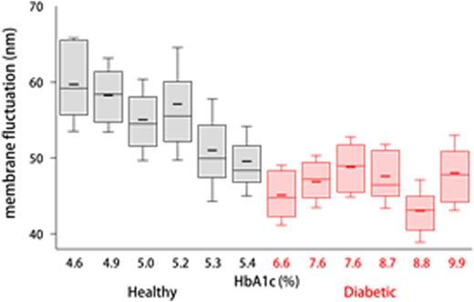
Results: HbA1c level is defined as a percentage of HbA1c, the majority of glycated Hbs in RBC, to total Hb contents in blood. Because HbA1c is formed through 120-days long, non-enzymatic glycation process of Hb, HbA1c level has been perceived as an effective mean to estimate mean glucose concentration of individuals for the last three months. In other words, HbA1c reflects the degree of hyperglycemia. The most noticeable alterations in diabetic RBCs can be found when mean RBC membrane fluctuations of individuals are rearranged in the increasing order of the measured HbA1c levels. The mean values of membrane fluctuations for RBCs are 59.7 ¡¾ 6.2, 58.3 ¡¾ 4.9, 55.0 ¡¾ 5.4, 57.1 ¡¾ 7.4, 51.0 ¡¾ 6.8, and 49.6 ¡¾ 4.6 nm for healthy volunteers, and 45.1 ¡¾ 4.0, 46.9 ¡¾ 3.4, 48.8 ¡¾ 4.0, 47.6 ¡¾ 4.2, 43.0 ¡¾ 4.1, and 48.0 ¡¾ 5.0 nm for diabetic patients, in order of increasing HbA1c. As shown in Fig. 4, mean membrane fluctuation of non-diabetic RBCs tends to decrease as the HbA1c level increases (Pearson correlation coefficient of -0.87 with a p-value of 0.02). Beyond this level of HbA1c, RBCs from diabetic patients exhibit significantly decreased and settled membrane fluctuation. This observed negative correlation between RBC membrane fluctuation and HbA1 is also conceptually in accordance with the report of the previous study.
Conclusions: We quantitatively and non-invasively characterize the morphological, biochemical, and mechanical alterations of individual RBCs by diabetes mellitus. HbA1c levels of subjects, which reflect one's mean blood glucose concentration of last three months, were also determined using hematology analyzers. Accordingly, correlative analyses among the retrieved individual RBC parameters and HbA1c level were performed to search unique features which cannot be addressed by conventional techniques with incapability of simultaneous measurement of those parameters.
Box plot of membrane fluctuation for measured RBCs from six healthy controls and diabetic patients in increasing order of HbA1c. Boxes, median values with upper and lower quartiles. Short horizontal lines and error bars in each box plot denote the mean values and standard deviations, respectively.
Box plot of membrane fluctuation for measured RBCs from six healthy controls and diabetic patients in increasing order of HbA1c. Boxes, median values with upper and lower quartiles. Short horizontal lines and error bars in each box plot denote the mean values and standard deviations, respectively.
No relevant conflicts of interest to declare.
Author notes
Asterisk with author names denotes non-ASH members.


