Abstract
Background: Routine bone marrow biopsy (BMB) for the initial staging of Hodgkin Lymphoma (HL) is not recommended in the era of FDG-PET/CT staging (J Clin Oncol. 2014;32(27):3059). However, patients from the Middle East and North Africa (MENA) region have epidemiologic and clinical features of lymphoma that are different from patients of other ethnicities (J Natl Compr Canc Net2010;8:S-29-S-35). Therefore, it is unknown whetherFDG-PET/CT can substitute for staging BMB in this population.At our center, we perform routine BMB for all newly diagnosed lymphoma cases.
Aim: To investigate whether routine BMB is essential in detecting bone marrow disease where FDG-PET/CT is used in initial staging for HL in patients from the MENA region.
Methods: Patients with HL at our institution between 2010 - 2015 were identified. Inclusion criteria included newly diagnosed patients who had BMB and FDG-PET/CT as part of initial staging. All baseline and laboratory features were retrospectively extracted. Pathology reports of bone marrow aspirate and trephine biopsies were reviewed by two independent Hematologists. All written FDG-PET/CT reports were retrieved and carefully reviewed and cases with positive skeletal uptake were re-interpreted by an experienced radiologist. Pattern of skeletal FDG-PET/CT uptake was determined and classified as unifocal or multifocal. Sensitivity and specificity was computed while defining bone marrow disease by positive BMB and / or focal skeletal uptake on FDG-PET/CT. Categorical and continuous variables were analyzed using Pearson's chi-squared and Student's t-test, respectively.
Results:
A. Baseline characteristics:
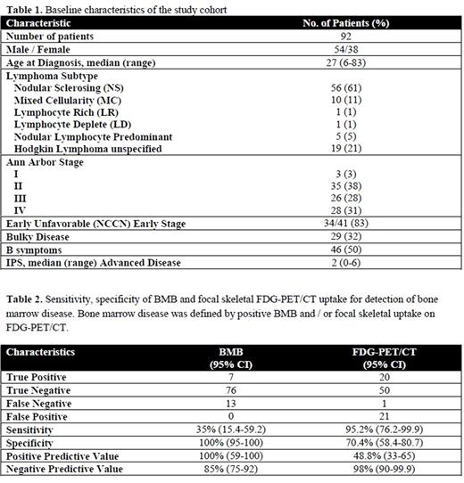
A total of92 patients met the inclusion criteria and were considered for this analysis. All patients were from the MENA region and > 90% were from the Arabian peninsula. From this cohort, bone marrow disease was detected in 7 (7.6%) patients using BMB while 20 (21.7%) patients had unifocal or multifocal bone marrow uptake on FDG-PET/CT. An additional 21 patients (23%) had diffuse homogenous FDG uptake and was not considered to represent HL. The cohort was characterized by a male to female ratio of 1.4 and a median age at diagnosis of 27 years (6-83). About two thirds of the cases were classical HL of the nodular sclerosis subtype (Table 1). Almost 60% of cases were stage III - IV with corresponding median IPS of 2 (0-6). Incidence of bulky disease and B-symptoms among the entire cohort was 32% and 50%, respectively.
B. Comparison of FDG-PET/CT and BMB
No patient with involved BMB (iBMB) had early stage disease on FDG-PET/CT and BMB identified only one patient with positive BM involvement yet negative skeletal uptake on FED-PET/CT. Involvement by BMB upstaged 3 patients previously assessed by CT scan as having stage III, however, none of the patients were allocated to a different treatment plan based on the BMB result. On the other hand, FDG-PET/CT upstaged 24 patients (26%); 9 patients from stage III to IV and 14 patients from early to advanced stage resulting in change of therapeutic plan in the latter group. Focal skeletal FDG-PET/CT lesions identified positive marrow disease with a sensitivity and specificity of 95.2% and 70.4%, respectively. On the other hand, sensitivity and specificity of BMB was 35% and 100%, respectively (Table 2).
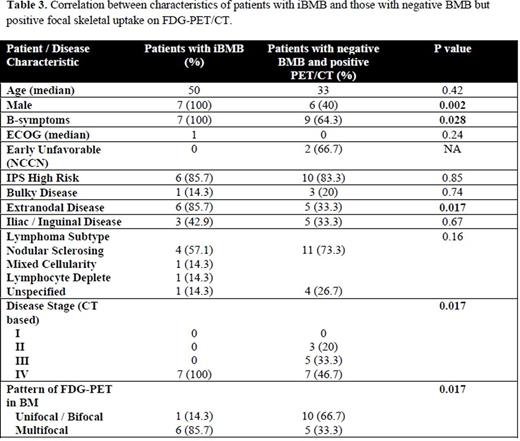
Abnormal skeletal FDG uptake was seen in a total of 20 patients (21.7%); 11 (55%) had unifocal / bifocal while 9 (45%) had multifocal disease of the axial skeleton. Patients with iBMB compared to those with negative BMB but positive unifocal / multifocal skeletal FDG-PET/CT lesions were more likely to be male (p = 0.002), have B-symptoms (p = 0.028), extranodal disease (p = 0.017) and more likely to have multifocal uptake on FDG-PET/CT (0.017) (Table 3).
Conclusion:To our knowledge, this is the first analysis to examine the role of FDG-PET/CT for detection of bone marrow involvement in HL in a patient cohort from the MENA region. We observed that FDG-PET/CT had a higher sensitivity and negative predictive value compared to BMB leading to a treatment change in a significant proportion of patients. This analysis highlightsthat FDG-PET/CT can substitute for BMB in routine staging for newly diagnosed patients with HL from the MENA region.
No relevant conflicts of interest to declare.
Author notes
Asterisk with author names denotes non-ASH members.


